Drugs are the major cause of liver failure that increasing every year (Lakshmi et al., 2018). Hepato-renal dysfunction is considered one of the major inevitable drugs complications (Cepa et al., 2018). Paracetamol is one of the most popular pain killer (analgesic) that used as an alternative for NSAIDs, which is commonly used without a prescription especially in children (Soha, 2017). It’s usually considered safe when used in its therapeutic dose, but overdose is justly common and frequently associated with hepatic and renal injury in both humans and experimental animals (Samuel et al., 2015). The present study attempted to explore the hepatoprotective activity of cardamom (Elettaria cardamomum) compared to silymarin against PCM-induced hepato-renal toxicity in adult male albino rats via the assessment of hematological, serum biochemical and histopathological changes associated with the administration of PCM and/or the herbal extracts to albino rats.
The main constituents of the cardamom oil were α-terpinenyl acetate (47.40%), 1,8-cineole (29.2%), α-terpineol (4.62%) linalyl acetate (2.09%) and D-limonene (1.66%) in cardamom. The results of GC-MS analysis indicated that chemical composition profiles obtained for cardamom oil were very similar to the previous results of different researchers with minor differences (Savan and Kucukbay, 2013). These five constituents of the essential oil possessed variable antioxidant activity when tested in the DPPH assay for non-specific hydrogen atom or electron donating activity (Mutlu-Ingok and Karbancioglu-Guler, 2017). Our data of present study strongly suggest that oral administration of cardamom essential oil might protect the liver against toxicity induced by PCM (500 mg/kg, p.o.) in male Sprague Dawley rats. The hepatotoxicity induced by PCM manifested biochemically by significant elevation of serum transaminases enzyme activities, urea and creatinine concentrations and decrease in TAC level.
Regarding the hemogram, PCM toxicity revealed normocytic normochromic anemia during the 1st week which may be related to destruction of mature RBC, reduction in the rate of erythropoiesis or inhibition of erythropoietin released from the kidney (Samuel et al., 2015).
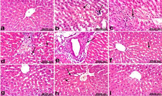
Serum biochemical results illustrated that the daily dosage of PCM (500 mg/kg) for two weeks caused elevations of serum liver enzymes (ALT & AST) and significant decrease in total protein, albumin in comparison to control group. These results are in agreement with the previous experiments (Fatemi et al., 2010; Dadkhah et al., 2015). Damaged structural integrity of the liver by PCM leads to release of cytoplasmic enzymes or to changes in hepatocyte membrane permeability and/or increased synthesis or decreased catabolism of aminotransferases (Lebda et al., 2013). Our results are confirmed by the histopathological findings which revealed remarkable hepatocellular vacuolization, sinusoidal leukocytosis and hepatocellular necrosis associated with inflammatory cells infiltration. Moreover, Kupffer cells activation, apoptosis of hepatocytes (Fig. 1d) and portal fibrosis were also recorded in all examined sections of liver in PCM treated rats. These alterations may be due to formation of large quantity of NAPQI, the PCM metabolites that promotes histopathological alteration of hepatocytes. These results were consistent with that reported by Hinson et al. (2010); Hamza and Al-Harbi, (2015).
Concerning protein profile our statistical analysis revealed significant decrease in both total protein and albumin in PCM treated rats, this is agreed with Lebda et al. (2013) and Hamza and Al-Harbi, (2015). They ascribed this to the decline in the number of cells responsible for albumin synthesis in the liver through necrosis. On the other hand, significant hyperglycemia was noticed in rats treated with acetaminophen. This is may be related to hepatotoxicity, or due to direct effect of PCM toxic metabolites on pancreatic beta cell ultrastructure or due to body response to stress enzymes (MacFie et al., 2009). Regarding TAC results PCM overdose leads to decreased TAC which may resulted from generation for more NAPQI, which is consequently adducted with mitochondrial proteins, leading to ROS formation and oxidant stress (Du et al., 2016; Ahmed et al., 2019). However, treatment with cardamom and silymarin improved TAC capacity which may be due to the ameliorating effect of both silymarin and cardamom as both have the antioxidant effect (Madrigal-Santillán et al., 2014; Aboubakr and Abdelazem, 2016; Ahmed et al., 2019).
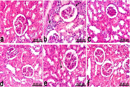
Rats treated with PCM showed significant increase in serum urea and creatinine comparable to control group and this is supported by histopathological picture of the kidneys that showed vacuolar degeneration of renal tubular epithelium, slight atrophy of glomerular tufts associated with distension of Bowman’s space, thickening of the parietal layer of Bowman’s capsule and focal inflammatory cells infiltration.
This may be due to nephrotoxicity that results from formation of reactive oxygen species and increased oxidative stress due to depletion of GSH levels (Canayakin et al., 2016). On the other hand, the administration of cardamom ameliorated PCM induced acute liver and kidney injuries in rats, as evidenced by histologic, hematologic and biochemical findings. Similar protective effects were also observed in rats receiving silymarin. The protective effects of cardamom and silymarin in the restoration of normal liver and kidney functions is confirmed by histopathological findings which revealed that the examined liver sections of rats co-treated with PCM + silymarin, showed no histopathological changes except Kupffer cells activation was noticed in some sections. Regression of the histopathological lesions was noticed in liver of rats co-treated with PCM + cardamom, mild changes were observed as slight activation of Kupffer cells and binucleation of hepatocytes. These effects could be attributed to protection of membrane integrity which may be due to the antioxidant potential of cardamom and silymarin (El-Segaey et al., 2007, Khare et al., 2012, Aboubakr and Abdelazem, 2016; Ahmed, et al. 2019). Moreover, kidneys of rats co-treated with PCM + silymarin showed restored histological structure and apparent normal renal tissue. On the other hand, improved histopathological picture was noticed in kidneys of rats co-treated with PCM + cardamom. Slight vacuolization of epithelial lining some renal tubules was the only histopathological finding observed in sections from this group. These effects could be attributed to improvement on glomerular function of kidney and maintaining positive nitrogen balance in addition to its antioxidant and free radical scavenging properties (Bektur et al., 2013; Elkomy et al., 2016).