
The liver is a multifunctional organ that metabolizes all forms of diets, and food ingredients can have beneficial or detrimental effects on liver cells during such a process. As we have already studied the components of chicken feed previously, the purpose of this study was to observe the effects of chicken meat consumption on liver cells of rats. One hundred and twenty female Albino Wistar rats were randomly divided into four groups and separately for standard chow rat, commercial chicken feed, commercial chicken meat and organic chicken meat over a six-week period. Plasma levels of tumor necrosis factor alpha (TNF-α) and alpha-fetoprotein (AFP) were estimated before and after the administration of different meals. After the experiment, the liver samples were weighed and histopathologically evaluated using a knodell score to assess portal hypertension and liver damage. Animals fed with commercial chicken meat and feed for six weeks showed development of inflammation, necrosis, apoptosis and cirrhosis on histopathological examination of the liver, and had raised plasma TNF-α and AFP-levels. This study shows that damage to liver cells is caused by consumption of commercial feed for chickens and their meat which is considered safe for human consumption.
Research Article · Volume 52, Issue 3, 27 February 2020 · Open Access
Effects of Feed Additives on Chicken Growth and Their Residues in Meat Instigating Deleterious Consequences on the Liver Health of Consumers - A Prospective Human Study
· · · · · · · · · ·
Affiliations & Notes ⌄
- 1Department of Biological and Biomedical Sciences, The Agha Khan University, Karachi, Pakistan 2Department of Biochemistry, Baqai Medical University, Karachi, Pakistan 3Neurochemistry and Biochemical Neuropharmacology Research Unit, Department of Biochemistry, University of Karachi, Karachi, Pakistan 4Dr. Panjwani Center for Molecular Medicine and Drug Research, International Center for Chemical and Biological Sciences, University of Karachi, Karachi, Pakistan 5Department of Opthalmology, Liaquat
Article Info ⌄
- Journal:
- Pakistan Journal of Zoology
- Published:
- 27 February 2020

From the article
- This is seen that the rats treated with the chicken feed and commercial chicken meat showed the maximum changes in terms of increase in TNF-α and AFP.
- The liver organ weight is seen to be increased significantly in the group C as compared to that of control group A rats.
- This study is therefore, revealed as an eye opener to the changes that may arise due to the persistent use of poultry fed on additive roxarsone, bringing the permanent damage to the liver cells.
- This study shows that the commercial poultry utilization may be swapped with organic chicken meat as it shows normal outcome of health status upon consumption in the rats in the study.
Selected from the article's own text.
Abstract
Introduction
Chicken meat is today one of the most popular diets because of its better taste and easy availability. However, chickens currently on the market are bred for commercial profit . These chickens can remain ad libitum in cages with excessive feeding. Foods offered consist of cereals that give volume to the food, as well as vitamins, minerals, calcium supplements and premixes. The constituents of the chicken feed has been studied previously by our group and showed that in addition to healthy ingredients, chicken feed also contains artificial sources of arsenic in the form of roxarsone, melamine and pesticides (Ahmad et al., 2017, 2018a,b). The basic utility of these components are to provide artificial nitrogenous back bone to the poultry so that they may generate and produce muscle mass along with the rapid weight gain . This is contrast to the free lander organic chickens. The organic chickens are fed upon whole grains and are allowed to roam freely on vast wilder lands. This helps them to amply digest their food without growing in shorter duration of time and take three months to grow to a consumable size of 1.5 kg, much longer than that of commercial chickens . Hence, the use of feed causing better muscle mass, taste, color and palatability bring commercial chicken meat on the top most choice for the consumers worldwide .
To scrutinize the scenario closely, it may be revealed that the additives for artificial nitrogen source is harmful for health. These sources of arsenic as roxarsone, melamine and pesticides concentrate in the chicken flesh and will be consumed by the humans ingesting affected flesh ; ; . The harmful effects of these deleterious components may be seen on skin, liver and kidneys . Therefore, chickens bred commercially are expectantly found to be the causative of liver inflammation, renal stones and multiple organ failure in human beings ; ; ; . This is the first ever study conducted to determine effects of feed components and their residual concentration in flesh. The presence of particular forms of inorganic arsenic roxarsone, melamine and pesticides in commercial chicken meat are compared with organic meat for their presence in them. This comparison is done to evaluate the health benefits of the chicken meat and the havoc that their presence may play in the body. This study has provided with the indication that the use of roxarsone in rearing of chickens pretenses public health risks. Our previous study showed that the use of roxarsone in chicken farming poses risks to public health (Ahmad et al., 2017, 2018a, b). It is noteworthy that the Food and Drug Administration (FDA), the agency responsible for regulating animal drugs, have also banned arsenicals compounds prior for use in chicken rearing . It is imperative that the use of melamine and pesticides may also be reviewed as they affect organs normal functioning leading to the organ shut down with inevitable and untimely demise of the consumer ; Pirarat et al., 2016).
The present was therefore designed to determine the effects of administration of commercial chicken feed, commercial chicken meat and organic chicken meat on livers of the Wistar rats. The liver size, TNF-α and AFP and knodell scoring were estimated in rats after the treatment for six weeks. These results were then finally compared.
Materials and methods
Animals
The experiment was carried out on 120 female rats of approximately 100 g. Rats were purchased from Dow University of Health Science, OJHA campus, Karachi. Pakistan. Three rats were housed in one cage for 12 h day and night cycle with ambient room temperature of 22±2°C. The experimental protocol was approved by the ethical committee of Baqai Medical University (BMU/BIOCHEM/19) and performed in line with National Institute of Health Guide for Care and Use of Laboratory Animals (Publication No. 85-23, revised 1985).
Experimental protocol
Organic chickens were raised in the animal house of the Baqai Medical University, Karachi, Pakistan. The commercial chickens were purchased from the poultry farm near the highway Karachi that supplies chickens and eggs to the suburbia of Karachi. The feed likewise was also purchased from the local market used for the rearing of the commercial broiler chickens between December 2013 and June 2014. The rats were randomly divided into four groups. These include group A standard rat chow treated control rats, group B commercial chicken feed treated rats, group C commercial chicken meat treated rats and group D organic chicken meat treated rats. All rats were fed for the period of six weeks with provision of the specified diet and water ad-libitum. Standard rat chow contained wheat flour (400 g), gram flour (171 g), barley flour (171 g), corn flour (100 g), vegetable oil (50 g), milk powder (100 g), vitamin mixture (2.5 g), iodized salt (NaCl; 5.5 g). Blood was collected before and after the experimental protocol from rat tails to assess the levels of TNF-α, AFP and alpha-FP. At the end of procedure rats were scarified to collect the liver samples for morphological and histopathological examinations.
TNF-α
The collected blood samples were used for TNF-α level by ELISA using a KHC3011 kit from Invitrogen. Results are expressed as pg/ml of plasma samples collected before and after the experiment.
Liver histopathology
Liver samples were excised out and fixed in 10% formalin for 24 h. Dehydration was carried out with the help of alcohols. The specimens were then embedded in paraffin bees wax tissue blocks to prepare sections of 4 microns. Hematoxylin and eosin dyes were used to stain the sections for histopathological examination using the light microscope. The degree of liver fibrosis was examined using the knodell scoring system . Arbitrary scoring scale from 0-4 (Table I) was used to examine the degree of periseptal interface hepatitis, confluent necrosis, spotty focal inflammation and apoptosis, and portal inflammation. Histopathological analysis was carried out by a pathologist who was blinded to the grouping and treatment.
Alpha-feto protein (AFP) test
The serum samples were also used to assess the presence of AFP by double diffusion method in 1% agarose .
Statistical analysis
Data for liver weight and TNF-α were statistically analyzed by one-way ANOVA followed by Tukey’s post-hoc test. To compare the levels of TNF-α before and after the experiments paired t-test was used. Whereas, nonparametric data were analyzed by Kruskal-Wallis with multiple-comparison post-hoc test using SPSS version 20.0. Data is presented as mean ± SEM. p values <0.05 were considered statistically significant.
Results
TNF-α
Statistical analysis of post-experimental TNF-α levels by one-way ANOVA showed significant effects of different diet treatment for six weeks. Post-hoc test revealed significantly increased levels of TNF-α in group B and C as compared to that of control group A rats (p<0.01). The levels of TNF-α after six week of treatment was also significantly higher than that of pre-treatment levels (p<0.01) in commercial chicken feed and meat treated animals (Fig. 1).
AFP
Plasma AFP was assessed by double diffusion method and the occurrence was recorded as shown in Table II. It was found that six and thirteen animals from group B and C, respectively showed the presence of AFP in plasma after six weeks of treatment.
Table I.- Knodell scoring system.
| Score | Piecemeal necrosis | Confluent necrosis | Spotty inflammation and apoptosis | Portal inflammation |
| 0 | Absent | Absent | Absent | Absent |
| 1 | Mild, few portal areas | Focal confluent necrosis | One focus or less per 10x objective | Mild, some or portal areas |
| 2 | Mild-moderate, most portal areas | Necrosis in some areas | Two to four foci per 10x objective | Moderate, some or all portal areas |
| 3 | Moderate, continues around <50% of tracts | Necrosis in most areas | Five to ten foci per 10x objective | Moderate-marked, all portal areas |
| 4 | Severe, continues around >50% of travel | Multiacinar necrosis | More than ten foci per 10x objective | Marked, all portal areas |
Table II.- Presence of alpha-feto protein using double diffusion method.
| Groups | Pre-treatment | Post-treatment | ||
| (+) | (-) | (+) | (-) | |
| Control | 0 | 25 | 0 | 25 |
| Commercial chicken feed | 0 | 25 | 6 | 19 |
| Commercial chicken meat | 0 | 25 | 13 | 12 |
| Organic chicken meat | 0 | 25 | 0 | 25 |
Liver weight
One-way ANOVA showed significant effects of different diet on liver weight of rats (Fig. 2). Intake of commercial chicken feed and meat significantly increased liver weight as compared to that of group A (p<0.01). Organic chicken meat administration in group D did not affect the liver weight six weeks of treatment.
Knodell scoring
Histopathological knodell scoring data were analyzed by Kruskal-Wallis test showed significant effects of treatment (Fig. 3). Post-hoc test revealed that all observed knodell scoring parameters were significantly increased to drastic values in groups B and C as compared to that of group A (p<0.01). Whereas, group D which was treated with organic chicken meat showed comparable results with that of group A rats (Fig. 3C). Morphological and histopathological changes can be observed in Figure 3A and 3B.
Discussion
As chicken meat is the one of the preferential diet nowadays and its consumption has amplified manifold throughout the world compared to red meat . However, its utilization may have detrimental effects that were never expected before due to the presence of the excess fats, proteins along with other toxicities that come from their commercially manufactured feed and concentrate in the flesh of the chickens. The proteins are used for the synthesis of the muscle and body proteins . However, the fats are consumed for the generation of energy and the excess of fats aids in weight gain and obesity including fat accumulation of fats in the livers of the rats . Other additives added in the chicken feed also concentrate in the chicken meat , 2018b) augmenting inflammatory insult to the liver cells of the rats . Such components include roxarsone, an artificial arsenic compound, pesticides and melamine . The purpose of such additions are to offer nitrogenous compounds to the chickens for the better growth and development as well as keep the diet free of pests upon storage but they do accumulate in the body causing damage to the organs like liver of the rats, which is the prime focus of the present study.
The pre-experiment base line values of TNF-α and AFP values were within normal range. However, after the experiment the values of these were increased drastically. The TNF-α demonstrates the inflammatory changes taking place in the body. The histopathology of the liver demonstrated via the Knodell scoring showed inflammatory changes, portal hypertension and necrosis in the livers of the rats as shown in Table I and Figure 3C. This is seen that the rats treated with the chicken feed and commercial chicken meat showed the maximum changes in terms of increase in TNF-α and AFP. The liver organ weight is seen to be increased significantly in the group C as compared to that of control group A rats. This was aligned by the increase in the liver organ weight of the group B fed by chicken feed. The significant rise in the organ weight of these groups as per the previous studies reflects upon the ingredients added and supplementations resulting in drastically augmenting weight leading to obesity ; ; . Our previous studies have shown the extent to which the commercial chicken feed reaches the score of fulfilling the vigorous weight gain within a short duration of time. In such scenarios the fat accumulates around the abdomen and the visceral organs as well as cause hepato-steatosis increasing the risk of developing diabetes and heart diseases . Besides inflammation fats also damage to surrounding liver tissue leading to cirrhosis, an irreversible condition, where the normal architecture of the liver is replaced by fibrotic tissue leading to a complete failure of the liver functions . Cirrhosis is also associated with an increased risk for developing liver cancer . These factors promote atherosclerotic process damaging the insides of arteries making blood to clot, a combination that can lead to heart attack or stroke .
The increased TNF-α in group B and C after six week of treatment also revealed the fact of rising accumulation of fats in the hepatocytes aids in the inflammation and a reason of augmented inflammatory markers in the plasma. The same may be associated to the injurious effects of the chicken feed and meat comprising of the elements of roxarsone and pesticides. Previously it is documented that roxarsone is present in commercial chicken feed and meat which may bring about deleterious effects on the health of consumers . Arsenic has lethal effects on liver essentially in form of hepatomegaly and non-cirrhotic portal fibrosis in population. Hepatic fibrosis has also been demonstrated in the population due to long term arsenic toxicity in an animal model . The initial biochemical evidence of the liver damage secondary to roxarsone ingestion includes hepatic membrane damage. This is probably due to reduction of glutathione and antioxidant enzymes activity . Continued arsenic feeding may result in long term effects such as the development of fatty liver, elevated serum amino-transferase levels and hepatic fibrosis (Liu et al., 2015a, b). The same was observed in the present study when the rats showed gross and histopathological changes in the liver of affected rats. The alterations in the livers of the rats were more pronounced in the groups B and C rats as compared to groups A and D rats. The periportal or periseptal interface hepatitis also known as piecemeal necrosis was highest in the rats of group C and the overall significant rise in the piecemeal necrosis was maximum in groups B and C as compared to all the other groups in the study. This again estimates the presence of inorganic arsenic roxarsone in the feed and its concentrates in the meat leading to such liver damage. However, such additions inadvertently bring about harmful effects to the livers of the consumers when fed upon such provisions . These effects may manifest in humans consuming commercial chicken meat on a routine basis. The early injury through roxarsone ingestion may get unnoticed but this chemical over a period of time may manifest fatty changes, portal hypertension, necrosis, cirrhosis and even cancer development of the liver cells . The present study also revealed the extensively high areas of confluent necrosis, focal spotty lytic necrosis, apoptosis, focal inflammation and portal inflammation to be at their peak in the livers of the rats of group B and C. These alarming results show the possibility of developing such damaging effects in a short span of just six weeks. This is concluded that the consumption of such poultry that is frequently and freely given such additives is crippling and detrimental for the young consumers . Arsenic is not only documented to play harmful roles in liver but also cause skin, lung and liver cancer. It is also documented that during chronic arsenic exposure, adaptation to the effects of arsenic occurs. Such remodeling includes apoptosis that frequently ensues towards a generalized tolerance for apoptosis in the body (Liu et al., 2015a, b). Apoptotic resistance is a common phenomenon in cells malignantly transformed by arsenic, including rat liver epithelial cells. Tolerance to apoptosis may be a hallmark for arsenic carcinogenesis, as it allows the damaged cells that otherwise would be eradicated, to endure and transmit genetic or epigenetic lesions . Arsenic frequently provokes overexpression of cell proliferation-related genes, such as cyclin D1 and growing cell nuclear antigen. This is commonly seen in arsenic-treated mouse liver, in arsenite-distorted liver cells and in methylated arsenical-stimulated liver preneoplasia (Liu et al., 2015a, b).
To finally support the harmful effects of commercial chicken feed and meat on the body, the presence of alpha feto protein (AFP), an essential marker for the hepatic carcinoma was done . 24% of rats from group B and 52% from group C showed the presence of AFP in blood after six weeks of treatment. This showed the anticipated effects of roxarsone and pesticides on the liver. The histopathology also exposed a fully-fledged development of the liver cancer in some of the rats. The association between environmental arsenic exposure and human liver cancers has been repeatedly reported in previous studies ; . It was observed that the cancer development was highest in the groups C and B. Hence, further acknowledging the possibility of the changes of the liver cells to morphological abnormal cancerous cells. A recent data indicate that fetal exposure to inorganic arsenic in mice produces tumors in a number of organs during adulthood, including the liver . Several mechanisms that possibly concerned in arsenic-induced hepato-carcinogenesis are enlisted. A few of these include oxidative DNA breaks, acquired resistance to apoptosis with better cell proliferation, misrepresented DNA methylation, genomic unsteadiness and abnormal atypical estrogen signaling (Liu et al., 2015a, b).
Conclusion
This study is therefore, revealed as an eye opener to the changes that may arise due to the persistent use of poultry fed on additive roxarsone, bringing the permanent damage to the liver cells. These injuries may lead initially from hepatomegaly and fatty liver to hepatitis and then eventually to the end point as cancer development in the livers. This study may stand alone shows the harmful effects of poultry that is commercially bred on supplemented feeds and its consumption with health implications on the consumers. This study shows that the commercial poultry utilization may be swapped with organic chicken meat as it shows normal outcome of health status upon consumption in the rats in the study.
Acknowledgement
Authors are thankful to Baqai Medical University, Karachi, Pakistan for providing research facilities.
Statement of conflict of interest
None to declare.
Figures
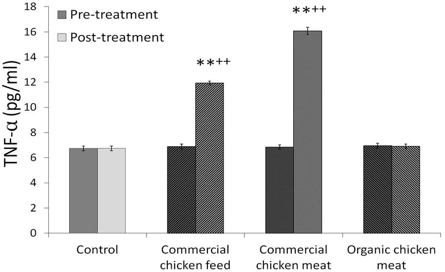
Effects of six weeks administration of commercial chicken feed, meat and organic meat on the levels of THF-alpha. Levels were estimated before and after the experiment. Values are mean ± SEM (n=25). **p<0.05 from respective group A rats; ++p<0.01 from pre-treatment values.
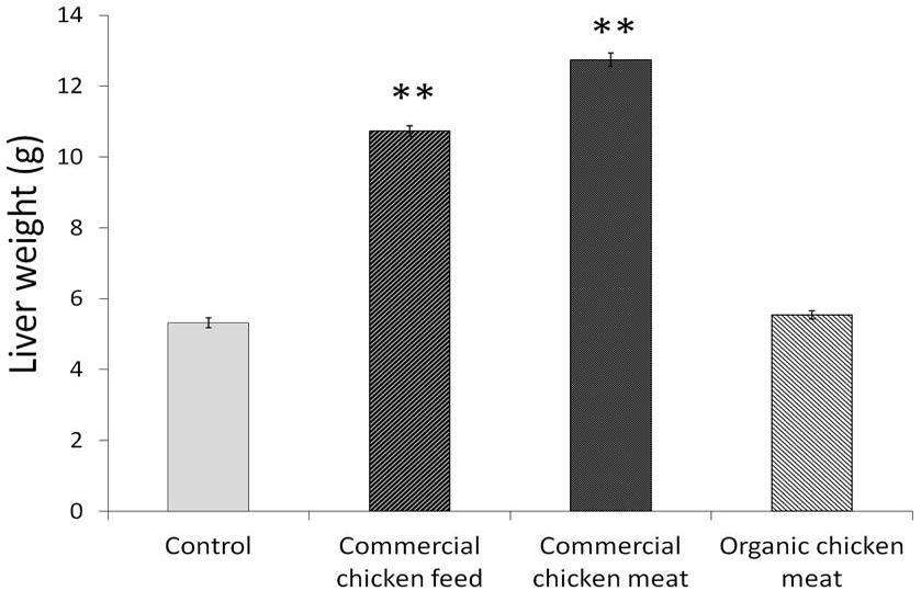
Liver weight of animals following the administration of commercial chicken feed, meat and organic chicken meat. Values are mean ± SEM (n=25). **p<0.05 with respect to group A.
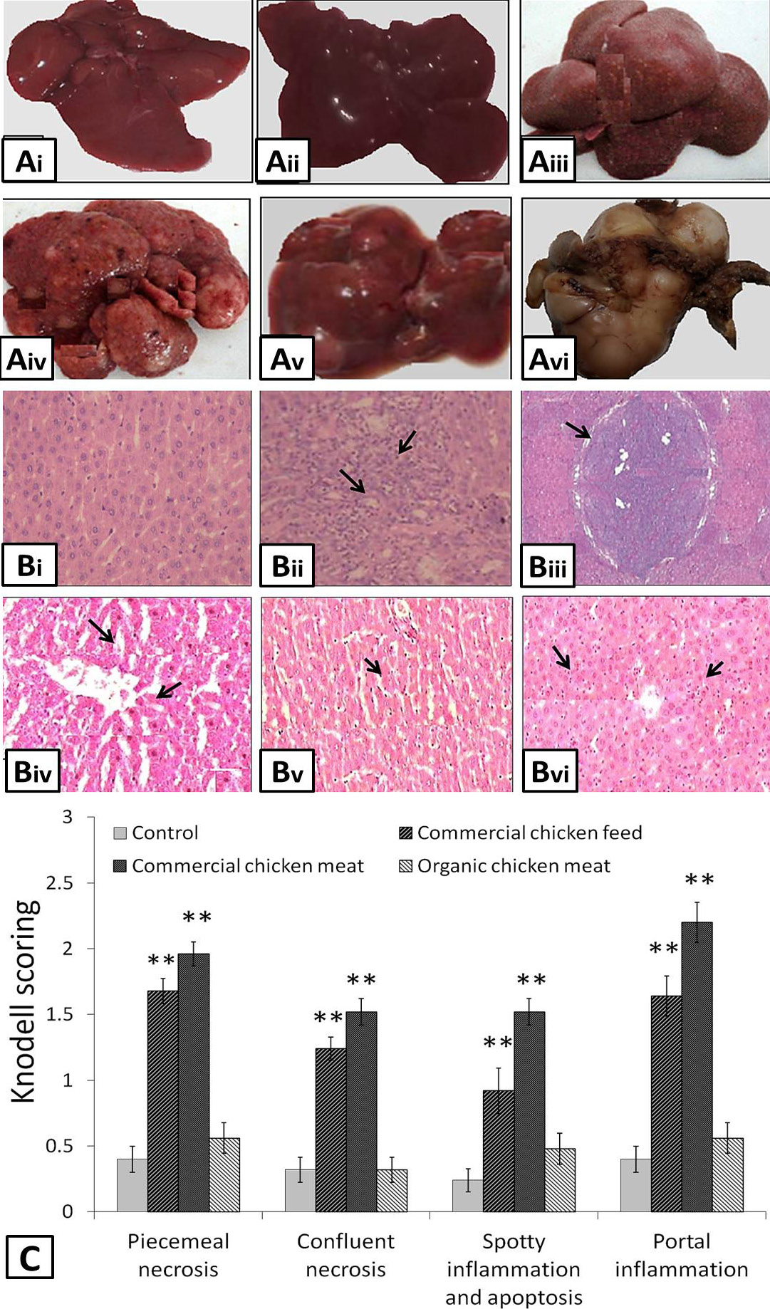
A, morphology of liver observed after the intake of commercial chicken feed, meat and organic meat. (i) and (ii) showed normal morphology of liver isolated from control and organic meat fed animals, (iii) inflamed, (iv) inflamed and nodular, (v) nodular and (vi) cirrhotic liver were seen in animals fed on commercial chicken feed and meat for six weeks. B, histopathological examination of liver samples showing the presence of (i) normal cells in control group A rats and organic chicken meat fed animals, (ii) Inflamed cells, (iii) cirrhotic nodule (iv) portal inflammation (v) confluent necrosis and (vi) spotty inflammation, necrosis with apoptosis were observed in liver cells isolated from the animals fed on commercial chicken feed and meat. C, Knodell scoring was done to assess the portal inflammation, necrosis, apoptosis and cirrhosis in liver samples. Values are mean ± SEM (n=25). **p<0.05 with respect to controls.
References
- Ahmad, S. and Ahmed, I., 2014. Response of Wistar rats to broiler chicken feed and soybean on body weight, obesity and weight of selected visceral organs. Pak. J. Biochem. mol. Biol., 47: 137-140.
- Ahmad, S. and Ahmed, I., 2015. Behavioral deficits in Wistar rats secondary to altered androgen levels after treatment with chicken feed and raw soybean. Int. J. Sci. Eng. Res., 6: 807-811.
- Ahmad, S., Omm-e-Hany, Ahmad, I., Ahmed, S.A., Alamgir, A. and Neelam, A., 2016. Potential effect of chicken boneless meat on the body weight and serum cholesterol levels of the female Albino Wistar rats: In direct human prospective studies. American-Eurasian J. Agric. enviorn. Sci., 16: 466-469.
- Ahmad, S., Ahmed, I., Haider, S., Batool, Z. and Ahmed, S.B., 2017. Daily consumption of commercial chicken feed and meat lead to alterations in serum cholesterol and steroidal sex hormones in female rats. Pak. J. Pharm. Sci., 30: 257-261.
- Ahmad, S., Ahmed, I., Haider, S., Batool, Z., Ahmed, F., Tabassum, S., Madiha, S., Perveen, T. and Ahmed, S.B., 2018a. Effects of consumption of caged and un-caged chicken meat on ovarian health of female Wistar rats. Pakistan J. Zool., 50: 487-493. https://doi.org/10.17582/journal.pjz/2018.50.2.487.493[Full text][CrossRef]
- Ahmad, S., Rehman, R., Haider, S., Batool, Z., Ahmed, F., Ahmed, S.B., Perveen, T., Rafiq, S., Sadir, S. and Shahzad, S., 2018b. Quantitative and qualitative assessment of additives present in broiler chicken feed and meat and their implications for human health. J. Pak. med. Assoc., 68: 876-881.
- Amin, M.M. and Arbid, M.S., 2017. Estimation of ellagic acid and/or repaglinide effects on insulin signaling, oxidative stress, and inflammatory mediators of liver, pancreas, adipose tissue, and brain in insulin resistant/type 2 diabetic rats. Appl. Physiol. Nutr. Metab., 42: 181-192. https://doi.org/10.1139/apnm-2016-0429[CrossRef]
- Balaha, M., Kandeel, S. and Barakat, W., 2016. Carvedilol suppresses circulating and hepatic IL-6 responsible for hepatocarcinogenesis of chronically damaged liver in rats. Toxicol. appl. Pharmacol., 311: 1-11. https://doi.org/10.1016/j.taap.2016.10.012[CrossRef]
- Chastre, A., Bélanger, M., Beauchesne, E., Nguyen, B.N., Desjardins, P. and Butterworth, R.F., 2012. Inflammatory cascades driven by tumor necrosis factor-alpha play a major role in the progression of acute liver failure and its neurological complications. PLoS One, 7: e49670. https://doi.org/10.1371/journal.pone.0049670[CrossRef]
- Choiniere, J. and Wang, L., 2016. Exposure to inorganic arsenic can lead to gut microbe perturbations and hepatocellular carcinoma. Acta Pharm. Sin. B., 6: 426-429. https://doi.org/10.1016/j.apsb.2016.07.011[CrossRef]
- Chovanec, P., Stolz, J.F. and Basu, P., 2010. A proteome investigation of roxarsone degradation by Alkaliphilus oremlandii strain OhILAs. Metallomics, 2: 133-139. https://doi.org/10.1039/B915479E[CrossRef]
- Dong, Y., Liu, Y., Kou, X., Jing, Y., Sun, K., Sheng, D., Yu, G., Yu, D., Zhao, Q., Zhao, X., Li, R., Wu, M. and Wei, L., 2016. The protective or damaging effect of Tumor necrosis factor-α in acute liver injury is concentration-dependent. Cell Biosci., 6: 8. https://doi.org/10.1186/s13578-016-0074-x[CrossRef]
- Fisher, D.J., Yonkos, L.T. and Staver, K.W., 2015. Environmental concerns of roxarsone in broiler poultry feed and litter in Maryland, USA. Environ. Sci. Technol., 49: 1999-2012. https://doi.org/10.1021/es504520w[CrossRef]
- Kroes, R., Sontag, J.M., Sell, S., Williams, G.M. and Weisburger, J.H., 1975. Elevated concentrations of serum alpha-fetoprotein in rats with chemically induced liver tumors. Cancer Res., 35: 1214-1217.
- Liang, T., Ke, Z., Chen, Q., Liu, L. and Chen, G., 2014. Degradation of roxarsone in a silt loam soil and its toxicity assessment. Chemosphere, 112: 128-133. https://doi.org/10.1016/j.chemosphere.2014.03.103[CrossRef]
- Liu, Q., Peng, H., Lu, X. and Le, X.C., 2015a. Enzyme-assisted extraction and liquid chromatography mass spectrometry for the determination of arsenic species in chicken meat. Anal. Chim. Acta, 888: 1-9. https://doi.org/10.1016/j.aca.2015.05.001[CrossRef]
- Liu, S.H., Yang, R.S., Yen, Y.P., Chiu, C.Y., Tsai, K.S. and Lan, K.C., 2015b. Low-concentration arsenic trioxide inhibits skeletal myoblast cell proliferation via a reactive oxygen species-independent pathway. PLoS One, 10: e0137907. https://doi.org/10.1371/journal.pone.0137907[CrossRef]
- Miyauchi, S., Oshima, S., Asaka, M., Kawano, H., Torii, S. and Higuchi, M., 2013. Organ size increases with weight gain in power-trained athletes. Int. J. Sport Nutr. Exerc. Metab., 23: 617-623. https://doi.org/10.1123/ijsnem.23.6.617[CrossRef]
- Nordborg, M., Davis, J., Cederberg, C. and Woodhouse, A., 2017. Freshwater ecotoxicity impacts from pesticide use in animal and vegetable foods produced in Sweden. Sci. Total Environ., 581-582: 448-459. https://doi.org/10.1016/j.scitotenv.2016.12.153[CrossRef]
- Pirarat, N., Katagiri, T., Chansue, N., Ponpornpisit, A., Endo, M. and Maita, M., 2012. The pathological effects of melamine and cyanuric acid in the diet of walking catfish (Clarius batrachus). J. comp. Pathol., 147: 259-266. https://doi.org/10.1016/j.jcpa.2011.12.008[CrossRef]
- Polat, B., Halici, Z., Cadirci, E., Karakus, E., Bayir, Y., Albayrak, A. and Unal, D., 2017. Liver 5-HT7 receptors: A novel regulator target of fibrosis and inflammation-induced chronic liver injury in vivo and in vitro. Int. Immunopharmacol., 43: 227-235. https://doi.org/10.1016/j.intimp.2016.12.023[CrossRef]
- Rizwan-ul-Haq, M., Zhenling, Z., Yongxue, S. and Wenguang, X., 2012. Evaluation of glutathione s-transferase as toxicity indicator for roxarsone and arsanilic acid in Eisenia fetida. J. appl. Toxicol., 32: 731-738. https://doi.org/10.1002/jat.2726[CrossRef]
- Shen, L., Zhang, G., Lou, Z., Xu, G. and Zhang, G., 2017. Cryptotanshinone enhances the effect of Arsenic trioxide in treating liver cancer cell by inducing apoptosis through down regulating phosphorylated- STAT3 in vitro and in vivo. Complement. Altern. Med., 17: 106. https://doi.org/10.1186/s12906-016-1548-4[CrossRef]
- Standish, R.A., Cholongitas, E., Dhillon, A., Burroughs, A.K. and Dhillon, A.P., 2006. An appraisal of the histopathological assessment of liver fibrosis. Gut, 55: 569-578. https://doi.org/10.1136/gut.2005.084475[CrossRef]
- Tolba, A.M. and Salama, A.A.A., 2016. Adverse effects of melamine formaldehyde on the liver, kidney and brain in rats. Der. Pharma. Chem., 8: 398-409.
- Tsang, D.P., Wu, W.K., Kang, W., Lee, Y.Y., Wu, F., Yu, Z., Xiong, L., Chan, A.W., Tong, J.H., Yang, W., Li, M.S., Lau, S.S., Li, X., Lee, S.D., Yang, Y., Lai, P.B., Yu, D.Y., Xu, G., Lo, K.W., Chan, M.T., Wang, H., Lee, T.L., Yu, J., Wong, N., Yip, K.Y., To, K.F. and Cheng, A.S., 2016. Yin Yang 1-mediated epigenetic silencing of tumour-suppressive microRNAs activates nuclear factor-κB in hepatocellular carcinoma. J. Pathol., 238: 651-664. https://doi.org/10.1002/path.4688[CrossRef]
- Uygur, R., Aktas, C., Caglar, V., Uygur, E., Erdogan, H. and Ozen, O.A., 2016. Protective effects of melatonin against arsenic-induced apoptosis and oxidative stress in rat testes. Toxicol. Ind. Hlth., 32: 848-859. https://doi.org/10.1177/0748233713512891[CrossRef]
- Yao, L., Huang, L., He, Z., Zhou, C., Lu, W. and Bai, C., 2016. Delivery of roxarsone via chicken diet chicken manure soil rice plant. Sci. Total Environ., 566-567: 1152-1158. https://doi.org/10.1016/j.scitotenv.2016.05.157[CrossRef]
- Zhang, F.F., Wang, W., Yuan, S.J. and Hu, Z.H., 2014. Biodegradation and speciation of roxarsone in an anaerobic granular sludge system and its impacts. J. Haz. Mater., 279: 562-568. https://doi.org/10.1016/j.jhazmat.2014.07.047[CrossRef]
Services
Article usage statistics
Loading…