
The aim of this study was to investigate the effect of epidermal growth factor (EGF) on ASCT2 expression in IPEC-J2 cells challenged by lipopolysaccharide (LPS). Cells were treated with: (1) EGF (0 ng / mL) + LPS (0 μg / mL) (Control group), (2) EGF (100 ng / mL) + LPS (0 μg / mL) (EGF group), (3) EGF (0 ng / mL) + LPS (1 μg / mL) (LPS group), and (4) EGF (100 ng / mL) + LPS (1 μg / mL) (EGF+LPS group) for 24 h. The gene expression of glutamine transport protein Na+-dependent neutral amino acid transporter (ASCT2) and protein expression of ASCT2 were measured. The results showed that LPS significantly (P<0.05) decreased the gene and protein expression of ASCT2 in IPEC-J2 cells. EGF significantly (P<0.05) promoted the gene and protein expression of ASCT2. EGF plus LPS group had a higher (P < 0.05) gene and protein expression of ASCT2 than that LPS treated group, although significantly (P < 0.05) lower than EGF treated group, but had no difference between the Control group. It can speculate that EGF promoted the absorption of glutamine through activating ASCT2 expression in injured intestinal epithelial cells.
Short Communication · Volume 54, Issue 4, 26 April 2022 · Open Access
Effects of Epidermal Growth Factor on ASCT2 Expression in IPEC-J2 Cells Challenged by Lipopolysaccharide
· ·
Affiliations & Notes ⌄
- 1State Key Laboratory Cultivation for Karst Mountain Ecology Environment of Guizhou Province, School of Karst Science, Guizhou Normal University, Guiyang, China 2Hunan Co-Innovation Center of Animal Production Safety, College of Animal Science and Technology, Hunan Agricultural University, Changsha, China * Corresponding author: tangxiaopeng110@126.com, fangrj63@126.com
Article Info ⌄
- Journal:
- Pakistan Journal of Zoology
- Published:
- 26 April 2022

From the article
- The results of the present study suggest that EGF could promot the gene and protein expression of ASCT2 in IPEC-J2 cells induced by LPS.
- It can be speculated that EGF promot the absorption of Gln in injured intestinal epithelial cells by activating ASCT2 expression.
Selected from the article's own text.
Abstract
Materials and methods
IPEC-J2 were obtained from Institute of Subtropical Agriculture, Chinese Academy of Science (Changsha, China) and cultured in DMEM / F12 medium (GE Healthcare life sciences, South Logan, Utah, USA) containing 10% FBS (GIBCO, Carlsbad, CA, USA), 1% antibiotics (Penicillin-Streptomycin) (GIBCO, Carlsbad, CA, USA), and grown in a humidified incubator at 37°C with 5 % CO2 and 95 % air as previously described . After 80% of fusion, cells were digested with 0.25% trypsin-EDTA (GIBCO, Carlsbad, CA, USA). The cells were seeded in 6-well plates (1×10⁵ / well) and cultured in DMEM / F12 with 10% FBS and 1% antibiotics for 24 h and then treated with (1) EGF (0 ng / mL) + LPS (0 μg / mL) (Control group), (2) EGF (100 ng / mL) + LPS (0 μg / mL) (EGF group), (3) EGF (0 ng / mL) + LPS (1 μg / mL) (LPS group), and (4) EGF (100 ng / mL) + LPS (1 μg / mL) (EGF+LPS group) for 24 h. EGF was purchased from Peprotech (Rocky Hill, NJ, USA). LPS was purchased from Sigma-Aldrich (Saint Louis, MO, USA). The EGF and LPS concentrations were adopted according to Tang et al. (2018).
Total cell RNA was extracted and purified using TRIzol Reagent (Invitrogen, Carlsbad, CA, USA) for Real-time PCR analysis of ASCT2 mRNA. The PCR procedure was performed as described previously (Tang et al., 2018). Briefly, the total RNA was reverse transcribed to cDNA. Then the RT-PCR was performed using the SYBR® Premix Ex TaqTM (Takara, Dalian, China) on an Applied Biosystems 7500 Fast Real-Time PCR System (Foster City, CA, USA). All PCRs were performed in triplicate on a 96-well RT-PCR plate under the following conditions: 95°C for 10 min followed by 40 cycles of 95°C for 15s, 60°C for 30s and 72°C for 60s. The primers used in this study were synthesized by Sangon Biotech (Shanghai, China), which listed in Table I.
Table I. Primers used for quantitative reverse transcription PCR.
| Genes | Primers squence | Product length |
| β-actin | F: 5’- GTTCGAGACCTTCAACACCCC -3’ R:5’- CCGGCCAGCCAGGTCCAGA -3’ | 181 bp |
| ASCT2 | F:5’- GATCCCCATTGGCACCGAGA -3’ R:5’- CATGACACCAGCACCATCGTT -3’ | 159 bp |
Total protein was extracted using RIPA Lysis Buffer R2220 (containing 1% PMSF) (Solarbio, Beijing, China) for Western blot analysis of ASCT2 prorein. Western blot was performed as described previously . The antibody information was listed in Table II. Briefly, loaded the protein samples for SDS-PAGE and subsequently transferred to PVDF membrane. The membrane was blocked with PBST buffer containing 5% skim-milk for 1 h at room temperature followed by overnight hybridization at 4°C with the indicated primary anti-bodies of anti-ASCT2 and anti-β-actin. After incubation with secondary antibody, HRP goat anti-rabbit IgG (Proteintech, Rosemont, IL, USA) for 1 h, signals were detected using enhanced chemiluminescence kits (ECL-Plus, Thermo, Waltham, MA, USA), and then scanned for detection of fluorescence using the BioRad gel detection system. Experiments were performed in triplicate.
For data analysis, all data were expressed as mean ± standard deviation (SD). Data were performed by one-way ANOVA procedure of SPSS 21.0 software (SPSS, Inc., Chicago, IL, USA). Differences among treatment mean were determined using Duncan’s multiple comparison test. P < 0.05 was considered significant.
Table II. Antibodies message used for western blot.
| Antibodies | Catalog number | Source | Dilution | Company |
| anti-ASCT2 | 20350-1-AP | Rabbit | 1:8000 | Proteintech, USA |
| anti-β-actin | 60008-1-Ig | Mouse | 1:4000 | Proteintech, USA |
Results and discussion
Gln is an important nutrient in the intestinal repair process, since it directly provides energy to the epithelial cells ; . Since ASCT2 is the major Gln transporter in epithelial cells ; ; , the regulation of ASCT2 expression to mediate Gln absorption across intestinal epithelium is particularly important for intestinal development and intestinal repair. The present study has investigated the effect of epidermal growth factor (EGF) on ASCT2 expression in IPEC-J2 cells challenged by LPS. The gene expression of ASCT2 mRNA is presented in Figure 1, and the protein expression of ASCT2 is presented in Figure 2. The results showed that IPEC-J2 cells challenged with LPS, the gene and protein expression of ASCT2 both decreased significantly (P<0.05) compared with control group. EGF significantly (P<0.05) promoted the gene and protein expression of ASCT2. EGF plus LPS group had a higher (P < 0.05) gene and protein expression of ASCT2 than that LPS treated group, although significantly (P < 0.05) lower than EGF treated group, but had no difference compared with the control group. It indicated that EGF had a protective effect on IPEC-J2 cells challenged by LPS through promoting ASCT2 expression.
Small intestine is the main site of the nutrient absorption and transport in the digestive tract, the integrity of intestinal mucosa is very important to maintain the digestion and absorption function of animals . Disruption of the intestinal epithelial homeostasis would cause a decline in the intestinal absorption function. Previous studies had demonstrated that LPS has a strong cytotoxicity which can induced serious oxidative damage ; , immunological stress and barrier function damage . The intestinal injury induced by LPS can lead to intestinal nutrient absorption disorders, which is demonstrated by the present study that LPS significantly inhibited ASCT2 expression.
EGF had established as a trophic factor for the intestinal mucosa homeostasis ; ; , which is benefit for nutrients absorption. Previous studies have demonstrated that EGF could promote nutrients absorption, including Na+ , Cl- , Ma2+ , glucose , peptide and Gln ; ; . Huang et al. (2007) reported that EGF could reverse ischemic injured ASCT2 down-expression in human intestinal epithelial cells. It indicated that EGF could through increasing the absorption of Gln repair injured intestinal epithelial cells. However, whether EGF has a positive effect on Gln absorption in injured porcine intestinal epithelial cells has not yet been reported. The present study used IPEC-J2 as a cell model and challenged by LPS to investigate the effect of EGF on ASCT2 expression in injured IPEC-J2 cells. The results also showed EGF could reverse LPS injured ASCT2 down-expression in IPEC-J2 cells, which are similar to those for ischemic injured human intestinal epithelial cells Huang et al. (2007).
Conclusions
The results of the present study suggest that EGF could promot the gene and protein expression of ASCT2 in IPEC-J2 cells induced by LPS. It can be speculated that EGF promot the absorption of Gln in injured intestinal epithelial cells by activating ASCT2 expression.
Acknowledgements
This research was supported by grants from the Key Project of Science and Technology Program of Guizhou Province (No. 5411 2017 Qiankehe Pingtai Rencai); the World Top Discipline Program of Guizhou Province (No.125 2019 Qianjiao Keyan Fa); the Natural Science Research Project of Education Department of Guizhou Province (Qianjiaohe KY Zi [2021] 294); and the Doctoral Launched Scientific Research Program of Guizhou Normal University (GZNUD(2018)26).
statement of conflict of interest
The authors have declared no conflict of interest.
Figures
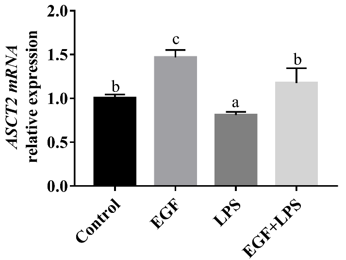
Effects of EGF on ASCT2 mRNA expression in IPEC-J2 cells challenged by LPS. Densitometric values were normalized to β-actin and expressed as a relative level to control values. Data are presented as mean ± SD, n=3, a, b, c means different lowercase letters indicate significant differences (P < 0.05).
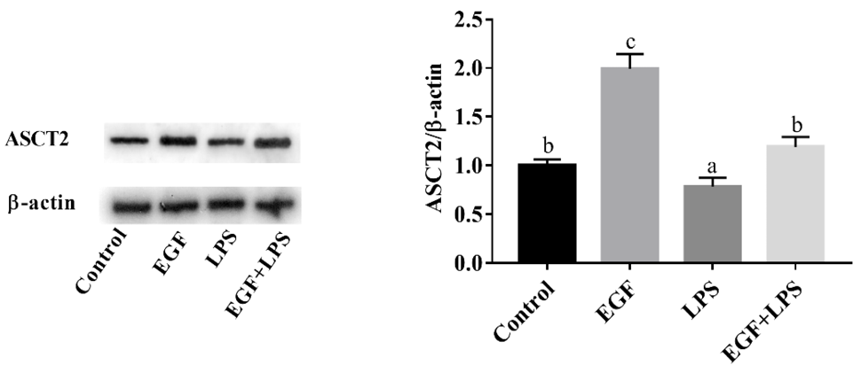
Effects of EGF on ASCT2 protein expression in IPEC-J2 cells challenged by LPS. Densitometric values were normalized to β-actin and expressed as a relative level to control values. Data are presented as mean ± SD, n=3, a, b, c means different lowercase letters indicate significant differences (P < 0.05).
References
- Avissar, N.E., Sax, H.C. and Tola, L., 2008. Dig. Dis. Sci., 53: 2113-2125. https://doi.org/10.1007/s10620-007-0120-y[CrossRef]
- Bedford, A., Chen, T., Huynh, E., Zhu, C., Medeiros, S., Wey, D., de Lange, C. and Li, J., 2015. J. Biotechnol., 196-197: 9-19. https://doi.org/10.1016/j.jbiotec.2015.01.007[CrossRef]
- Huang, Q., Li, N., Zhu, W., Li, Q. and Li, J., 2007. J. Parenter. Enter. Nutr., 31: 86-93. https://doi.org/10.1177/014860710703100286[CrossRef]
- Ledeganck, K.J., Boulet, G.A., Bogers, J.J., Verpooten, G.A. and De Winter, B.Y. 2013. PLoS One, 8: e57016. https://doi.org/10.1371/journal.pone.0057016[CrossRef]
- O’Mahony, F., Toumi, F., Mroz, M.S., Ferguson, G. and Keely, S.J., 2008. Am. J. Physiol. Cell Physiol., 294: C1362-C1370. https://doi.org/10.1152/ajpcell.00256.2007[CrossRef]
- Pochini, L., Scalise, M., Galluccio, M. and Indiveri, C., 2014. Front. Chem., 2: 61. https://doi.org/10.3389/fchem.2014.00061[CrossRef]
- Ray, E.C., Avissar, N.E. Salloum, R. and Sax, H.C., 2005. J. Nutri., 135: 14-18. https://doi.org/10.1093/jn/135.1.14[CrossRef]
- Ruhul, A., Temitope, O., Sangeeta, T., Dudeja, P.K., Krishnamurthy, R. and Jaleh, M., 2011. Inflamm. Bowel Dis., 17: 720-731.
- Talavera, M.M., Nuthakki, S., Cui, H., Jin, Y., Liu, Y. and Nelin, L.D., 2017. Front. cell. dev. Biol., 5: 15. https://doi.org/10.3389/fcell.2017.00015[CrossRef]
- Tang, X., Liu, B., Wang, X., Yu, Q. and Fang, R., 2018. Int. J. mol. Sci., 19: 848. https://doi.org/10.3390/ijms19030848[CrossRef]
- Tang, X., Liu, H., Yang, S., Li, Z., Zhong, J. and Fang, R., 2016. Mediators Inflamm., 2016: 1927348. https://doi.org/10.1155/2016/1927348[CrossRef]
- Wang, L., Zhu, F., Yang, H., Zhong, J., Li, Y., Ding, X., Xiong, X. and Yin, Y., 2019. J. Anim. Physiol. Anim. Nutri., 103: 618-625. https://doi.org/10.1111/jpn.13059[CrossRef]
- Xu, S., Wang, D., Zheng, P., Lin, Y., Fang, Z., Che, L. and Wu, D. 2015. J. appl. Microbiol., 119: 225-235. https://doi.org/10.1111/jam.12833[CrossRef]
- Yang, F., Wang, A., Zeng, X., Hou, C., Liu, H. and Qiao, S., 2015. BMC Microbiol., 15: 32. https://doi.org/10.1186/s12866-015-0372-1[CrossRef]
- Yi, D., Hou, Y., Wang, L., Ouyang W., Long, M., Zhao, D., Ding, B., Liu, Y. and Wu, G., 2015. Amino Acids, 47: 65-78. https://doi.org/10.1007/s00726-014-1842-8[CrossRef]
- Zhou, Y., Yuan, H., Cui, L., Ansari, A.R., Xiao, K., Luo, Y., Wu, X.T., Guo, L., Khan, F.A., Yang, Z. and Song, H., 2017. Acta Histochem., 119: 26-31. https://doi.org/10.1016/j.acthis.2016.11.002[CrossRef]
Cited by 1 article
In journals published by ResearchersLinks.
Services
Article usage statistics
Loading…