Special Issue: Veterinary Medicine between Sustainable Development and Public Health to Confront Global Changes · Volume 12, Issue 1, 14 November 2024 · Open Access
Effective Methods for Characterizing Genetically Unique Avian Reovirus Variants Responsible for Disease Outbreaks in Broiler Chickens in Egypt
· · · · · · ·
Affiliations & Notes ⌄
1Avian and Rabbit Medicine Department, Faculty of Veterinary Medicine, Suez Canal University, Ismailia, Egypt
22MEVAC for Vaccines, Second Industrial Area, El-Salihya El-Gededa, El-Sharkia, 44671, Egypt
33Reference Laboratory for Veterinary Quality Control on Poultry Production (RLQP), Animal Health Research Institute, Agriculture Research Center (ARC), Egypt
44 Faculty of Veterinary Medicine, Cairo University, Giza, Egypt
55Faculty of Veterinary Medicine, Zagazig University, Egypt. *Corresponden
The ARV isolation in ECEs revealed embryonic deaths within 72 to 96 HPI (chorio-allantoic membrane isolation) while deaths were recorded from 48-96 HPI in yolk sac isolation (Table 1).
This research revealed the existence of mutant strains belonging to genogroup 5 in Egypt, that can overcome maternal immunity conferred by all currently utilized conventional vaccines, resulting in significant economic losses and a high rate of morbidity.
The ARVs can be detected through RT-PCR employing a universal primer that targets sigma C within the S1 gene, and isolated in SPF-ECE, particularly using the YS method, resulting in various pathological lesions.
It is advisable to conduct further epidemiological investigations on the various ARV genogroups present and to enhance specialized autovaccines to effectively manage the reovirus situation in Egyptian poultry populations.
Selected from the article's own text.
Abstract
In recent years, Egypt has witnessed a resurgence of running-stunting syndrome (RSS) and viral arthritis cases attributed to a novel strain of variant avian reovirus (ARV) that could bypass the protection conferred by traditional vaccines. Consequently, our study focused on identifying and characterizing the circulating ARV strain among various broiler flocks in Egypt. Out of 73 suspected reovirus examined samples, 23% tested positive for reovirus PCR at a fragment of 1088 base pairs using primers P1 and P4. These samples were collected from broiler flocks that originated from vaccinated breeders and were experiencing dwarfism, viral arthritis, and abnormal feathering. The full sequencing of the sigma C gene “partial S1 segment” from two samples indicated that they belong to variant ARV cluster 5, distinct from the vaccine strain, that showed only 41-47% amino acid similarity. The virus was isolated in specific pathogen-free embryonated chicken eggs (SPF-ECE) via the yolk sac and chorioallantoic membrane routes, resulting in embryonic mortality and hemorrhage with congested internal organs. Survivors exhibited liver necrosis. Hepatitis, myocarditis, and infiltration of heterophilic diverse inflammatory cells were recorded upon histological examination. These findings confirm that the mutant ARV can skip maternal immunity induced by conventional vaccines due to the mismatch between vaccine strains and circulating variants. It is advisable to conduct further epidemiological research on various circulating genotypes and develop targeted autovaccines to address the reovirus situation in Egyptian flocks effectively.
Keywords: ARV Isolation, Viral arthritis, Running-stunting syndrome, ARV vaccine, ARV Egypt
INTRODUCTION
The first detection and isolation of avian reovirus (ARV) was in 1954 , and from tenosynovitis in 1957 . In 1967, ARV alterations in tendon and tendon sheath were termed viral arthritis or tenosynovitis and caused an enteric disease in chickens and turkeys . Later in 1972, the virus became known as a reovirus . ARV became controllable when the first classical S1133 live attenuated vaccine was released in 1983 (Van der Heide et al., 1983). Viral arthritis ; Johnson and Van der Heide, 1971; ; ; Van der Heide et al., 1983; ; and runting-stunting syndrome (RSS) were the main forms of ARV infection, also reported with respiratory ; ; ; , nervous (Van de Zande and Kuhn, 2007), osteoporosis (brittle bone disease) (Van der Heide et al., 1981), hepatitis, myocarditis, and immunosuppression ; symptoms.
ARVs are members of the genus orthoreovirus, which is part of the reoviridae family. They have ten segments (3 large “L1-L3”; 3 medium “M1-M3”; 4 small “S1-S4”). The S1 segment encodes three viral proteins, P10, P17 and sigma c “σC” . Sigma C protein plays an important role in the induction of neutralizing antibodies, cell attachment, and virus-cell interaction ; ; . Seven genotypes were identified based on phylogenetic analysis of partial sequencing of the S1 segment and full sequencing of the σC gene (De la Torre et al., 2021; . However, most published data revealed the existence of 6 genogroup clusters with sub-lineage ; ; ; ; ; .
ARV can be isolated in SPF ECE via yolk sac (YS) and chorioallantoic membrane (CAM) routes for 3-8 days of incubation based on the concentration of inoculated virus. Embryo hemorrhages, liver necrosis, and splenic necrosis can be observed after reovirus inoculation ; ; . However, YS appears to be better than CAM in terms of higher virus titer . Histological examination shows accumulation of macrophages, heterophils, and lymphocytes can be detected in synovial fluids. These inflammatory responses can result in immobility and fibrosis of the tendon. Myocarditis, hepatitis, splenitis, and hydro-pericarditis are observed ; ; . Commercial seed-derived ARV vaccines (S1133-S1733-2408) belong to genotype cluster 1 and exhibit limited amino acid (aa) identity with a common field strain that falls within the same cluster (sub-lineage), along with their differences with other clusters, indicating their ineffectiveness in preventing infection and outbreaks of ARVs since 2011 ; ; . So, to select vaccinal seeds, it’s important to choose isolates with high aa identities with the circulating challenging viruses. Additionally, justifies are required to establish a general, uniform, molecular ARV classification method, including various genogroup clusters and sub-clusters .
The first report of ARV in Egypt was from synovial fluids and tendon sheath in 1984 . Since that discovery in Egypt, only a few studies have been reported in cases associated with arthritis, enteric and respiratory manifestations without reference to the global genetic classification of ARVs based on partial S1 sequencing ; ; ; . Currently, limited published research addresses the prevalence of genotypes 4 and 5 ; ; while reported the existence of all five genogroups in Egypt, encompassing clusters 1 through 5. So, this study aimed to understand the ARV situation in broiler flocks, detection, virus isolation, phylogenetic analysis, and histopathological examination of variant ARV.
MATERIALS AND METHODS
Examined Birds
Seventy-three broiler flocks of various ages (5-38 days) from 5 different Egyptian provinces (40 Giza- 27 Sharqia- 4 Ismailia- 1 Behira and 1 Menoufia) were examined from 2020 to 2023 for avian reovirus infection. These birds showed significant weight loss with variable size, decreased feed intake, abnormal feathering, pale bird syndrome, and joint problems. Postmortem examinations were recorded.
Sampling and Sample Processing
Tissue samples were obtained from the duodenum, pancreas, proventriculus, and liver, along with tendons and tendon sheaths. These specimens were preserved in sterile phosphate buffer solution (PBS), appropriately labeled, and subsequently stored in a -80°C freezer for PCR and isolation.
Molecular Detection of ARV and Phylogenetic Analysis
Viral RNA was extracted utilizing the GeneJET viral DNA/RNA purification Kit Catalog number K0821, following the manufacturer’s instructions. Specific primers P1 5’-AGTATTTGTGAGTACGATTG-3’ and P4 5’-GGCGCCACACCTTAGGT-3’ were employed for the detection and sequencing of the ARV virus, targeting an amplicon size of 1088 bp with an annealing temperature of 58°C. This primer set is designed for the partial amplification of S1 segment “σC”. The amplification process was conducted using the SuperScript™ III One-Step RT-PCR System along with Platinum™ Taq DNA Polymerase, Catalog Number 12574-018, as follows: cDNA synthesis step at 45℃ for 30 min, pre-denaturation step at 94℃ for 2 min, 40 cycles as denaturation 94℃ for 15 sec, annealing at 58℃ for 30 sec. and extension at 68℃ for 1 min, then the final extension at 68℃ for 5 min. Data analysis was performed using BioEdit version 7.2 and MEGA7 software, with reference samples sourced from NCBI.
Virus Isolation
A total of 200 μl of positive ARV PCR homogenate (1:10 w/v) was inoculated into 5–7-day-old specific pathogen-free embryonated chicken eggs (SPF ECEs) via the yolk route (YS) and into 11-12-day-old SPF ECEs through the chorioallantoic membrane (CAM) route, undergoing three blind passages with 10 eggs per sample. The inoculated eggs were incubated at 60% relative humidity (RH) and 37°C, with daily candling for a duration of up to 7 days. Any deaths occurring within the first 48 hours were deemed non-specific. Eggs containing dead embryos, as well as those with live embryos up to 7 days post-inoculation, were chilled at 4 ºC for a maximum of 24 hours. Allantoic fluids and tissue homogenates were collected and re-inoculated for subsequent passages ; ; .
Histopathological Examination
Samples from the proventriculus, heart, liver, spleen, and bursa of Fabricius were obtained and then fixed in buffered neutral formalin at a 10 % concentration. Standard procedures for tissue processing, including dehydration, clearing, embedding, and casting, were subsequently carried out. A microtome was employed to section the paraffin blocks into slices measuring 4–5 µm. These sections were then affixed to glass slides and stained using hematoxylin and eosin (H&E) ; .
RESULTS
Clinical Signs and Post-mortem Lesions
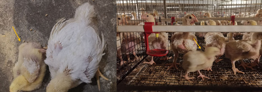
The examined broiler flocks exhibited marked weight inconsistency including stunting, reduced feed consumption, abnormal plumage, pale bird syndrome, along with locomotory disturbances with joint enlargements, and arthritis. Post-mortem lesions revealed pancreatic atrophy and proventriculitis (Figure 1).
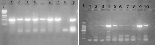
The ARV PCR analysis targeting the 1088 bp fragments with primers P1 and P4 revealed that out of 73 samples, 17 tested positive, at 23%. The highest positivity rate was observed in chicks aged 29-38 days, at 41%, while the lowest rates were recorded at 29.5% in chicks aged 16-21 days and 22-28 days. No virus detection was observed in chicks younger than 15 days (Figure 2).
Phylogenetic Analysis Results
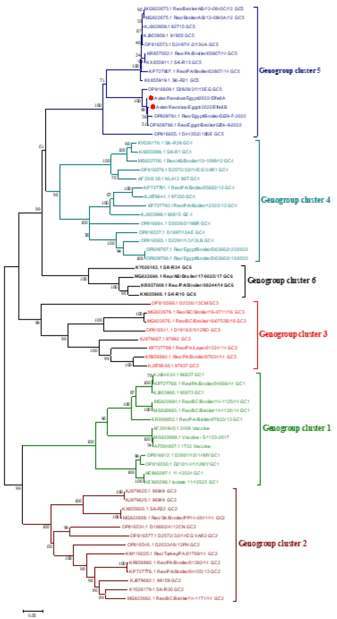
Phylogenetic analysis conducted on our isolates beside various representative samples from GeneBank identified the classification of avian reovirus into six distinct genogroups. The complete sigma c gene sequence analysis for the two samples (accession numbers PQ139057 and PQ139058) demonstrated their affiliation with the variant ARV within genogroup cluster 5, showing a divergence of 53-59% in amino acid identity from the conventional vaccine strains classified within genogroup cluster 1 (Figure 3).
Virus Isolation Results
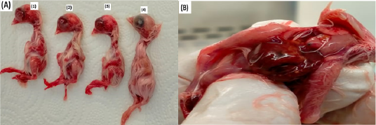
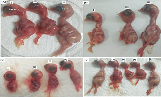
The ARV isolation in ECEs revealed embryonic deaths within 72 to 96 HPI (chorio-allantoic membrane isolation) while deaths were recorded from 48-96 HPI in yolk sac isolation (Table 1). Dead embryos were hemorrhagic with congested internal organs. Liver necrosis was observed in the surviving embryos (Figure 4 and 5) (Table 1).
Histopathological Examination
Inflammation and infiltration of different mononuclear cells
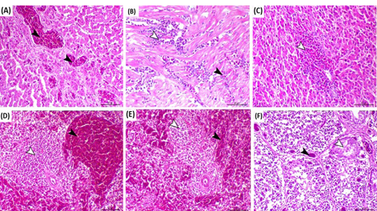
specifically macrophages and lymphocytes are the primary pathological findings in the examined organs. Lymphocytic depletion in the spleen and the bursa of Fabricius was identified along with the formation of an intrafollicular cyst in the bursa of Fabricius. A detailed histopathological examination of the infected chickens is presented in Figure 6.
Table 1: Results of ARV isolation on ECEs alongside PCR findings.
Flock No.
Passage No.
Egg isolation route
ECE age
Egg mortality in hours
Total embryo mortality
ARV RT-PCR result
24 hr
48 hr
72 hr
96 hr
120 hr
8
1
Yolk
5 days
2
2
1
5
-
10/10
-
8
2
Yolk
7 days
2
4
3
1
-
10/10
-
8
3
Yolk
6 days
0
4
3
3
10/10
Positive
8
1
CAM
12 days
1
0
1
4
0
6/10
-
8
2
CAM
12 days
2
0
3
5
10/10
-
8
3
CAM
12 days
0
0
6
4
10/10
Positive
DISCUSSION
A crucial approach for managing clinical diseases linked to reovirus infections involves properly vaccinated breeders with effective vaccines. This practice diminishes the likelihood of vertical transmission and equips offspring with specific maternal antibodies that offer protection against virulent field strains . The mutation nature characteristics of avian reovirus resulted in novel variants not effectively controlled by traditional vaccines attributable to mismatching, leading to substantial outbreaks across different bird populations and causing considerable economic impacts ; ; . Determining the status of circulating reovirus strains in our flocks is crucial to facilitate the development of effective autogenous vaccinations and mitigate their transmission and spread. Consequently, conducting clinical investigations, molecular characterization, virus isolation, and histopathological examination of the currently circulated reoviruses, along with their classification into their related groups is essential, and holds considerable significance.
Our study revealed the detection of reovirus by RT-PCR in 17 broiler flocks (23%) of different ages, obtained from five governorates in Egypt, between 2020 and 2023. The findings align closely with those presented by , who reported an ARV detection rate of 20%. However, they contrast with the 100% detection of reovirus observed by . This divergence may be due to our study focusing exclusively on broiler chicks across ages (5-38 days), with the virus not being detected in chicks younger than 15 days. These samples were collected from vaccinated flocks, suggesting that the existing vaccines fail to offer sufficient protection against the disease ; . Common symptoms detected due to the reovirus variants were malabsorption syndrome characterized by stunting, and abnormal feathers, as well as pancreatic atrophy and inflamed proventriculus in addition to joint enlargement and arthritis agreement with ; ; ; . Reovirus is recognized for its role in inducing viral arthritis, it has also been identified in chickens that exhibit no symptoms of arthritic lesions, indicating that not all avian reoviruses
possess arthritic characteristics ; . Typical signs were reported in this study, including stunting, reduced feed consumption, abnormal plumage, pale bird syndrome, along with locomotory disturbances with joint enlargements, and arthritis. Similar symptoms and PM lesions were also reported by ; ; , ; ; . Conversely, other samples displayed identical ARV signs and PM lesions while testing negative for reovirus, supporting the notion of a multifactorial origin for the infection (Van der Heide, 2000).
There exists a limited number of studies published concerning the circulation of ARV variant clusters in Egypt, along with their isolation and identification. To enhance the molecular understanding and detection of reovirus in cases associated with viral arthritis and runting-stunting syndrome, which are the primary disorders, a universal primer set (P1-P4), in use since 2003 , was employed for viral detection and sequencing, specifically targeting the sigma C gene “partial S1 segment” differs from the primers that were predominantly utilized in Egyptian researches until 2023, which focused on the S2 segment for both detection and sequencing ; ; ; .
In recent developments in Egypt, genetic clusters 4 and 5 have been identified in different chicken flocks and spread across various localities including Gharbia, Giza, Al Menofia, Al Qalyoubia, and Al Beheira ; ; which is consistent with our results. Additionally, by the conclusion of 2023, genotypes 1 to 5 were identified throughout a broad geographic region in the Middle East, particularly in Egypt . This widespread confirmation can be attributed to the international nature of the study, which encompassed multiple countries, including Egypt, and involved the collection of a substantial number of samples on a large scale.
To build the phylogenetic tree, we utilized various representative samples from GeneBank that correspond to the six genogroup clusters and adopted the idea of six different globally published papers ; Troxler et al., 2013; ; ; ; and we agreed with their classification, which stands in contrast to that of and who altered this global classification substituting the fourth gene cluster with the fifth and vice versa.
Phylogenetic analysis of two samples identified by accession numbers PQ139057 and PQ139058 indicated their placement within genogroup cluster 5. The sequence analysis revealed a significant amino acid variation ranging from 53% to 59% compared to the vaccine strains, aligning closely with the findings of which reported a diversity of 55.09% to 56.23% also come constant with results of low sequence identity (43–55%) obtained by . This discovery underscores the potential for isolated reovirus variants to bypass maternal immunity gained from standard vaccines designed to prevent the disease. Most examined samples in these studies originated from vaccinated breeders, suggesting that a mismatch between vaccine strains and the circulating ARV variants may contribute to the manifestation of disease symptoms.
Although virus isolation can be a lengthy process, it is still the most favored approach for diagnosing viruses and is essential for detecting new strains or emerging variants. This research demonstrated that isolating reoviruses in SPF ECE resulted in embryonic deaths within 72 to 96 hours post-inoculation (HPI) when utilizing the CAM method, while the YS method recorded deaths within a shorter timeframe of 48 to 96 HPI. These findings indicate that the YS approach is more sensitive for virus isolation than the CAM method, as noted by . The enhanced sensitivity of the YS method may be linked to the age of the embryos at the time of injection; it utilizes very young embryos (5-7 days old), which allows for more efficient viral replication and growth, unlike the CAM method, which involves older embryos (11-12 days old) that are closer to hatching.
ARVs are known to induce atrophy of lymphoid tissues, particularly affecting the bursa of Fabricius ; ; . In our cases, lymphoid depletion was evident not only in the bursa of Fabricius but also in the spleen, accompanied by the development of an intrafollicular cyst within the bursa, as noted by ; ; . The most prevalent pathological findings linked to ARV in the birds we found were myocarditis and hepatitis, consistent with the reports of ; ; ; . Additionally, we observed infiltration of mononuclear cells, particularly macrophages, which play a crucial role in the replication and transmission of ARV, as highlighted by across various examined organs.
Our findings demonstrate the circulation of ARV genotype 5 variant strains in Egypt with different clinical and histopathological lesions in different organs and a standard technique for the detection and viral isolation of different variants of ARVs.
Additional investigation is required on the ARV variants we have presented to assess their pathogenicity, antigenicity, and the potential for cross-protection among them.
CONCLUSIONS And RECOMMENDATIONS
This research revealed the existence of mutant strains belonging to genogroup 5 in Egypt, that can overcome maternal immunity conferred by all currently utilized conventional vaccines, resulting in significant economic losses and a high rate of morbidity. The ARVs can be detected through RT-PCR employing a universal primer that targets sigma C within the S1 gene, and isolated in SPF-ECE, particularly using the YS method, resulting in various pathological lesions. Additionally, histopathological analysis proves to be an effective technique for virus identification. It is advisable to conduct further epidemiological investigations on the various ARV genogroups present and to enhance specialized autovaccines to effectively manage the reovirus situation in Egyptian poultry populations.
Ethics Approval and Consent to Participate
The ethical and research committee at the Faculty of Veterinary Medicine, Suez Canal University, approved the study protocol under reference number 2022020.
ACKNOWLEDGMENTS
We express our gratitude to the original and submitting laboratories that contributed the sequences accessible in the NCBI database.
NOVELTY STATEMENT
Current traditional ARV vaccines cannot deliver adequate protection for the challenging circulating variant viruses in Egypt and autogenous vaccine is recommended.
AUTHOR’S CONTRIBUTIONS
Each author has made an equal contribution in offering their technical expertise and insights to develop this article.
Conflict of Interest
All authors affirm that the research was carried out without any commercial or financial affiliations that might be interpreted as a possible conflict of interest.
Figures
Figure 1:.
Broiler flocks at 23 days old (A) and 18 days old (B) exhibiting stunted and irregular growth patterns.
Figure 2:.
RT-PCR results of ARV-positive samples, (A): L: 15K DNA Marker (500, 1000, 1500,3000, 5000, 7500, 10000, 15000bp), 1-7 ARV isolates, 8: negative control, 9: Positive control. (B): L: 1500 bp DNA Marker (100, 200, 300, 400, 500, 600, 700, 800, 900, 1000, 1500 bp), 2-9 ARV isolates, 1: negative control, 10: Positive control.
Figure 3:.
Phylogenetic tree of ARV isolates based on the nucleotide sequence of sigma C encoding gene used neighborhood likelihood method along with 1,000 bootstrap replicates resulted in sequences were clustered into six distinct genogroups (genogroup cluster 1–6). Our isolates represented by red circles, are clustered within variant genogroup cluster 5 and all vaccinal strains located in genogroup cluster 1.
Figure 4:.
(A) The chicken embryos inoculated through the CAM route following the third passage and a four-day incubation period exhibited stunted growth and significant hemorrhaging in samples 1, 2, and 3 and necrotic foci on the liver (B). In contrast, sample number 4, which was inoculated with a sterile phosphate buffer solution, displayed normal growth.
Figure 5:.
Inoculation of chicken embryos through the YS route is depicted, with panels (A) and (B) showing that samples 1 and 2 demonstrated dwarfism and considerable hemorrhaging after the initial passage and a four-day incubation period, whereas sample 3 functioned as a negative control. Panel (C) reveals that samples 1, 2, and 3 exhibited dwarfism and pronounced hemorrhaging following the second passage and a three-day incubation, with sample 4 serving as a negative control. In panel (D), samples 2, 3, and 4 showed signs of dwarfism and severe hemorrhaging after the third passage and a four-day incubation, while samples 1 and 5 were utilized as negative controls.
Figure 6:.
Birds infected with reovirus showed several pathological changes: (A) The proventriculus exhibited proventriculitis characterized by necrosis and hemorrhage in the central areas of the proventricular glands (indicated by arrowheads), H&E, scale bar=50 µm. (B) The heart displayed severe myocarditis, marked by significant myolysis and infiltration of mononuclear cells, including macrophages and lymphocytes (white arrowhead), and heterophilic cells (black arrowhead), H&E, scale bar= 100 µm. (C) The liver showed features of hepatitis, associated with periportal infiltration of heterophilic inflammatory cells (arrowhead), H&E, scale bar= 50 µm. (D) The spleen exhibited congestion of blood capillaries in the red pulp (black arrowhead) alongside lymphoid depletion (white arrowhead), H&E, scale bar=50 µm. (E) The spleen demonstrated congestion of blood capillaries in the red pulp (black arrowhead) and lymphoid depletion (white arrowhead), H&E, scale bar= 50 µm. (F) The bursa of Fabricius revealed vascular congestion (black arrowhead) and marked lymphoid depletion within the germinal area of the lymphoid follicles along with the formation of an intrafollicular cyst (white arrowhead), H&E, scale bar= 50 µm.
References
Abd El-Samie, L.(2014). Some causes of chicken’s growth retardation in sharkia, Egypt. Assiut Veterinary Medical Journal, 61, 32-37
Al-Ebshahy, E., Mohamed, S. & Abas, O.(2020). First report of seroprevalence and genetic characterization of avian orthoreovirus in Egypt. Tropical animal health and production, 52, 1049-1054.https://doi.org/10.1007/s11250-019-02100-z[CrossRef]
Amer, M. M., Mekky, H. M. & Fedawy, H. S.(2019). Molecular identification of Mycoplasma synoviae from breeder chicken flock showing arthritis in Egypt. Veterinary World, 12, 535.https://doi.org/10.14202/vetworld.2019.535-541[CrossRef]
Ayalew, L. E., Gupta, A., Fricke, J., Ahmed, K. A., Popowich, S., Lockerbie, B., Tikoo, S. K., Ojkic, D. & Gomis, S.(2017). Phenotypic, genotypic and antigenic characterization of emerging avian reoviruses isolated from clinical cases of arthritis in broilers in Saskatchewan, Canada. Scientific reports, 7, 1-13.https://doi.org/10.1038/s41598-017-02743-8[CrossRef]
Bányai, K., Dandár, E., Dorsey, K. M., Mató, T. & Palya, V.(2011). The genomic constellation of a novel avian orthoreovirus strain associated with runting-stunting syndrome in broilers. Virus genes, 42, 82-89.https://doi.org/10.1007/s11262-010-0550-z[CrossRef]
Chénier, S., Boulianne, M. & Gagnon, C. A.(2014). Postvaccinal reovirus infection with high mortality in breeder chicks. Avian diseases, 58, 659-665.https://doi.org/10.1637/10860-050914-Case.1[CrossRef]
Czekaj, H., Kozdruń, W., Styś-Fijoł, N., Niczyporuk, J. S. & Piekarska, K.(2018). Occurrence of reovirus (ARV) infections in poultry flocks in Poland in 2010–2017. Journal of Veterinary Research, 62, 421.https://doi.org/10.2478/jvetres-2018-0079[CrossRef]
Dalton, P. J. & Henry, R.(1967). TENO SYNOVITIS IN POULTRY. Veterinary Record, 80, 638-&
De Carli, S., Wolf, J. M., Gräf, T., Lehmann, F. K., Fonseca, A. S., Canal, C. W., Lunge, V. R. & Ikuta, N.(2020). Genotypic characterization and molecular evolution of avian reovirus in poultry flocks from Brazil. Avian Pathology, 49, 611-620.https://doi.org/10.1080/03079457.2020.1804528[CrossRef]
De La Torre, D., Astolfi-Ferreira, C. S., Chacón, R., Puga, B. & Piantino Ferreira, A.(2021). Emerging new avian reovirus variants from cases of enteric disorders and arthritis/tenosynovitis in Brazilian poultry flocks. British Poultry Science, 62, 361-372.https://doi.org/10.1080/00071668.2020.1864808[CrossRef]
Egana-Labrin, S. & Broadbent, A. J.(2023). Avian reovirus: a furious and fast evolving pathogen. Journal of Medical Microbiology, 72, 001761.https://doi.org/10.1099/jmm.0.001761[CrossRef]
Egana-Labrin, S. C. (2022).Molecular, Pathogenic, and Antigenic Characterization of Emerging Avian Reovirus Variant Strains. University of California, Davis.
Fahey, J. & Crawley, J.(1954). Studies on chronic respiratory disease of chickens II. Isolation of a virus. Canadian journal of comparative medicine and veterinary science, 18, 13
Guneratne, J., Jones, R. & Georgiou, K.(1982). Some observations on the isolation and cultivation of avian reoviruses. Avian Pathology, 11, 453-462.https://doi.org/10.1080/03079458208436117[CrossRef]
Hieronymus, D. R., Villegas, P. & Kleven, S.(1983). Identification and serological differentiation of several reovirus strains isolated from chickens with suspected malabsorption syndrome. Avian Diseases, 246-254
Hoerr, F. J.(2010). Clinical aspects of immunosuppression in poultry. Avian diseases, 54, 2-15.https://doi.org/10.1637/8909-043009-Review.1[CrossRef]
Johnson, D. & Van Der Heide, L.(1971). Incidence of tenosynovitis in Maine broilers. Avian diseases, 829-834
Jones, R.(2000). Avian reovirus infections. Revue Scientifique et Technique-Office International des Epizooties, 19, 614-619.https://doi.org/10.20506/rst.19.2.1237[CrossRef]
Jones, R. & Guneratne, J.(1984). The pathogenicity of some avian reoviruses with particular reference to tenosynovitis. Avian Pathology, 13, 173-189.https://doi.org/10.1080/03079458408418522[CrossRef]
Jones, R. C.(2013). Reovirus infections. Diseases of poultry, 351-373
Kant, A., Balk, F., Born, L., Van Roozelaar, D., Heijmans, J., Gielkens, A. & Ter Huurne, A.(2003). Classification of Dutch and German avian reoviruses by sequencing the $\sigma $ C protein. Veterinary research, 34, 203-212.https://doi.org/10.1051/vetres:2002067[CrossRef]
Kawamura, H.(1965). Avian reovirus: its properties and serological classification. Natl. Inst. Anim. Health Q.(Jpn.), 5, 115-124
Kerr, K. M. & Olson, N. O.(1969). Pathology of chickens experimentally inoculated or contact-infected with an arthritis-producing virus. Avian Diseases, 729-745
Kovács, E., Varga-Kugler, R., Mató, T., Homonnay, Z., Tatár-Kis, T., Farkas, S., Kiss, I., Bányai, K. & Palya, V.(2023). Identification of the main genetic clusters of avian reoviruses from a global strain collection. Frontiers in Veterinary Science, 9, 1094761.https://doi.org/10.3389/fvets.2022.1094761[CrossRef]
Liu, H. J., Lee, L. H., Hsu, H. W., Kuo, L. C. & Liao, M. H.(2003). Molecular evolution of avian reovirus:: evidence for genetic diversity and reassortment of the S-class genome segments and multiple cocirculating lineages. Virology, 314, 336-349.https://doi.org/10.1016/S0042-6822(03)00415-X[CrossRef]
Liu, Q., Zhang, G., Huang, Y., Ren, G., Chen, L., Gao, J., Zhang, D., Han, B., Su, W. & Zhao, J.(2011). Isolation and characterization of a reovirus causing spleen necrosis in Pekin ducklings. Veterinary microbiology, 148, 200-206.https://doi.org/10.1016/j.vetmic.2010.09.016[CrossRef]
Lu, H., Tang, Y., Dunn, P. A., Wallner-Pendleton, E. A., Lin, L. & Knoll, E. A.(2015). Isolation and molecular characterization of newly emerging avian reovirus variants and novel strains in Pennsylvania, USA, 2011–2014. Scientific reports, 5, 14727.https://doi.org/10.1038/srep14727[CrossRef]
Mansour, S. M., Elbakrey, R. M., Orabi, A., Ali, H. & Eid, A. A.(2018). Isolation and detection of avian reovirus from tenosynovitis and malabsorption affected broiler chickens with involvement of vertical transmission. Journal of Virological Sciences, 4, 24-32
Mills, J. & Wilcox, G.(1993). Replication of four antigenic types of avian reovirus in subpopulations of chicken leukocytes. Avian Pathology, 22, 353-361.https://doi.org/10.1080/03079459308418926[CrossRef]
Mohamed, S., Al-Ebshahy, E. M., Khalil, S. A., Abdelhady, H. A. & Elbestawy, A. R.(2020). Molecular identification and pathogenicity assessment of avian reovirus in Egypt.https://doi.org/10.5455/ajvs.52329[CrossRef]
Montgomery, R., Villegas, P., Dawe, D. & Brown, J.(1985). Effect of avian reoviruses on lymphoid organ weights and antibody response in chickens. Avian diseases, 552-560.https://doi.org/10.2307/1590520[CrossRef]
Mor, S. K., Sharafeldin, T. A., Porter, R. E., Ziegler, A., Patnayak, D. P. & Goyal, S. M.(2013). Isolation and characterization of a turkey arthritis reovirus. Avian diseases, 57, 97-103
Mosad, S. M., Elmahallawy, E. K., Alghamdi, A. M., El-Khayat, F., El-Khadragy, M. F., Ali, L. A. & Abdo, W.(2023). Molecular and pathological investigation of avian reovirus (ARV) in Egypt with the assessment of the genetic variability of field strains compared to vaccine strains. Frontiers in Microbiology, 14.https://doi.org/10.3389/fmicb.2023.1156251[CrossRef]
No, O. & Da, M.(1957). Infectious synovitis control by medication; effect of strain differences and pleuropneumonia-like organisms. American journal of veterinary research, 18, 735-739
Olson, N. & Solomon, D.(1968). A natural outbreak of synovitis caused by the viral arthritis agent. Avian Diseases, 12, 311-316
Olson, N. & Weiss, R.(1972). Similarity between arthritis virus and Fahey-Crawley virus. Avian diseases, 535-540
Page, R., Fletcher, O., Rowland, G. N., Gaudry, D. & Villegas, P.(1982). Malabsorption syndrome in broiler chickens. Avian Diseases, 618-624
Palomino-Tapia, V., Mitevski, D., Inglis, T., Van Der Meer, F. & Abdul-Careem, M. F.(2018). Molecular characterization of emerging avian reovirus variants isolated from viral arthritis cases in Western Canada 2012–2017 based on partial sigma (σ) C gene. Virology, 522, 138-146.https://doi.org/10.1016/j.virol.2018.06.006[CrossRef]
Petek, M., Felluga, B., Borghi, G. & Baroni, A.(1967). The Crawley agent: an avian reovirus. Archiv für die gesamte Virusforschung, 21, 413-424
Pitcovski, J. & Goyal, S. M.(2020). Avian reovirus infections. Diseases of poultry, 382-400
Ramzy, N. M., Ibrahim, H. N. & Elhadad, S. F.(2016). Molecular Characterization and Hemato Biochemical Studies of Reovirus in Ismailia Farms. Egyptian Journal of Chemistry and Environmental Health, 2, 167-182.https://doi.org/10.21608/ejceh.2016.247635[CrossRef]
Reck, C., Menin, Á., Canever, M. F., Pilati, C. & Miletti, L. C. J. J. O. T. S. A. V. A.(2019). Molecular detection of Mycoplasma synoviae and avian reovirus infection in arthritis and tenosynovitis lesions of broiler and breeder chickens in Santa Catarina State, Brazil. 90, 1-5.https://doi.org/10.4102/jsava.v90i0.1970[CrossRef]
Robertson, M. A.(1986). Avian reovirus. Vet. Bull., 56, 155-174
Rosenberger, J. (1983). Published. Characterization of reoviruses associated with a runting syndrome in chickens. Proc, 1983. 141-152.
Rosenberger, J., Sterner, F., Botts, S., Lee, K. & Margolin, A.(1989). In vitro and in vivo characterization of avian reoviruses. I. Pathogenicity and antigenic relatedness of several avian reovirus isolates. Avian diseases, 535-544
Safwat, M., Adel, A., Rady, M., Hamoud, M. H. & Kilany, W. H.(2024). Identification and genetic correlation of avian reoviruses to the currently used vaccines in Egypt. German Journal of Veterinary Research, 4, 1-8.https://doi.org/10.51585/gjvr.2024.1.0068[CrossRef]
Sahu, S. & Olson, N.(1975). Comparison of the characteristics of avian reoviruses isolated from the digestive and respiratory tract, with viruses isolated from the synovia. American journal of veterinary research, 36, 847-850
Sánchez-Cordón, P., Hervás, J., De Lara, F. C., Jahn, J., Salguero, F. & Gómez-Villamandos, J.(2002). Reovirus infection in psittacine birds (Psittacus erithacus): morphologic and immunohistochemical study. Avian diseases, 46, 485-492.https://doi.org/10.1637/0005-2086(2002)046[0485:RIIPBP]2.0.CO;2[CrossRef]
Sellers, H. S.(2017). Current limitations in control of viral arthritis and tenosynovitis caused by avian reoviruses in commercial poultry. Veterinary microbiology, 206, 152-156.https://doi.org/10.1016/j.vetmic.2016.12.014[CrossRef]
Song, Z., Bi, X., Meng, F., Yang, Q., Cheng, X., Zhou, D., Zhang, X., Ding, L., Lang, F. & Deng, B.(2024). Research Note: Genetic characterization and pathogenicity of an epidemic variant strain of avian reovirus. Poultry Science, 103, 103370.https://doi.org/10.1016/j.psj.2023.103370[CrossRef]
Souza, S. O., De Carli, S., Lunge, V. R., Ikuta, N., Canal, C. W., Pavarini, S. P. & Driemeier, D. J. P. S.(2018). Pathological and molecular findings of avian reoviruses from clinical cases of tenosynovitis in poultry flocks from Brazil. 97, 3550-3555.https://doi.org/10.3382/ps/pey239[CrossRef]
Spackman, E., Day, J. M. & Pantin-Jackwood, M. J.(2010). Astrovirus, reovirus, and rotavirus concomitant infection causes decreased weight gain in broad-breasted white poults. Avian diseases, 54, 16-21.https://doi.org/10.1637/8986-070909-Reg.1[CrossRef]
Tantawi, H., Amina, N., Youssef, Y., Fawzia, M., Al-Abdulla, J., El-Batrawi, A., El-Ghawas, A., Nasser, A. & Reda, I.(1984). Infectious tenosynovitis in broilers and broiler breeders in Egypt. Veterinary research communications, 8, 229-235.https://doi.org/10.1007/BF02214716[CrossRef]
Troxler S, Rigomier P, Bilic I et al. (2013) Identification of a new reovirus causing substantial losses in broiler production in France, despite routine vaccination of breeders. Veterinary Record 172:556-556. https://doi.org/10.1136/vr.101262[CrossRef]
Van De Zande, S. & Kuhn, E.-M.(2007). Central nervous system signs in chickens caused by a new avian reovirus strain: a pathogenesis study. Veterinary Microbiology, 120, 42-49
Van Der Heide, L.(2000). The history of avian reovirus. Avian diseases, 44, 638-641
Van Der Heide, L., Kalbac, M. & Brustolon, M.(1983). Development of an attenuated apathogenic reovirus vaccine against viral arthritis/tenosynovitis. Avian diseases, 698-706
Van Der Heide, L., Lütticken, D. & Horzinek, M.(1981). Isolation of avian reovirus as a possible etiologic agent of osteoporosis (“ brittle bone disease”;” femoral head necrosis”) in broiler chickens. Avian Diseases, 847-856
Walker, E. R., Friedman, M. & Olson, N.(1972). Electron microscopic study of an avian reovirus that causes arthritis. Journal of ultrastructure research, 41, 67-79
Wickramasinghe, R., Meanger, J., Enriquez, C. E. & Wilcox, G. E.(1993). Avian reovirus proteins associated with neutralization of virus infectivity. Virology, 194, 688-696.https://doi.org/10.1006/viro.1993.1309[CrossRef]
Wooley, R. E., Dees, T., Cromack, A. & Gratzek, J.(1972). Infectious enteritis of turkeys: characterization of two reoviruses isolated by sucrose density gradient centrifugation from turkeys with infectious enteritis. American Journal of Veterinary Research, 33, 157-164
Zaher, K. & Mohamed, S.(2009). Diagnosis of avian reovirus infection in local Egyptian chicks. Global Veterinaria, 3, 227-231
Zanaty, A., Mosaad, Z., Elfeil, W. M., Badr, M., Palya, V., Shahein, M. A., Rady, M. & Hess, M.(2023). Isolation and Genotypic Characterization of New Emerging Avian Reovirus Genetic Variants in Egypt. Poultry, 2, 174-186.https://doi.org/10.3390/poultry2020015[CrossRef]
Zhang, X., Lei, X., Ma, L., Wu, J. & Bao, E.(2019). Genetic and pathogenic characteristics of newly emerging avian reovirus from infected chickens with clinical arthritis in China. Poultry Science, 98, 5321-5329.https://doi.org/10.3382/ps/pez319[CrossRef]
Effective Methods for Characterizing Genetically Unique Avian Reovirus Variants Responsible for Disease Outbreaks in Broiler Chickens in Egypt · ResearchersLinks