
Abnormalities of intestines in carnivores such as blockage, peritonitis, tumors and diverse local injuries and lesions are corrected by resection of the intestine. Twelve cats were subjected to 3 variable degrees of jejunal resection, i.e. group A (20%) and group B (40%), while group C (0%). Sampling was performed on days 0, 15, and 30 to test various clinical and biochemical parameters. Results showed weight loss in all groups until day 15 post-surgery while in group A and C gradual weight gain was noted. Fecal consistency was observed in all the groups and noticed moist till day 15 post-surgery, which gradually turned to normal except in group B. The electrolyte parameters showed a significant difference (P<0.05) in the concentration of sodium ions in group C compared to group A and B on day 15 and 30 of post-surgery. In contrast, the concentration of chloride, potassium, and bicarbonate ions was found to be insignificant. A significant difference (P<0.05) was reported in various blood parameters in group A and B when compared with group C. However, all the observed values were within the normal range. It was concluded that resection of jejunum up to 20% in cats makes natural body weight gain while ≥40% jejunal resection increases morbidity and mortality risk.
Short Communication · Volume 54, Issue 5, 19 July 2022 · Open Access
Effect of Variable Degrees of Jejunal Resection on Clinical and Haematological Parameters of Cats
· · · · · · · ·
Affiliations & Notes ⌄
- 1Department of Clinical Medicine and Surgery, University of Veterinary and Animal Sciences, Lahore 54000, Pakistan 2Department of Parasitology, University of Veterinary and Animal Sciences, Lahore 54000, Pakistan 3National Veterinary Laboratories, Ministry of National Food Security and Research, Islamabad 44000, Pakistan 4University of Turin, Italy * Corresponding author: ayoob.faisal@yahoo.com
Article Info ⌄
- Journal:
- Pakistan Journal of Zoology
- Published:
- 19 July 2022

From the article
- Loss in weight (Debilitation) was observed in all the groups (Fig. 1), but gradually weight gain was noted except B group in which 40% jejunal resection was performed.
- Gradual healing was observed in all animals except for two cats.
- WBCs and granulocytic count were significantly higher in A and B group from the control group on different study intervals (Fig. 2B).
- It was concluded in the present research that 20% jejunectomy had shown no injurious effect on the health of cats while 40% jejunal resection seriously affects physiology, clinical and biochemical parameters in cats.
Selected from the article's own text.
Abstract
Materials and methods
The study was conducted on a total of 12 cats of both sexes, ranging from 1- 2 years of age and was purchased from the local market<market (Lahore-Pakistan). All cats were shifted to cages, ten days before the start of surgeries to acclimatize them friendly handling and physical examination. Cats were examined thoroughly to rule out any GIT abnormality and systemic ailment before the surgery. All cats were maintained on the same nutritional regimen, allowing the water ad libitum, and housed in a stress-free aseptic environment. The cats were allocated into three different groups viz A, B, and C having four cats in each group. Resection of jejunum was performed with varying percentages as in group A 20% and in group B 40%. Whereas group C serves the control group with only end to end anastomosis without resection, and all the parameters compared with resected groups. The surgical intervention was performed at the operation theatre of Clinical Medicine and Surgery (CMS) Department of University of Veterinary Sciences Lahore- Pakistan.
All the procedures in the present work were approved and performed in compliance with the institutional guidelines of the Ethical Review Committee. All the surgical equipment was autoclaved and sterilized one day before performing surgery. Cats off fed 10-12 hours before performing surgery. The surgical site of the patient was clipped and shaved with a razor to minimize the contamination, and then scrubbed with an antiseptic (Pyodine). Sterile surgical drapes were positioned around the surgical vicinity. Sterilized gown, disposable gloves, masks, and caps worn to carry out proper aseptic surgery.
A combination of anesthetic drugs Ketamine, Xylazine, and Atropine sulfate in a ratio of 7:2:1 (0.7 ml + 0.2 ml + 0.1 ml) used for the anesthesia, injected intramuscularly at the hind leg by using 1ml disposable insulin syringe. The dose rate was planned according to the body mass of the animal . The patient was positioned in dorsal recumbency for the ‘midline laparotomy incision’ to expose the jejunum. An incision of 5-8 cm was made at the surgical site with the help of a surgical blade (No. 24), considered umbilicus as a landmark. Undermining was performed till an incision was made to ‘linea alba’ and reached into the abdominal cavity. Retractors were used to see the abdominal visceral organs and the jejunum was manually exposed and handled gently to avoid shock by keeping moistened through the normal saline sponge. The jejunum in each cat ascertained through distinguishing coils of it. The length of jejunum was measured and then exteriorized in each group as mentioned (i.e. 20% Group A, 40% Group B, 0 % Group C) through the celiotomy incision. After performing jejunal resection, the incision site closed in three layers from inward to outward, i.e. linea alba with simple interrupted suture technique using 2/0 absorbable suture material, Sub-cut tissue with the simple continuous pattern using 3/0 chromic catgut absorbable suture material, and skin with simple interrupted suture technique using 3/0 non-absorbable silk material . After the surgery, all the cats were kept on intravenous nutrition for 2-3 days, and after that soft oral feed was given.
A physical examination was performed to judge health with weight loss or gain at day 0, 15, and 30 postoperatively. Blood was collected in EDTA vacutainers at 0, 15, and 30 days post-surgery and was analyzed for various hematological parameters by auto hematological analyzer (SPHERA), and for
sodium, potassium, chloride, and bicarbonate ions concentration in serum by atomic absorption spectrometer (PerkinElmer Analyst 400) on day 0, 15 and 30 post-surgery. The consistency of feces was evaluated by grading as 1) watery 2) moist and 3) soft and firm to see the activity status of small intestines . Wound healing was evaluated by scoring it as score 1 (poor healing), score 2. (partial healing) and score 3 (properly healing). The data analysis was done through a one-way analysis of variance (ANOVA) using IBM SPSS, ver.23 .
Results and discussion
Loss in weight (Debilitation) was observed in all the groups (Fig. 1), but gradually weight gain was noted except B group in which 40% jejunal resection was performed
; . Fecal consistency remains moist in pets after having intestinal surgery and improved gradually over a period , which is in line with the results of the present study except in group B where fecal consistency remained moist till day 30 post-surgery. Gradual healing was observed in all animals except for two cats. All cats utterly healed at the end of the research. Resection showed no significant change in potassium, chloride, and bicarbonate ion’s value throughout the study (Fig. 2A) which is in line with the studies of Fabritius et al. (1986) and Vazquez et al. (1988). The hematocrit of A and group B was significantly lower from control after surgery. WBCs and granulocytic count were significantly higher in A and B group from the control group on different study intervals (Fig. 2B). RBCs count of both groups A and B was substantially lesser than the control group, and the number increased over time (Fig. 2C). For platelets count, there was a significant difference in all the groups, all observed values; were within the normal range as also reported by Astre et al. (1992) and Dilawer et al. (2011). Hemoglobin, MCHC, and MCV values of both groups A and B were observed significantly lower than control at different intervals (Fig. 2D). At the same time, MCH was lower throughout the research than group C. The sodium-ion concentration of A group was considerably higher from group B and C after jejunal resection .
It was concluded in the present research that 20% jejunectomy had shown no injurious effect on the health of cats while 40% jejunal resection seriously affects physiology, clinical and biochemical parameters in cats.
Statement of conflict of interest
The authors have declared no conflict of interest.
Figures
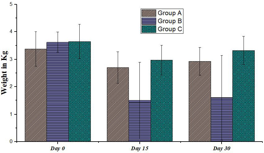
Bodyweight of the studied groups at different days.
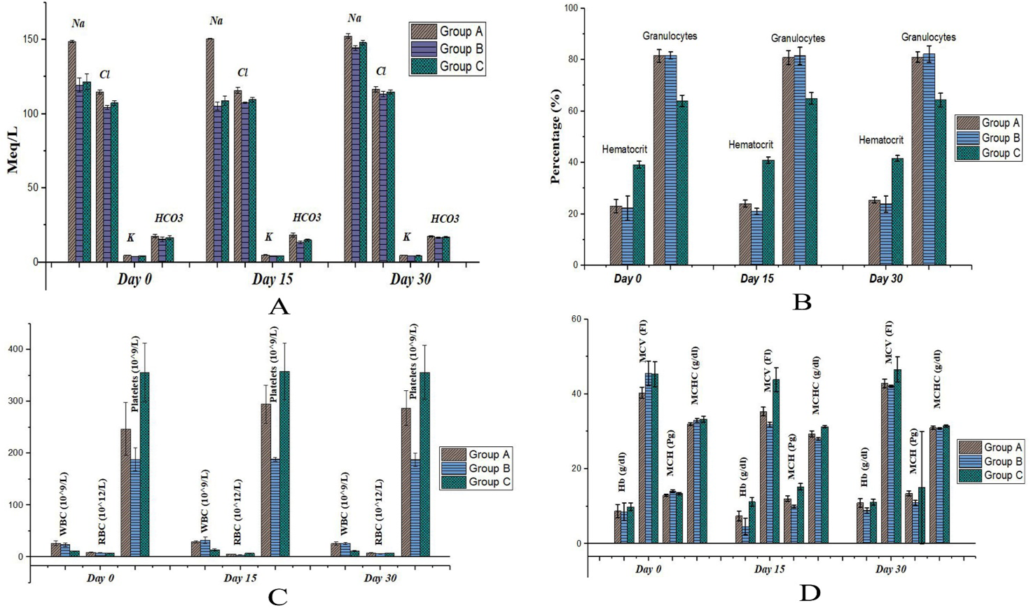
Effect of jejunal resection on the (A) electrolytes, (B) hematocrit and granulocytes, (C) WBC, RBC and platelets, (D) hemoglobin, MCV, MCH and MCHC on different days after resection.
References
- Astre, C., Gouttebel, M.C., Saint-Aubert, B., Girardot, P.M., Briand, D. and Joyeux, H., 1992. Clin. Nutr., 11: 30–34. https://doi.org/10.1016/0261-5614(92)90060-4[CrossRef]
- Bojrab, M.J., Ellison, G.W. and Slocum, B., 1998. Current techniques in small animal surgery. 4th eds., Williams and Wilkins, Baltimore, Md, London.
- Dilawer, M.S., Khan, M.A., Abidin, Z.U., Azeem, S., Majeed, K.A., Shahbaz, A. and Khan, A.R., 2011. Korean J. Vet. Res., 51: 309–313.
- Fabritius, J., Nell, G. and Loeschke, K., 1986. Pfl. Arch. Eur. J. Physiol., 406: 328–332. https://doi.org/10.1007/BF00640923[CrossRef]
- Guyton, A.C. and Hall, J.E., 2005. Textbook of medical physiology. W.B. Saunders, Philadelphia.
- Konig, H.E., Liebich, H.G. and Bragulla, H., 2004. Veterinary anatomy of domestic mammals: Textbook and colour atlas. Schattauer. https://books.google.com.pk/books?id=54RVGMmon10C
- Nightingale, J.M.D., 2001. World J. Gastroenterol., 7 (6): 741–751. https://doi.org/10.3748/wjg.v7.i6.741[CrossRef]
- Slatter, D.H., 2002. Textbook of small animal surgery. PA, Saunders, Philadelphia.
- Vazquez, C.M., Molina, M.T. and Ilundain, A., 1988. Rev. Esp. Fisiol., 44: 141–145. http://www.ncbi.nlm.nih.gov/pubmed/3194570
- Wolf, S.A., Telander, R.L., Go, V.L.W. and Dozois, R.R., 1979. J. Pediat. Surg., 14: 441–445. https://doi.org/10.1016/S0022-3468(79)80011-1[CrossRef]
- Yamada, S., Mayahara, T., Maruyama, I., Shibanoki, S., Tokuyoshi, K. and Iso, A., 1978. Nih. Yakuri. Zass., 74: 49–60. https://doi.org/10.1254/fpj.74.49[CrossRef]
- Zar, J.H., 1999. Biostatistical analysis. Upper Saddle River, N.J, Prentice Hall.
Services
Article usage statistics
Loading…