Research Article · Volume 11, Issue 2, 1 February 2023 · Open Access
MicroRNA-122 Expression Versus ALT for Liver Injury Detection Involved in Feline Infectious Peritonitis
· ·
Affiliations & Notes ⌄
1Department of Internal Medicine and Infectious Diseases, Faculty of Veterinary Medicine, Cairo University, Giza, Egypt. *Correspondence | Eman S. Ramadan, Department of Internal Medicine and Infectious Diseases, Faculty of Veterinary Medicine, Cairo University, Giza, Egypt
This study was focused on the measurement of miR-122 as a recently valuable indicator for liver injury versus to ALT activity in cats.
This study showed a significant increase of serum miRNA-122 in cats with effusive form of feline infectious peritonitis including liver by as 11.31-fold change compared to control healthy cats.
Serum biochemistry evaluation of these diseased cats showed a significant increase in ALT and AST.
This study suggests that serum miR-122 might be used as non-invasive, stable, diagnostic biomarker for hepatic injury detection involved in feline infectious peritonitis and consequently might be added to routine and traditional diagnostic methods of hepatic diseases in cats.
Selected from the article's own text.
Abstract
This study was carried out to evaluate the diagnostic utility of microRNA-122 (miR-122) for liver injury detection involved in feline infectious peritonitis (FIP). Twelve cats were enrolled in this study. Cats were admitted to small animal hospital, Faculty of veterinary medicine, Cairo University. Cats were classified into six apparently healthy cats as a control group and six clinically diseased cats. All cats were exposed to clinical examination, abdominal ultrasonography, serum biochemical analysis as well as rapid feline infectious peritonitis (FIP) test and Rivalta test for the diseased cats. On basis of these results, six cats diagnosed with feline infectious peritonitis (effusive form) involving liver injury. Serum microRNA-122 was estimated by real-time polymerase chain reaction in all cats. Effusive form of FIP with liver injury was manifested by abdominal distension with fluid, anorexia, icteric buccal mucous membrane, lethargy. Biochemical analysis showed significant elevation in alanine aminotransferase (ALT), aspartate aminotransferase (AST), and Globulin with significant reduction in albumin and A/G ratio. Abdominal ultrasonography revealed anechoic ascetic fluid in between internal organs, abdominal lymphadenopathy, and peritoneal adhesion. Serum microRNA-122 analysis showed significant elevation by 11.31 fold change compared to control cats. In conclusion, microRNA-122 is of diagnostic value and stable biomarkers for liver injury detection in cats.
One of the most fatal illnesses in cats is feline infectious peritonitis (FIP), a virus belongs to Feline Coronavirus . The virus has two forms in cats: Dry and effusive forms .
In clinical setting, effusive form is the most encountered with up to 85.8% of cats presented with ascites . Internal organs that are mostly affected are liver, kidney, intestine, and lymph nodes, with subsequent elevation in AST and ALT because of liver cell damage ; . As the liver is a vital organ, its affection is a major cause of mortality and morbidity in cats .
Owing to the non-specific nature of signs associated with hepatic disorders in cats, the diagnostic procedure might be challenging. Preliminary screening for hepatic injury depends on liver enzymes measurement as alanine aminotransferase (ALT) and aspartate transaminase (AST) ; ; .
In dogs and cats, the ALT estimation has been shown to have suboptimal sensitivity for hepatocellular injury detection and extrahepatic affections in cats causing hepatic enzymes elevation without primary hepatic disease ; ; .
MiRNAs are class of non-coding RNAs that have vital roles in liver as imperative regulators of post-transcriptional gene expression . MicroRNAs (miRNAs) are evolving as potential serum biomarkers for different affections in human and veterinary patients ; . Their specificity and stability in all body fluids makes them excellent biomarker ; Wang et al., 2016; .
Hepatocyte-derived miRNAs are the recently utilized in detection of early liver cell injury . miRNAs are advocated a stable and sensitive biomarker for liver injury detection in human and animal medicine ; ; ; Van der Meer et al., 2013; . MiRNA-122 is One of the most important miRNAs for liver cell injuries as it represents 72% of all miRNAs population in the liver ; , b). Circulating miR-122 has been shown to be a sensitive and specific indicator for liver injury in humans and dogs ; ; ; ; ; .
In human with liver injury, ALT elevation usually subsequent to miR-122 elevation, this early expression of miR-122 poised it as an early marker for hepatic injuries and disorders . Reports dealing with expression of miR-122 are scarce in cats, with one report showing elevation in miR-122 expression in diabetic cats and it may be related to diabetic ketoacidosis hepatocyte damage and hepatic lipidosis . Another recent report highlights the potential diagnostic capability of miR-122 as indicator of liver injury compared to ALT in cats . Therefore, this study aimed to evaluate the expression of miRNA-122 as indicator for liver injury detection involved in feline infectious peritonitis.
MATERIALS AND METHODS
Study design, animal examination and blood sample collection
This study employed twelve cats (n=12) admitted to teaching hospital of small animal, faculty of veterinary medicine, Cairo University. According to physical examination, the cats were classified into clinically diseased cats; (n=6) and apparently healthy cats; (n=6). All cats were subjected to complete clinical, abdominal ultrasonography examination, and laboratory analyses. Clinical signs were recorded at admission time. Blood sample was withdrawn from cephalic vein in each animal on plain tube for serum separation to estimate ALT, AST, total proteins, albumin, globulin, and A/G ratio using specific kits according to manufacturer instructions (Spectrum Diagnostic Kits, Egypt) (Eccls, 1989; ; . Remaining serum was stored at −20 °C till estimation of miRNA-122.
Abdominal fluid sampling and rivalta test
Abdominal fluid was aspirated under complete aseptic condition by abdominocentesis method, butterfly needle connected to a closed system and syringe (a 10-ml) was used for fluid collection. Fluid was placed in plain tubes and EDTA tubes. Fluid was examined for physical characters (color, viscosity, aspect) and the Rivalta test was performed on the collection day according to ; ; .
Feline infectious peritonitis (FIP) rapid test
Rapid test is immunochromatographic assay for the qualitative detection of FIP viral antigens in ascetic fluid (Diagnostic Megacor, AUSTRIA).
Abdominal ultrasonography examination
Abdominal ultrasonography (Mindray) was performed on each cat according to previously stated method . The ultrasound was performed to evaluate the echogenicity of hepatic parenchyma, liver size, kidney echogenicity, kidney shape, abdominal lymph nodes, the gastrointestinal tract, presence of free abdominal fluid and splenic echogenicity .
Determination of hepatocyte-derived miRNA-122
RNA extraction from blood samples was performed according to manufacture instructions of QIAamp RNeasy Mini kit (Qiagen, Germany, GmbH).
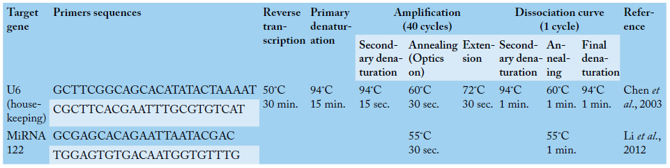
Primers used were supplied from Metabion (Germany) were miRNA-122 (GCGAGCACAGAATTAATACGAC, TGGAGTGTGACAATGGTGTTTG) and U6 (housekeeping) (GCTTCGGCAGCACATATACTAAAAT, CGCTTCACGAATTTGCGTGTCAT) are shown in Table 1. Primers were utilized in a 25- µl reaction containing 12.5 µl of the 2x Quanti Tect SYBR Green PCR Master Mix (Qiagen, Germany, GmbH), 0.25 µl of RevertAid Reverse Transcriptase (200 U/µL) (Thermo Fisher), 0.5 µl of each primer at a concentration of 20 pmol, 8.25 µl of water, and 3 µl of RNA template was used. A Stratagene MX3005P real-time PCR machine was used to carry out the reaction. The stratagene MX3005P software was used to calculate the amplification curves and CT values. The CT of each sample was compared to the CT of the positive control group according to the “ΔΔCt” method stated by Yuan et al. (2006) using the following ratio: (2-∆∆ct).
Statistical analysis
All data of serum biochemistry were presented as mean ± standard error. The comparison between control and the diseased group was made using SPSS statistic program version 16.0 (independent-samples T test), p value ≤ 0.05 considered significant.
RESULTS
Clinical examination and biochemical analyses findings
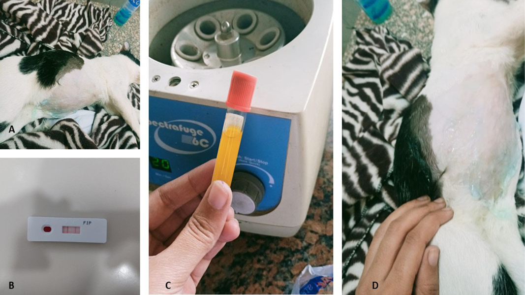
In diseased cats, abdominal distension with fluid, icteric buccal mucous membrane, anorexia, depression, lethargy and recumbent were recorded (Figure 1a, c, d). Results of biochemical analysis showed significant elevation in ALT, AST and Globulin associated with significant reduction in albumin and A/G ratio in diseased group compared to control group (Table 2).
Rivalta test finding of abdominal fluid
Rivalta test showed clump which reflected a positive result.
Feline infectious peritonitis (FIP) rapid test finding
FIP rapid test showed a colored line in the test region representing a positive result (Figure 1b).
Data are represented as mean ±SE; P value ≤ 0.05 considered significant.
Abdominal ultrasonography finding
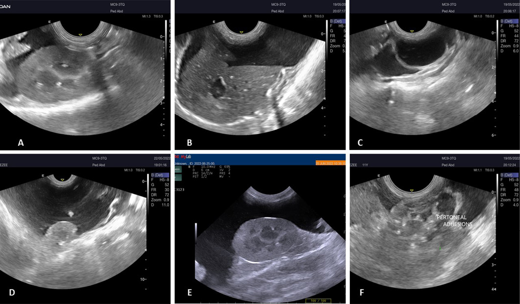
Abdominal ultrasonography revealed free anechoic fluid in the peritoneal cavity (in between liver lobes, separating right kidney and quadate liver lobe, abdominal cavity), abdominal lymphadenopathy, intestinal thickening as well as peritoneal adhesion was recorded (Figure 2a –f).
Serum hepatocyte derived miRNA-122 finding
The serum miRNA-122 analysis showed significant increase by 11.31-fold change in cats with liver injury involved in feline infectious peritonitis when compared to control healthy cat group (Table 3). miR- 122 elevated by 11.31-fold change but ALT level elevated two-fold change when compared to control group.
Feline infectious peritonitis (FIP) is a fatal viral illness currently incurable by medications. Also, there are no effective vaccines ; ; . It is immune-mediated infection caused by feline coronavirus ; . There are two forms effusive and non-effusive form . Effusive form of it progresses faster and more lethal than dry form .
Owing to non-specificity of signs, the diagnostic procedure could be exhaustive and challenging. The ALT is the gold standard marker for liver cell damage , though, it has limitations ; . Moreover, the elevated serum ALT activity does not always correlate with histopathological findings of hepatic tissue ; ; . For that, there was an increase on the demand in veterinary medicine for more specific and more sensitive biomarkers for early recognition of cellular injury in liver
This study was focused on the measurement of miR-122 as a recently valuable indicator for liver injury versus to ALT activity in cats. MiRNA-122 is One of the most important miRNAs for hepatic cell injuries as it represents 72% of all miRNAs population in the liver ; , b).
This study showed a significant increase of serum miRNA-122 in cats with effusive form of feline infectious peritonitis including liver by as 11.31-fold change compared to control healthy cats. These findings come in accordance with human studies where serum miR-122 level elevated due to liver cell injury of different etiology ; Van der Meer et al., 2013; ; . Vliegenthart et al. (2015) reported in peoples with acetaminophen-induced hepatic injury that miR-122 had better specificity than other existing biomarkers and predicted the course of illness than ALT alone. Veterinary studies on canine have shown that serum miRNA-122 level elevates due to hepatic injury , b; ; . To date there are few papers available regarding miR-122 in feline hepatic diseases. In one study related to diabetes in cats, there was increase in miR-122 expression in diabetic cats . The more than 40-fold change increase was assumed to be a result of hepatocyte damage from diabetic ketoacidosis or hepatic lipidosis ; . Another study reported that miR-122 was higher in cats with increased ALT level than in cats with normal serum ALT level and this study highlights the potential diagnostic utility of miR-122 as an indicator of liver injury in cats and encourages further investigation to be applied in this species . In the current investigation, 11.31-fold change increase in miR-122 was reported while elevation in ALT and AST was estimated to be about two-fold change, these findings may support that miR-122 might be valuable indicator for hepatic injuries detection .
The current study recorded abdominal distension with fluid, icterus, weight loss and anorexia in the diseased cats. These signs are comparable with those recorded in the effusive FIP ; . Abdominal fluid aspiration revealed viscous fluid, which could be attributed to high protein content ; . Positive result of Rivalta test obtained from diseased cats. It is a simple test that distinguishes between exudate and transudate , it was used in diagnosis of feline infectious peritonitis .
Serum biochemistry evaluation of these diseased cats showed a significant increase in ALT and AST. Nururrozi et al. (2022) reported that ALT and AST were elevated in effusive form of FIP by two-fold than the normal. These elevations of ALT and AST may be related to the changes in liver cell membrane permeability and hepatocytes damage ; . Advanced peritonitis in association with FIP was implicated as a cause of hepatic inflammation ; .
Hypoalbuminemia and hyperglobulinemia were recorded in this study which comes in accordance with other reports ; . The reduction of albumin production by affected liver in FIP infection could be implicated . Hyperglobulinemia was reported in nearly 89.1% of cats with FIP regardless its type , because of specific anti-FCoV immune response . Low albumin: globulin ratio (A/G ratio) was recorded in this study which agreed with other reports . A/G ratio was argued to be of higher diagnostic value compared to other hematological tests, ; ; ; ; .
Abdominal ultrasonography in this study revealed anaechoic asitiec fluid in abdomen and in between internal organs as liver, kidney, and intestines confirming the effusive form which is characterized by ascites ; . Furthermore, Yin et al., (2021) reported ascites and few cats showed loss of corticomedullary distinction of the kidneys in cats with FIP.
CONCLUSIONS AND RECOMMENDATION
This study suggests that serum miR-122 might be used as non-invasive, stable, diagnostic biomarker for hepatic injury detection involved in feline infectious peritonitis and consequently might be added to routine and traditional diagnostic methods of hepatic diseases in cats.
ACKNOWLEDGEMENTS
Authors would like to thank small animal teaching hospital, Faculty of veterinary medicine, Cairo University for facilitating this work.
Novelty Statement
To our knowledge, this is the third study in cats to evaluate the diagnostic utility of microRNA-122 for liver injury detection.
Author’s Contribution
All authors contributed equally according to their tasks and approved the final manuscript.
Ethical approval
All procedures performed in this study were in accordance with the ethical standards of the institution at which the study was conducted “Institutional Animal Care and Use Committee, Cairo University” and was allotted permit number (Vet Cu 2009 2022491).
Conflict of interest
The authors have declared no conflict of interest.
Figures
Figure 1:.
Clinical examination of cat suffering from feline infectious peritonitis (effusive form) revealed abdominal distension with fluid (a, c, d). Rapid FIP test showed positive result (b).
Table 1:.
Primers sequences, target genes, amplicon sizes and cycling conditions for SYBR green RT-PCR.
Figure 2:.
Abdominal ultrasonography of cats with feline infectious peritonitis involving liver (effusive form) showed free anechoic fluid present in the peritoneal cavity, abdominal lymphadenopathy and peritoneal adhesion.
References
Addie D, Belák S, Boucraut-Baralon C, Eqberink H, Frymus T, Gruffydd-Jones T, Hartmann K, Hosie MJ, Lloret A, Lutz H, Marsilio F, Pennisi MG, Radford AD, Thiry E, Truyen U, Horzinek MC (2009). Feline infectious peritonitis. ABCD guidelines on prevention and management. J. Feline Med. Surg. 11: 594–604. https://doi.org/10.1016/j.jfms.2009.05.008[CrossRef]
Antoine DJ, Dear JW, Lewis PS, Platt V, Coyle J, Masson M, Thanacoody RH, Gray AJ, Webb DJ, Moggs JG, Bateman DN, Goldring CE, Park BK (2013). Mechanistic biomarkers provide early and sensitive detection of acetaminophen-induced acute liver injury at first presentation to hospital. Hepatology, 58: 777–787. https://doi.org/10.1002/hep.26294[CrossRef]
Armstrong SK, Oosthuyzen W, Gow AG, Schmitz SS, Dear JW, Mellanby RJ (2022). Investigation of a relationship between serum concentrations of microRNA-122 and alanine aminotransferase activity in hospitalised cats. J. Feline Med. Surg., 24(8): 289-294. https://doi.org/10.1177/1098612X221100071[CrossRef]
Breuer J (1996). Report on the symposium drug effects in clinical chemistry methods. Eur. J. Clin. Chem. Clin. Biochem., 34: 385-386.
Bruskiewicz KA, Nelson RW, Feldman EC, Griffey SM (1997). Diabetic ketosis and ketoacidosis in cats: 42 cases (1980–1995). J. Am. Vet. Med. Assoc., 211: 188–192.
Cermelli S, Ruggieri A, Marrero JA, Loannou GN, Beretta L (2011). Circulating microRNAs in patients with chronic hepatitis C and Non-alcoholic fatty liver disease. PLoS One, 6: e23937. https://doi.org/10.1371/journal.pone.0023937[CrossRef]
Chen Y, Sun Y, Chen L, Xu X, Zhang X, Wang B, Min L, Liu W (2003). MiRNA-200c increases the sensitivity of breast cancer cells to doxorubicin through the suppression of E-cadherin-mediated PTEN/Akt signaling. Mol. Med. Rep., 7(5): 1579-1584. https://doi.org/10.3892/mmr.2013.1403[CrossRef]
Condrat CE, Thompson DC, Barbu MG, Bugnar OL, Boboc A, Cretoiu D, Suciu N, Cretoiu SM, Voinea SC (2020). miRNAs as biomarkers in disease: Latest findings regarding their role in diagnosis and prognosis. Cells, 9: 276. https://doi.org/10.3390/cells9020276[CrossRef]
Dear JW, Antoine DJ (2014). Stratification of paracetamol overdose patients using new toxicity biomarkers: current candidates and future challenges. Expert. Rev. Clin. Pharmacol., 7: 181–189. https://doi.org/10.1586/17512433.2014.880650[CrossRef]
Diaz JV, Poma R (2009). Diagnosis and clinical signs of feline infectious peritonitis in the central nervous system. Can. Vet. J., 50: 1091-1093.
Dirksen K, Burgener IA, Rothuizen J (2017). Hepatocyte derived microRNAs as sensitive serum biomarkers of hepatocellular injury in Labrador retrievers. J. Vet. Intern. Med., 31: 1017–1027.
Dirksen K, Verzijl T, Grinwis GC, Favier RP, Penning LC, Burgener IA, van der Laan LJ, Fieten H, Spee B (2016b). Use of serum MicroRNAs as biomarker for hepatobiliary diseases in dogs. J. Vet. Intern. Med. 30(6): 1816–1823. https://doi.org/10.1111/jvim.14602[CrossRef]
Dirksen K, Verzijl T, van den Ingh TS, Vernooij JC, van der Laan LJ, Burgener IA, Spee B, Fieten H (2016a). Hepatocyte-derived microRNAs as sensitive serum biomarkers of hepatocellular injury in Labrador retrievers. Vet. J., 211: 75–81. https://doi.org/10.1016/j.tvjl.2016.01.010[CrossRef]
Eccl S (1989). Determination of the catalytic activity concentration in serum on L-alanine aminotransferase (EC 2.6.1.2, ALAT). Clin. Chem., 20: 204-211.
Eman SR, Kubesy AA, Baraka TA, Torad FA, Shaymaa IS, Mohammed FF (2018). Evaluation of hepatocyte-derived microRNA-122 for diagnosis of acute and chronic hepatitis of dogs. Vet. World, 11: 667–673. https://doi.org/10.14202/vetworld.2018.667-673[CrossRef]
Ettinger SJ, Feldman EC (2017). Textbook of veterinary internal medicine, 8th ed. St Louis, MO, Elsevier.
Felten S, Hartmann K (2019). Diagnosis of feline infectious peritonitis: A review of the current literature. Viruses, 11(11): 1068. https://doi.org/10.3390/v11111068[CrossRef]
Fischer Y, Sauter-Louis C, Hartmann K (2012). Diagnostic accuracy of the rivalta test for feline infectious peritonitis. Vet. Clin. Pathol., 41: 558–567. https://doi.org/10.1111/j.1939-165X.2012.00464.x[CrossRef]
Fleischhacker SN, Bauersachs S, Wehner A, Hartmann K, Weber K (2013). Differential expression of circulating microRNAs in diabetic and healthy lean cats. Vet. J., 197: 688–693. https://doi.org/10.1016/j.tvjl.2013.03.027[CrossRef]
Goodson TL, Randell SC, Moore LE (2009). Feline infectious peritonitis. Compend. Contin. Educ. Pract. Vet., 31: 1-9.
Grasedieck S, Schöler N, Bommer M, Niess JH, Tumani H, Rouhi A, Bloehdorn J, Liebisch P, Mertens D, Döhner H, Buske C, Langer C, Kuchenbauer F (2012). Impact of serum storage conditions on microRNA stability. Leukemia, 26: 2414. https://doi.org/10.1038/leu.2012.106[CrossRef]
Guo Z, Maki M, Ding R, Yang Y, zhang B, Xiong L (2014). Genome-wide survey of tissue-specific microRNA and transcription factor regulatory networks in 12 tissues. Sci. Rep., 4: 5150. https://doi.org/10.1038/srep05150[CrossRef]
Harrill AH, Eaddy JS, Rose K, Cullen JM, Ramanathan L, Wanaski S, Collins S, Ho YU, Watkins PB, LeCluyse EL (2014). Liver biomarker and in vitro assessment confirm the hepatic origin of aminotransferase elevations lacking histopathological correlate in beagle dogs treated with GABAA receptor antagonist NP260. Toxicol. Appl. Pharmacol., 277: 131–137. https://doi.org/10.1016/j.taap.2014.03.015[CrossRef]
Hartmann K, Ritz S (2008). Treatment of cats with feline infectious peritonitis. Vet. Immunol. Immunopathol. 123: 172–175. https://doi.org/10.1016/j.vetimm.2008.01.026[CrossRef]
Hasan KMDM, Tamanna N, Haque MDA (2018). Biochemical and histopathological profiling of Wistar rat treated with Brassica napus as a supplementary feed. Food Sci. Hum. Wellness, 7: 77–82. https://doi.org/10.1016/j.fshw.2017.12.002[CrossRef]
Hirschberger J, Hartmann K, Wilhelm N, Frost J, Lutz H, Kraft W (1995). Clinical symptoms and diagnosis of feline infectious peritonitis. Tierarztl. Prax., 23: 92–99.
Jeffery U, Deitz K, Hostetter S (2012). Positive predictive value of albumin: Globulin ratio for feline infectious peritonitis in a mid-western referral hospital population. J. Feline Med. Surg., 14: 903–905. https://doi.org/10.1177/1098612X12454862[CrossRef]
Kipar A, Meli ML (2014). Feline infectious peritonitis: Still an enigma? Vet. Pathol., 51: 505–526. https://doi.org/10.1177/0300985814522077[CrossRef]
Krol J, Loedige L, Filipowicz W (2010). The widespread regulation of microRNA biogenesis, function and decay. Nat. Rev. Genet., 11: 597-610. https://doi.org/10.1038/nrg2843[CrossRef]
Kundrotas LW, Clement DJ (1993). Serum alanine aminotransferase (ALT) elevation in asymptomatic US Air Force basic trainee blood donors. Digest. Dis. Sci., 38: 2145–2150. https://doi.org/10.1007/BF01299887[CrossRef]
Laterza OF, Lim L, Garrett-Engele PW, Vlasakova K, Muniappa N, Tanaka WK, Johnson JM, Sina JF, Fare TL, Sistare FD, Glaab WE (2009). Plasma microRNAs as sensitive and specific biomarkers of tissue injury. Clin. Chem., 55: 1977-1983. https://doi.org/10.1373/clinchem.2009.131797[CrossRef]
Lewis KM, O’Brien RT (2010). Abdominal ultrasonographic findings associated with feline infectious peritonitis: A retrospective review of 16 cases. J. Am. Anim. Hosp. Assoc., 46(3): 152-160. https://doi.org/10.5326/0460152[CrossRef]
Lewis PJS, Dear J, Platt V, Simpson KJ, Craig DG, Antoine DJ, French NS, Dhaun N, Webb DJ, Costello EM, Neoptolemos JP, Moggs J, Goldring CE, Park BK (2011). Circulating microRNAs as potential markers of human drug-induced liver injury. Hepatology, 54: 1767-1776. https://doi.org/10.1002/hep.24538[CrossRef]
Li s, Zhu J, Fu H, Wan J, Hu Z, Liu S, Li J, Tie Y, Xing R., Zhu J, Sun Z, Zheng X (2012). Hepato-specific microRNA-122 facilitates accumulation of newly synthesized miRNA through regulating PRKRA. Nucl. Acids Res., 40(2): 884-891. https://doi.org/10.1093/nar/gkr715[CrossRef]
Lidbury JA, Suchodolski JS (2016). New advances in the diagnosis of canine and feline liver and pancreatic disease. Vet. J., 215: 87–95. https://doi.org/10.1016/j.tvjl.2016.02.010[CrossRef]
Malbon AJ, Fontara S, Meli ML, Hahn S, Egberink H, Kipar A (2019). Feline infectious peritonitis as a systemic inflammatory disease: Contribution of liver and heart to the pathogenesis. Viruses, 11(12): 1144. https://doi.org/10.3390/v11121144[CrossRef]
Miretti S, Lecchi C, Ceciliani F, Baratta M (2020). MicroRNAs as biomarkers for animal health and welfare in livestock. Front. Vet. Sci., 7: 985. https://doi.org/10.3389/fvets.2020.578193[CrossRef]
Norris JM, Bosward KL, White JD, Baral RM, Catt MJ, Malik R (2005). Clinico- pathological findings associated with feline infectious peritonitis in Sydney, Australia: 42 cases (1990–2002). Aust. Vet. J., 83: 666–673. https://doi.org/10.1111/j.1751-0813.2005.tb13044.x[CrossRef]
Nururrozi A, Ramandani D, Wasissa M, Yanuartono, Indarjulianto S (2022). Serum biochemistry profiles in confirmed effusive feline infectious peritonitis cats. Adv. Anim. Vet. Sci., 10(1): 126-130. https://doi.org/10.17582/journal.aavs/2022/10.1.126.130[Full text][CrossRef]
Nyland TG, Larson MM, Mattoon JS (2015). Liver. In: Mattoon JS, Nyland TG (ed). Small animal diagnostic ultrasound, 3rd edn. St. Louis, MO: Elsevier: 332–399. https://doi.org/10.1016/B978-1-4160-4867-1.00009-X[CrossRef]
Oosthuyzen W, Ten Berg PWL, Francis B, Campbell S, Macklin V, Milne E, Gow AG, Fisher C, Mellanby RJ, Dear JW (2018). Sensitivity and specificity of microRNA-122 for liver disease in dogs. J. Vet. Intern. Med., 32: 1637–1644. https://doi.org/10.1111/jvim.15250[CrossRef]
Ozer J, Ratner M, Shaw M, Bailey W, Schomaker S (2008). The current state of serum biomarkers of hepatotoxicity. Toxicology, 245: 194–205. https://doi.org/10.1016/j.tox.2007.11.021[CrossRef]
Paltrinieri S, Comazzi S, Spagnolo V (2002). Laboratory changes consistent with feline infectious peritonitis in cats from multicat environments. J. Vet. Med. Physiol. Pathol. Clin. Med., 49: 503–510. https://doi.org/10.1046/j.1439-0442.2002.00494.x[CrossRef]
Pedersen NC (2009). A review of feline infectious peritonitis virus infection: 1963–2008. J. Feline Med. Surg. 11: 225–258. https://doi.org/10.1016/j.jfms.2008.09.008[CrossRef]
Pedersen NC (2014). An update on feline infectious peritonitis: Diagnostics and therapeutics. Vet. J., 201: 133–141. https://doi.org/10.1016/j.tvjl.2014.04.016[CrossRef]
Pedersen NC, Eckstrand C, Liu H, Leutenegger C, Murphy B (2015). Levels of feline infectious peritonitis virus in blood, effusions, and various tissues and the role of lymphopenia in disease outcome following experimental infection. Vet. Microbiol., 175: 157–166. https://doi.org/10.1016/j.vetmic.2014.10.025[CrossRef]
Ramadan ES, Kubesy AA, Baraka TA, Torad FA, Shaymaa IS, Salem NY (2019). Expression of blood hepatocyte-derived microRNA-122 in canine multicentric lymphoma with hepatic involvement. Vet. Res. Commun., 43: 231–238. https://doi.org/10.1007/s11259-019-09764-w[CrossRef]
Riemer F, Kuehner KA, Ritz S, Sauter-Louis C, Hartmann K (2016). Clinical and laboratory features of cats with feline infectious peritonitis-a retrospective study of 231 confirmed cases (2000–2010). J. Feline Med. Surg., 18: 348–356. https://doi.org/10.1177/1098612X15586209[CrossRef]
Sparkes AH, Gruffydd-Jones TJ, Harbour DA (1991). Feline infectious peritonitis: A review of clinico- pathological changes in 65 cases, and a critical assessment of their diagnostic value. Vet. Rec., 129: 209–212. https://doi.org/10.1136/vr.129.10.209[CrossRef]
Tanaka Y, Sato Y, Takahashi D, Matsumoto H, Sasaki T (2015). Treatment of a case of feline infectious peritonitis with cyclosporine A. Vet. Rec. Case Rep., 3: e000134. https://doi.org/10.1136/vetreccr-2014-000134[CrossRef]
Tietz NW (1994). Fundamentals of clinical chemistry: 2nd ed. NW Tietz, editor, pp. 692.
Tsai HY, Chueh LL, Lin CN, Su BL (2011). Clinicopathological findings and disease staging of feline infectious peritonitis: 51 cases from 2003 to 2009 in Taiwan. J. Feline Med. Surg., 13: 74–80. https://doi.org/10.1016/j.jfms.2010.09.014[CrossRef]
Van der Meer AJ, Farid WRR, Sonneveld MJ, de Ruiter PE, Boonstra A, van Vuurren AJ, Verheij J, Hansen BE, de Knegt RJ, van der Laan LJW (2013). Sensitive detection of hepatocellular injury in chronic hepatitis C patients with circulating hepatocyte-derived microRNA-122. J. Viral Hepat., 20: 158-166. https://doi.org/10.1111/jvh.12001[CrossRef]
Vliegenthart ADB, Shaffer JM, Clarke JI, Peeters LEJ, Caporali A, Bateman DN, Wood DM, Dargan PI, Craig DG, moore JK, Thompson AI, Henderson NC, Webb DG, Sharkey J, Antoine DJ, Park BK, Bailey MA, Lader E, Simpson KJ, Dear JW (2015). Comprehensive microRNA profiling in acetaminophen toxicity identifies novel circulating biomarkers for human liver and kidney injury. Sci. Rep., 5: 1–13. https://doi.org/10.1038/srep15501[CrossRef]
Wang H, Naghavi M, Allen C, Barber RM, Bhutta ZA, Carter A (2016). Global, regional, and national life expectancy, all-cause mortality, and cause specific mortality for 249 causes of death, 1980–2015: A systematic analysis for the global burden of disease study. Lancet, 388: 1459–1544.
Wang K, Zhang S, Marzolf B, Troisch P, Brightman A, Hu Z, Hood LE, Galas DJ (2009). Circulating microRNAs, potential biomarkers for drug-induced liver injury. Proc. Natl. Acad. Sci. USA, 106: 4402-4407. https://doi.org/10.1073/pnas.0813371106[CrossRef]
Yin Y, Li T, Wang C, Liu X, Ouyang H, Ji W, Liu J, Liao X, Li J, Hu C (2021). A retrospective study of clinical and laboratory features and treatment on cats highly suspected of feline infectious peritonitis in Wuhan, China. Sci. Rep., 11: 5208. https://doi.org/10.1038/s41598-021-84754-0[CrossRef]
Yuan JS, Reed A, Chen F, Stewart CN (2006). Statistical analysis of real-time PCR data. BMC Bioinf., 7: 85. https://doi.org/10.1016/j.csda.2005.11.017[CrossRef]
Zhang Y, Jia Y, Zheng R, Guo Y, Wang Y, Guo H, Fei M, Sun S (2010). Plasma microRNA-122 as a biomarker for viral-, alcohol-, and chemical related hepatic diseases. Clin. Chem., 56: 1830-1838. https://doi.org/10.1373/clinchem.2010.147850[CrossRef]