
The 25-hydroxyvitamin D [25(OH)D] level in serum is currently considered the best indicator of vitamin D supply to the body from cutaneous synthesis and nutritional intake. Few reports are available regarding the effects of vitamin D and its metabolites on heart development in swine. The effect of 25(OH)D3 and Ca2+ supplementation in diets on heart development in neonatal piglets during pregnancy were examined in this study. Total 40 sows of 7 gestational age with good health and nutritional condition were divided into four groups (n=10): the control group, low calcium, 25(OH)D3 group and low calcium with 25(OH)D3 group. Each groups consist of 5 piglets, randomly selected and euthanized within 2 h after birth. After euthanasia, heart was collected for histopathological examination and IGF, IGF1, IGF2, IGFBP3, PCNA and ki67 expression were analyzed. The results showed that low calcium supplementation decreased the cardiac index and development of myocardial muscle fiber, while vitamins D3 can improve the decrease of cardiac index and development of myocardial fiber due to low calcium supplementation. The expression profile of IGF1, IGF2, IGFBP3, PCNA and ki67 genes were down regulated in LCa group as compared to control group; while the vitamin D3 supplementation significantly up regulated the above genes expression. In conclusion, our findings suggested that 25(OH)D3 and Ca2+ supplementation in sows’ diets has an important role in the development of heart and differentiation of myocardial fibers in neonatal piglets by changing the expression of the IGF pathway and myocardial cells proliferation.
Short Communication · Volume 55, Issue 1, 14 November 2022 · Open Access
Effect of 25(OH)D3 Supplementation in Sows’ Diets on Heart Development in Neonatal Piglets
· · · · · · · · · · · ·
Affiliations & Notes ⌄
- 1College of Veterinary Medicine, South China Agricultural University, Guangzhou 510642, China. 2Huizhou Engineering Vocational College, Huizhou, China. 3Faculty of Veterinary and Animal Sciences, Islamia University of Bahawalpur, Bahawalpur 4Lasbela University of Agriculture Water and Marine Sciences, Uthal, Balochistan * Corresponding author: tangzx@scau.edu.cn
Article Info ⌄
- Journal:
- Pakistan Journal of Zoology
- Published:
- 14 November 2022

From the article
- In our study, the results indicated that the daily weight gain was increased in vitamin D3 and LCa+VD3 groups as compared with control and LCa groups from day 0-7d and 15-21d.
- In our study, the effects of 25(OH)D3 and Ca2+ on cardiac index of piglets showed that low calcium supplementation decreased the index of cardiac compared to control group, while VD3 can improve the decrease of cardiac index, caused by low calcium.
- H and E stained histopathology micrograph showed that low calcium supplementation decreased the development of myocardial fiber, and VD3 can promote the development of myocardial fibers (Fig. 2).
- However, the expression profile of IGF1, IGF2 and IGFBP3 genes were decreased in LCa group as compared with control group; while the VD3supplementation can significantly up regulated the IGF1, IGF2 and IGFBP3 gene expression (Fig. 3).
Selected from the article's own text.
Abstract
Materials and methods
Total 40 sows of 7 gestational age with good health and nutritional condition, similar genetic background and due date were selected, and randomly divided into 4 groups (n=10). The sows were kept and raised under the recommended temperature conditions. The feed was prepared as per recommendations of NRC while nutrient composition of the sow’s diet is shown in Supplementary Table S1. The contents of calcium, phosphorus and 25(OH)D3 in the diet were adjusted in accordance with the experimental design, and were divided into control group, low calcium group, 25(OH)D3 group and low calcium with 25(OH)D3 group each of 10 animals. The calcium, phosphorus and 25(OH)D3 were supplied as shown in Supplementary Table S2.
These four groups, were raised from day 85 to day 110; after day 110, all the sows were fed in the delivery room with recommended temperature (20 oC). During the experimental period, the sows were feeding, drinking and immunization according to the husbandry procedures of the pig farm; meanwhile, the weight, feed intake and mortality in each group were recorded. A total of 5 piglets in each groups were randomly selected and euthanized within 2 h after birth. After euthanasia, the heart was collected and fixed in 4% paraformaldehyde; the body weight and the weight of the heart were measured for cardiac index analysis, and then the heart samples were dissected, immediately frozen in liquid nitrogen for RT-qPCR analysis.
The heart samples were fixed into 4% neutral buffered paraformaldehyde at 4 °C, and then tissue was dehydrated in graded (70%, 80%, 90%, 95%, and 100%) ethanol solutions, removed in xylene and embedded in paraffin. Sections were cut with 4-5μm thickness and placed on polylysine-coated slides for hematoxylin and eosin staining.
For qRT-PCR all the heart samples from each group were frozen in liquid nitrogen and homogenized in TRIzol reagent (TaKaRa) to extract total RNA, and reverse transcribed to cDNA by using a commercial kit (TransScript First-Strand cDNA Synthesis) with the recommended reaction temperatures. The qRT-PCR was performed five times by using the Step One-PlusTM Real-Time PCR System for IGF, IGF1R, IGF2, IGFBP3, PCNA, ki67 and GAPDH genes. The primers were synthesized by Shenggong (Shanghai) and are shown in Table I. The PCR reaction were performed with following thermal cycling parameters; denaturing at 98 °C for 30s, 40 amplification cycles at 98 °C for 10s, 57 °C for 40s and 72 °C for 40s.The relative quantification
The data were expressed as mean ± standard deviation (SD) by using SPSS 19.0 software. P<0.05, statistically significant.
Table I. Primers used for the qRT-PCR.
| Primers | Primer sequence (5' to 3') | Product size (bp) |
| IGF1 | F: TTCTACTTGGCCCTGTGCTT | 222 |
| R: CTCCAGCCTCCTCAGATCAC | ||
| IGF2 | F: ACACCCTCCAGTTTGTCTGC | 212 |
| R: GGGGTATCTGGGGAAGTTGT | ||
| IGF1R | F: ACTGTATGGTGGCCGAAGAC | 163 |
| R: TCAGAGTGCGTGGTGAAGAC | ||
| IGFBP3 | F: TCTGTCCACACCAAGATGGA | 181 |
| R: GGAACTTGAGGTGGTTCAGC | ||
| PCNA | F: TGTGCTGGCAATGAAGACAT | 209 |
| R: TCTCGGCATATACGTGCAAA | ||
| Ki67 | F: CACCAGGCTTTACGGAAGAA | 189 |
| R: AGATACGGGCTGCTTGAAAA | ||
| GAPDH | F: GTCGGTTGTGGATCTGACCT | 210 |
| R: AGCTTGACGAAGTGGTCGTT |
Results and discussion
Ca2+ is essential for a variety of vital physiological processes such as cell proliferation, differentiation, motility, secretion, excitation, and apoptosis. Adding suitable calcium and vitamin D3 in the diet can improve the growth performance of animals significantly, such as reduced feed-meat ratio, increased daily weight gain and daily feed intake ; . However, there were limited studies about the relationship between maternal 25(OH)D3 and Ca2+ supplementation on heart development of neonatal piglets during anaphase of embryonic development of maternal and neonatal has been reported. In our study, the results indicated that the daily weight gain was increased in vitamin D3 and LCa+VD3 groups as compared with control and LCa groups from day 0-7d and 15-21d. Whereas, the daily weight gain was decreased in VD3 and LCa+VD3 groups (Fig. 1A). However, the average daily weight gain was no significantly changed among those four different treatment groups during the experimental period (Fig. 1B), which indicated no correlation to the average daily weight gain between 25(OH)D3 and Ca2+ supplementation.
The main hormones that regulate calcium and calcium metabolism are active vitamin D, parathyroid hormone and calcitonin . Vitamin D promotes calcium absorption and bone salt deposition in the small intestine. The main target organs are the small intestine and bone (Yao et al., 2019; . The principal action of vitamin D in maintenance of calcium homeostasis is increased intestinal calcium absorption and mineralization of bone ; . In neonatal developing cardiac cells, the relative mature sarcoplasmic reticulum plays a role as the main source of Ca required for contraction compared to that in the fetus. Calcium homeostasis in cardiomyocytes shows central role in the development of the heart during its early development . In our study, the effects of 25(OH)D3 and Ca2+ on cardiac index of piglets showed that low calcium supplementation decreased the index of cardiac compared to control group, while VD3 can improve the decrease of cardiac index, caused by low calcium. H and E stained histopathology micrograph showed that low calcium supplementation decreased the development of myocardial fiber, and VD3 can promote the development of myocardial fibers (Fig. 2).
As an important endocrine hormone, IGF has been confirmed to participate in variety of cell proliferation process, is mainly responsible for tissue growth and development, such as muscle, bone, kidney, skin, lung, and liver, etc. Meanwhile, as a strong mitogen, IGF can promote cell growth, proliferation, differentiation, and inhibits apoptosis . The expression profile of IGF gene involved in the growth plates of piglets indicated that the mRNA expression of IGF was increased in LCa and LCa+VD3 treatment groups as compared to control group and VD3 group. However, the expression profile of IGF1, IGF2 and IGFBP3 genes were decreased in LCa group as compared with control group; while the VD3supplementation can significantly up regulated the IGF1, IGF2 and IGFBP3 gene expression (Fig. 3).
Previous studies have shown that PCNA and ki67 are considered as the most widely used markers of cell proliferation . PCNA has significant roles in the metabolism of nucleic acid in DNA replication, DNA excision repair, cell cycle control, chromatin assembly, and RNA transcription; while ki67 is a nuclear antigen that exists in proliferating cells, connected with chromatin and related to cell mitosis . The mRNA expression of PCNA and ki67 genes were confirmed through qRT-PCR in control, LCa, VD and LCa+VD groups. The expression of PCNA and ki67 was decreased in LCa group compared to control and VD groups. After the administration of VD, the expression of PCNA and ki67 was increased compared to LCa sows (Fig. 4).
In conclusion, our findings suggested that 25(OH)D3 and Ca2+ supplementation in sows’ diets shows key role in heart development in neonatal piglets, and 25(OH)D3 treatment of gestational Ca2+ deficiency reactivates the IGF system and cell proliferation and promotes heart development and differentiation of myocardial fibers in the fetal pigs during late-gestation sows.
Acknowledgment
The study was supported by National Key Research and Development Program of China (Project No. 2017YFD0502200).
Supplementary material
There is supplementary material associated with this article. Access the material online at: https://dx.doi.org/10.17582/journal.pjz/20200528020558
Statement of conflict of interest
The authors have declared no conflict of interest.
Figures
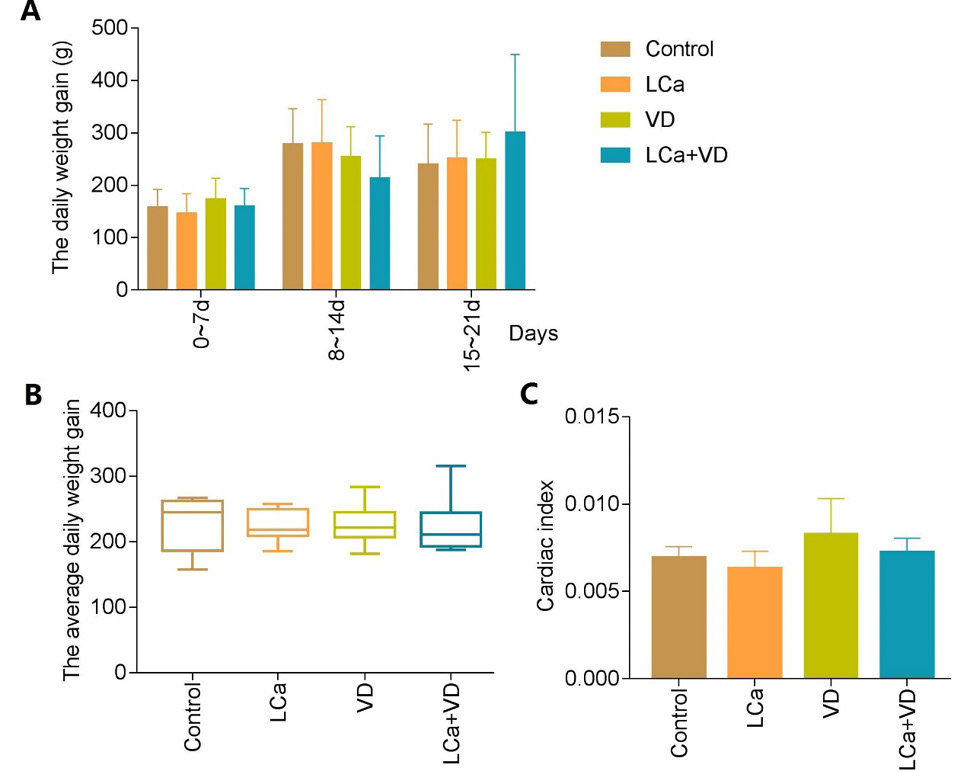
Performance and cardiac index analysis of different treatment groups. Daily weight gains of different groups piglets in day 7, 14, 21 (A); Average daily weight gain (B); Effects of 25(OH)D3 and Ca2+ supplementation in sows’ diets on cardiac index (C).
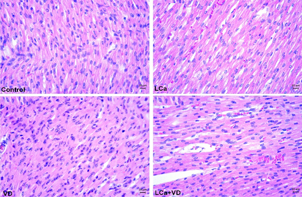
Effect of Vit D3 on histological structure of heart piglets. Note development of myocardial fiber A, control; B, low calcium treatment; C, Vit D3 treatment; D, Vit D3 + low calcium treatment. Stain: Hematoxilyn & Eosin.
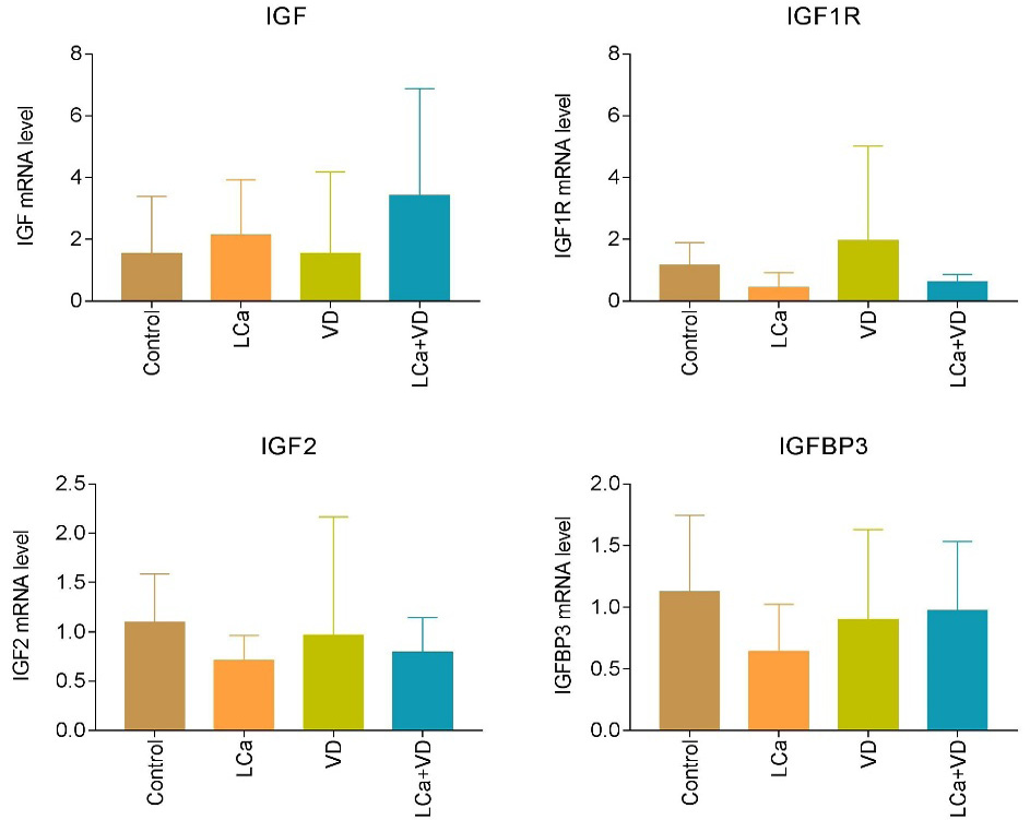
Quantitative RT-PCR analysis of cardiac insulin-like growth factor (IGF). The mRNA expressions levels of IGF, IGF1R, IGF2 and IGFBP3 genes in the control, low calcium group (LCa), 25(OH)D3 group (VD) and low calcium + 25(OH)D3 group (LCa+VD) groups in the cardiac from piglets as determined by qRT-PCR. The data represent the mean ± SD.
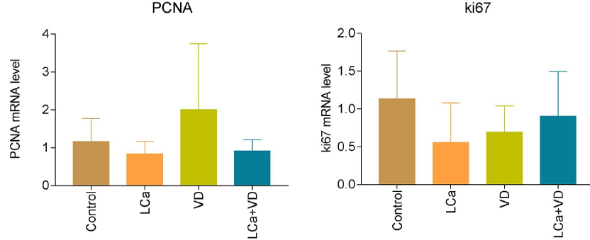
Effect of 25(OH) D3 and Ca2+ on PCNA and ki67 expression in neonatal piglets. *P< 0.05; expression levels were normalized to the levels of the geometric mean of GAPDH gene expression (mean ± SD).
References
- Amundson, L.A., Hernandez, L.L. and Crenshaw, T.D., 2017. Br. J. Nutr., 118: 30-40. https://doi.org/10.1017/S0007114517001751[CrossRef]
- Berchtold, M.W., Brinkmeier, H. and Muntener, M., 2000. Physiol. Rev., 80: 1215-1265. https://doi.org/10.1152/physrev.2000.80.3.1215[CrossRef]
- Braun, F., 1986. Wien. Klin. Wochenschr. Suppl., 166: 1-23.
- Du, X., Liu, Y., Zhao, C., Fang, J., Wang, X. and Wei, L., 2019. BMC Endocr. Disord., 19: 48. https://doi.org/10.1186/s12902-019-0376-1[CrossRef]
- Gao, J., Shi, X., He, H., Zhang, J., Lin, D., Fu, G. and Lai, D., 2017. J. Vis. Exp., 127: 55797. https://doi.org/10.3791/55797[CrossRef]
- Gonzalo, E., Letourneau-Montminy, M.P., Narcy, A., Bernier, J.F. and Pomar, C., 2018. Animal, 12: 1165-1173. https://doi.org/10.1017/S1751731117002567[CrossRef]
- Jurikova, M., Danihel, L., Polak, S. and Varga, I., 2016. Acta Histochem., 118: 544-552. https://doi.org/10.1016/j.acthis.2016.05.002[CrossRef]
- Peacock, M., 2010. Clin. J. Am. Soc. Nephrol., 5 (Suppl 1): S23-S30. https://doi.org/10.2215/CJN.05910809[CrossRef]
- Tan, F.P., Kontulainen, S.A. and Beaulieu, A.D., 2016. J. Anim. Sci., 94: 4205-4216. https://doi.org/10.2527/jas.2016-0298[CrossRef]
- Wullschleger, M., Blanch, J. and Egger, M., 2017. Cardiovasc. Res., 113: 542-552. https://doi.org/10.1093/cvr/cvx020[CrossRef]
- Yao, P., Bennett, D., Mafham, M., Lin, X., Chen, Z., Armitage, J. and Clarke, R., 2019. JAMA Netw. Open, 2: e1917789. https://doi.org/10.1001/jamanetworkopen.2019.17789[CrossRef]
- Zhang, H., Mehmood, K., Jiang, X., Yao, W., Iqbal, M., Li, K., Tong, X., Wang, L., Wang, M., Zhang, L., Nabi, F., Rehman, M. U. and Li, J., 2018. Biomed. Res. Int., 2018: 6796271. https://doi.org/10.1155/2018/6796271[CrossRef]
- Zhang, L., Hu, J., Li, M., Shang, Q., Liu, S. and Piao, X., 2019. J. Bone Miner. Metab., 37: 1083-1094. https://doi.org/10.1007/s00774-019-01020-0[CrossRef]
Supplementary material
Services
Article usage statistics
Loading…