
The present study was conducted to find out the effect of UV irradiation of Aspergillus niger at different concentrations of wheat bran as carbon source (2.5, 3.0, and 3.5 %) on the production of xylanase. Submerged fermentation was carried out in 500 ml Erlenmeyer flasks using vogel’s medium at 35°C for five days. UV irradiated A. niger has significantly increased the xylanase production as compared to parent strains. Twenty minutes exposure of A. niger was considered suitable for the production of xylanase. Wheat bran concentration (3.5%) was considered best substrate for xylanase production than corn cobs and sugarcane baggasse. Maximum xylanase production was observed at 30°C with pH 5.6. Molecular mass of the purified xylanase was found to be 27.2 KDa by SDS-PAGE. Total protein present in crude enzyme was calculated as 188 mg/ml. Crude xylanase showed specific activity of 41.22 IU/mg protein. Partially purified xylanase showed protein content of 80.6 mg/ml. The novelty of this study is basic sources used are indigenous and cheap for production of xylanase.
Short Communication · Volume 51, Issue 6, 23 September 2019 · Open Access
Effect of UV Irradiation of Aspergillus niger on the Production of Xylanase in the Presence of Wheat Bran as Carbon Source
· · ·
Affiliations & Notes ⌄
- 1Department of Chemistry, Women University of Azad Jammu and Kashmir, Bagh 2Department of Zoology, Women University of Azad Jammu and Kashmir, Bagh 3Department of Pathobiology, Faculty of Veterinary and Animal Sciences, PMAS Arid Agriculture University, Rawalpindi , * Corresponding author: muhammad.suleman78@yahoo.com
Article Info ⌄
- Journal:
- Pakistan Journal of Zoology
- Published:
- 23 September 2019

From the article
- It revealed that maximum xylanase production was observed at 35°C for wheat bran (Fig. 1).
- When activity of xylanase was observed at different temperatures, it showed maximum activity at 50°C. When the enzyme was subjected to higher temperatures, there is a negative effect on the enzyme performance.
- Crude xylanase have 45.65 IU/mg protein but purified enzyme showed specific activity of 619.31 IU/mg protein.
- Characterization of xylanase showed that pH 8.0 and 50°C are optimum conditions for maximum activity of xylanase.
Selected from the article's own text.
Abstract
Materials and methods
The indigenous carbon sources (wheat bran, corn cobs and sugarcane bagasse) were dried, ground to 40mm mesh and was treated with 2.0% NaOH, stored in air tight containers. Preisolated and purified culture of A. niger was stored in refrigerator at suitable temperature. The sporulation medium (100 ml) was prepared in 250 ml Erlenmeyer flask (4g of potato dextrose agar in distilled water (100 ml). The cotton plugged Erlenmeyer flasks were a placed in an autoclave at 115°C for 20 min for sterilization and transferred into sterilize test tubes to solidifying agar medium. The cotton plugged test tubes were inoculated from the mother culture of A.niger with a presterilized inoculation loop for 72 h at 37°C for germination (Ahmad et al., 2009). Inoculation medium was prepared at pH 5.6, at 37°C and was sterilized by autoclaving. Sporulation medium and inoculum medium was transferred aseptically in 500 ml flasks and shake at 37°C in orbital shaker at 130 rpm for three days. Medium composition was made by following reagents (NH4)2SO4 1.40 g/100ml, MgSO4 0.30 g/100ml, Urea 0.30 g/100ml, Proteose peptone 0.75 g/100ml, CaCl2 0.30 g/100ml, KH2PO4 2.0 g/100ml, Glucose 10.00 g/100ml, and yeast extract 0.25 g/100ml.
In each fermentation flask 3% inoculums medium was added containing substrate. After suitable interval of time, the biomass from these flasks was filtered and centrifuged at 4000 rpm for 10 min at 10°C. All these prepared samples were refrigerated in sterilized glass bottles.
Different indigenous carbon sources (corn cobs, wheat bran, and sugar bagasse) were used with different concentrations separately (2.5, 3.0 and 3.5%). fermentation was carried out for a period of 72 h for the optimization of substrate. To find out optimum pH for xylanase production, biosynthesis of enzyme was carried at different pH values like, 5.0, 5.6, 6.0, 6.6 and 7.0. To optimize temperature Xylanase production was carried at different temperature (20, 25, 30 and 35°C).To find out optimum time required for maximum xylanase activity, samples were harvested for period of 12, 24, 48, 72 and 96 h.
After incubation of medium at different time intervals, the medium from the flasks was filtered by whatman filter paper of suitable grade. The filtrate was centrifuged at 11000 rpm at 15°C for 15 min. Centrifugation was done to remove mycelia and spores of the organism. Supernatant was carefully refrigerated in sterilized glass bottles.
Crude enzyme solution (25 ml) was taken in 250 ml beaker. Different fractions of ammonium sulphate were added into the solution. These solutions were placed on a constant magnetic bar for constant slow stirring at 4°C. After the addition of each fraction, the solution was centrifuged at 4°C and 4000 rpm for 15 min. Supernatants were removed carefully and the pellets obtained were dissolved in 5ml of 0.1M phosphate buffers at pH 6.0.
Dialysis tubes were filled with 5 ml of the crud enzyme solution tight from both ends. 1000 ml beaker was filled with phosphate buffer at pH 6.0 and 4°C. The apparatus was left with constant slow stirring. Buffer solution was changed after regular interval of time for 24 by following
The precipitates of the dialyzed enzymes were purified by gel filtration. 5ml of enzyme solution was used. The column was equilibrated with phosphate buffer pH 6.0. After regular interval of time 5ml of enzyme sample was collected and used for xylanase activity. The fraction which gives better xylanase activity was collected while rest of the fraction was discarded .
Different concentrations of xylose (0.2, 0.4, 0.6, 0.8, 1.0, 1.2, 1.4 mg/ml) were prepared in distilled water. 0.5 ml of each concentration was added in test tubes along with 0.5 ml of DNS solutions and boiled for 5 minutes in boiling water. A blank was also prepared by dissolving 1ml each of DNS and distilled water. Absorbance of these solutions was determined by using spectrophotometer (Labomed) at 550 nm and graph was plotted. Xylanase hydrolyses the xylan in xylose the free xylose units thus react with DNS to form color complex. This color was detected by spectrophotometer at 550nm. Crude xylanase assays were performed using 0.5ml of 1% oat spelt xylan using citrate phosphate buffers of pH 5.6. The composition of reaction mixture was as 0.5 ml substrate and 0.5ml of crude enzyme. The reaction mixture was incubated in shaking water bath at 50°C for 15 min. After incubation 1ml of DNS was added. Reddish brown color was developed, when the reaction tubes were boiled in boiling water for 5 min. After cooling the reaction tubes, O.D’s was measured at 550 nm with xylose as standard material.
Protein was determined by Bradford with bovine serum albumin (BSA) as standard by comparison with standard curve .
The molecular weight was determined by sodium dodecyl sulphate, polyacrylamide gel electrophoresis (SDS-PAGE) .
Results and discussion
Xylanase production by A. niger was studied at 20, 25, 30, 35°C. At 20°C production of xylanase is 28.2, 18.4 and 14.3 IU/ml for wheat bran, corn cobs and sugarcane bagasse, respectively. At 35°C production of enzyme in carbon sources (wheat bran, corn cobs and sugarcane baggasse) were 80.04, 63.02 and 45.01 IU/ml, respectively. It revealed that maximum xylanase production was observed at 35°C for wheat bran (Fig. 1).
Xylanase production for three days incubation (72 h) that is 46.7, 35.6, 27.0 IU/ml of enzyme activities for wheat bran, corn cobs and sugarcane bagasse, respectively. With a 96 h incubation enzyme activities decreased for all three substrates due to nutrients depletion by the action of the organism in growth medium. These results are closely related the results of Haq et al. (2002), they reported maximum xylanase recovery for 72 h of incubation and presented in Figure 2.
Effect of different pH e.g. 5.0, 5.6, 6.0, 6.6 and 7.0 was studied on the enzyme production. Activities for wheat bran, corn cobs and sugarcane baggasse were 39.2, 33.7 and 23.0 IU/ml. It was concluded that 5.6 pH was most suitable to produce maximum xylanase activity by using above carbon sources (Fig. 3A).
After the purification, characterization of the xylanase was done to find optimum pH, temperature, heat stability and molecular mass. It is revealed that when enzyme assay was performed at different temperatures like 20°C to 100°C. Xylanase activities increase with rise in temperature up to 50°C and showed maximum activity at 50°C. When temperature was further increase, there is a decline in activity. Minimum activity was found at 100°C (Fig. 3B).
It is obvious from the results when xyalanse assay was carried out at 50°C maximum activity was found at pH 8.0 (Fig. 4A). Further increase in pH showed a gradual decline in xylanase activity. Thermal stability of xylanase is promising for its application in different industrial processes (Fig. 4B). When activity of xylanase was observed at different temperatures, it showed maximum activity at 50°C. When the enzyme was subjected to higher temperatures, there is a negative effect on the enzyme performance. The decrease in the heat stability is due to denaturation of the enzyme at higher temperature.
Molecular weight of the purified xylanase by SDS-PAGE was 27.2 kDa. The results found by the study were closely related with Arief et al. (2009) as 27 kDa. The molecular weight of purified xylanase was 24 kDa (Fig. 5).
Conclusion
It was concluded that maximum xylanase was produced using wheat bran (3.5%) at 35°C, 5.6 pH and incubation time 72 h by A. niger. Crude xylanase have 45.65 IU/mg protein but purified enzyme showed specific activity of 619.31 IU/mg protein. Characterization of xylanase showed that pH 8.0 and 50°C are optimum conditions for maximum activity of xylanase. SDS-PAGE analysis showed that purified xylanase has molecular weight of 27.2 kDa. This method of xylanase production was more feasible and economical because all raw materials were of indigenous and cheap.
Statement of conflict of interest
The authors declare no conflict of interest.
Figures
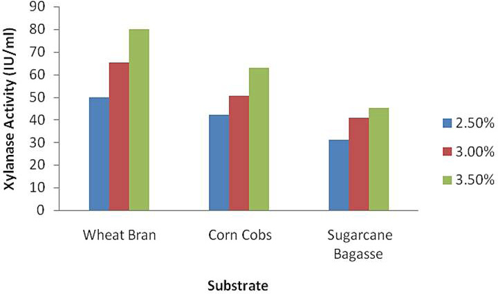
Xylanase production at different concentration of wheat bran, corn cobs and sugarcane bagasse by A. niger.
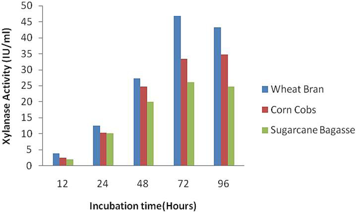
Xylanase production at different incubation and different substrate by A. Niger.
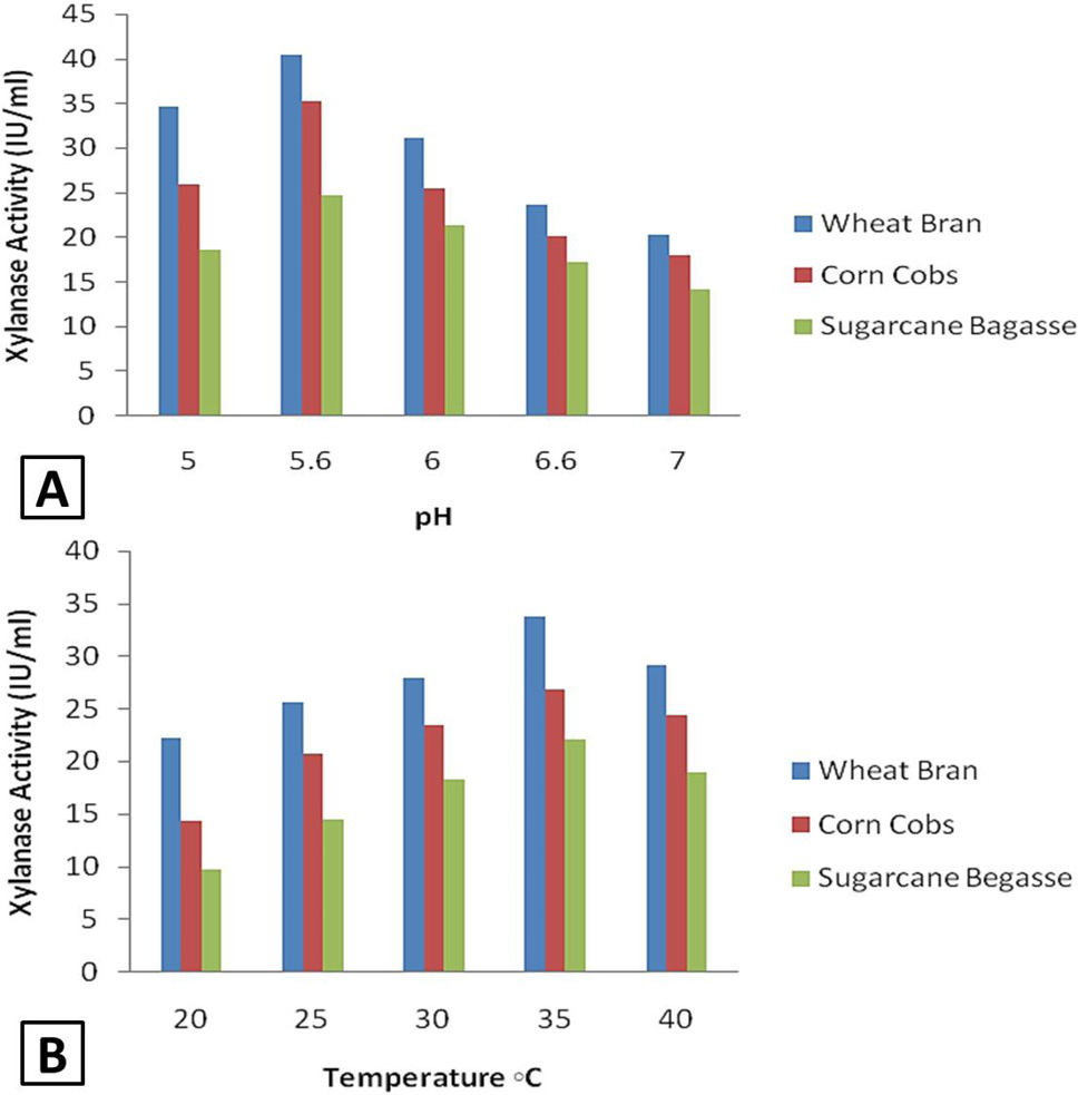
Effect of pH (A) and temperature (B) for xylanase production.
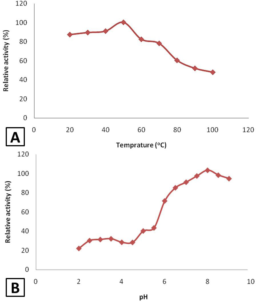
Effect of temperature (A) and pH (B) on relative activity of xylanase.
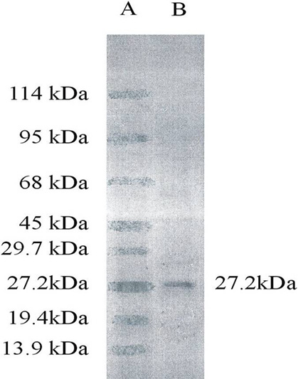
SDS-PAGE analysis for molecular weight determination.
References
- Ahmed, S., Riaz, S. and Jamil, A., 2009. Appl. Microbiol. Biotechnol., 1: 19-35.
- Arief, W., Lestari, E., Tanjung, A., Alfian, W. and Hiroyasu, O., 2009. J. appl. Sci. Environ. Sani., 4: 219-232.
- Baker, H.V., 1991. Proc. natl. Acad. Sci. U.S.A., 88: 9443-9447. https://doi.org/10.1073/pnas.88.21.9443[CrossRef]
- Bakri, Y., Jacques, P. and Thonart, J. 2003. Appl. Biochem. Biotechnol., 108: 737-748. https://doi.org/10.1385/ABAB:108:1-3:737[CrossRef]
- Beg, Q.K., Bhushan, B., Kapoor, M. and Hoondal, G.S., 2000. Enzyme Microb. Technol., 27: 459-466. https://doi.org/10.1016/S0141-0229(00)00231-3[CrossRef]
- Bradford, M.M., 1976. Anal. Biochem., 72: 248-257. https://doi.org/10.1016/0003-2697(76)90527-3[CrossRef]
- Carmona, E.C., Marcia, R.B.B., Aline, A.P.K. and Jao, A.J., 1998. FEMS Microbiol. Lett., 166: 311-315. https://doi.org/10.1111/j.1574-6968.1998.tb13906.x[CrossRef]
- Coral, G., Arikahn, B., Unaldi, M.N. and Guvenmez, H., 2002. Turk. J. Biol., 26: 209-213.
- Haq, I., Khan, A., Butt, W.A., Ali, S. and Qadeer, M.A., 2002. J. biol. Sci., 2: 143-144. https://doi.org/10.3923/jbs.2002.143.144[CrossRef]
- Michael, J.B., Peter, B. and Kaisa, P., 1992. J. Biotechnol., 23: 257-270. https://doi.org/10.1016/0168-1656(92)90074-J[CrossRef]
- Miller, G.L., 1959. J. anal. Chem., 31: 426-429. https://doi.org/10.1021/ac60147a030[CrossRef]
- Norazlina, I., Tanes, P.M., Halim, K.U. Miradatul, K.H., Najwa, M.R. and Mohibah, M., 2013. Int. J. Agric. Sci. Res., 3: 245-252.
- Sardar, M., Roy, I. and Gupta, M.N., 2000. Enzyme Microb. Technol., 27: 672-679. https://doi.org/10.1016/S0141-0229(00)00257-X[CrossRef]
- Zulfiqar, A., Masood, S.B., Faqir, M.A. and Asghar, M., 2009. Int. J. Agric. Biol., 11: 571-576.
Services
Article usage statistics
Loading…