
Many bacterial species are harbored by cockroaches on the external and internal surfaces. These harbored species have great public health concern. There is a variable tendency of these microbes to adhere to various abiotic and biotic surfaces showing chances of infection. Present study aims to check biofilm formation and auto aggregation potential of the bacteria associated with cockroaches. For this purpose, cockroaches were captured from house sewerage and two bacterial strains CXA1 (Planomicrobium chinense) and IM2 (Alkaligenes faecalis) were isolated from external surface and rectum portion of cockroach, respectively. Bacterial autoaggregation started increasing after 1 h of culture incubation and reached maximum after 4 h. Isolate CXA1 showed maximum (136 %) auto aggregation as compared to isolate IM2 (37 %) after 4 h. However, biofilm formation in the isolate IM2 was higher as compared to isolate CXA1 at non shaking conditions. The study showed microbes with a potential to form biofilm are carried by insects and therefore there should be proper hygienic conditions and control efforts for the growth of these vectors.
Short Communication · Volume 51, Issue 6, 22 September 2019 · Open Access
Biofilm Formation and Bacterial Aggregation Response of Planomicrobium chinense and Alkaligenes faecalis Associated with Periplaneta americana Found in Household Sewerage
· · · ·
Affiliations & Notes ⌄
- 1Department of Biology, Lahore Garrison University, Sector-C, Av-4, Phase VI, DHA Lahore , * Corresponding author: aishawaheedqureshi@lgu.edu.pk
- 2uzmazeeshan@lgu.edu.pk
Article Info ⌄
- Journal:
- Pakistan Journal of Zoology
- Published:
- 22 September 2019

From the article
- The percentages of auto aggregation increased with the passage of time and increased to 37 % after four hours of culture incubation in A. faecalis while in P. chinense auto aggregation increased to 136% after four hours of culture incubation (Fig. 1).
- Bacterial aggregation is a reversible process observed in bacterial cells and it is directly correlated with the bacterial adhesion and colonization.
- The biofiolm formation was highest in non-shaking condition in A. faecalis; however, biofilm formation was maximum in P. chinense as compared to other strain (Fig. 2).
- These microbes in the colon portion of cockroach and external cuticle have prevented themselves from external stress factors like enzymes or disinfectants by increasing the rate of auto aggregation.
Selected from the article's own text.
Abstract
Material and methods
Cockroaches were trapped from house sewerage of Lahore, Pakistan in the month of March, 2016 and transported to laboratory for bacterial isolation. For the isolation of CXA1 from external surface, cockroaches were surface swabbed using sterile swab, while for isolation of IM2 isolate from internal surfaces, cockroaches were dissected using sterile dissecting tools and the digestive part was separated and serially diluted to isolate bacteria. Samples were finally spread on LB agar media and plates were incubated at 37°C for 24 h for bacterial growth. Isolated colonies were purified by quadrant streaking and screened for biofilm formation by mucoidy colony appearance and crystal violet ring formation assay . Bacterial biofilm development in L-Broth culture was recorded after 48 h of culture incubation.
On the basis of their initial positive response towards biofilm formation bacterial isolates were characterized morphologically by following the methodology of Bergey’s Manual of Determinative Bacteriology and biochemically (starch hydrolysis test, Citrate utilization test, Litmus milk reaction, Methyl Red Test, Voges Proskauer test, Catalase test) by following Tittsler and Sandholzer (1936). Molecular identification of bacterial isolates was done by analysis of 16S rRNA gene sequences done by commercially available ABI sequencing service Malaysia. Bacterial phylogenetic tree was constructed using neighbor joining method from the obtained nucleotide sequence with the help of MEGA 6.0 software to observe phylogenetic association. The obtained sequences were submitted in gene bank and the accession number was obtained.
Auto-aggregation assay was performed by following the method of Abdulla et al. (2014). Briefly, bacterial cells were harvested from the cultures (centrifugation at 2000 rpm for 20 min) grown in LB-Broth at 37°C for 24 h in shaking incubator. Harvested cell pellet was repeatedly washed and finally suspended in phosphate buffered saline. Auto aggregation response of bacterial isolates was determined by measuring absorbance at 600 nm from upper suspension after of every hour interval till five hours of incubation at room temperature.
For biofilm formation assay, quantification of bacterial biofilm formation of both isolates was determined in terms of tightly bound cells following the method of Liaqat et al. (2009) under shaking and non-shaking conditions after 144 h of culture incubation. Biofilm formation was determined in liquid media in test tubes and fresh bacterial cultures of CXA1 and IM2 were inoculated in L-Broth at equal cell densities OD0.5 at 600 nm. Cultures were incubated at 37°C for 144 h and after incubation. The biofilm forming cells were stained using 0.01 % aqueous crystal violet solution after incubation. The stained cells were finally re dissolved in ethanol to get absorbance at 570 nm for biofilm formation. Bacterial biofilm was recorded as normalized values (OD 570/ OD 600) for excluding the difference in bacterial growth rate.
In all the experiments the data was analyzed statistically by calculating the mean values of replicates and measuring the standard errors of the means. The error bars are shown in each figure.
Results and discussion
The morphological characteristics of Planomicrobium chinense and Alkaligenes faecalis showed pigmented colonies, off white (A. faecalis) and orange (P. chinense), respectively. A. faecalis showed entirely flat colonies with Gram negative cocci cells. Cells showed no spores or capsules formation. P. chinense showed pin point colonies with smooth elevation (Supplementary Table I) while cells were gram positive, non-spore forming and non-capsulated rods (Supplementary Table II). A. faecalis showed positive result of catalase, starch, litmus milk reaction, methyl red while negative for Voges-Proskauer test. P. chinense showed positive results for catalase, litmus milk reaction while negative results were recorded for starch test, methyl red test and Voges-Proskauer test. On the basis of 16 S rRNA gene sequence homology with data base of Genebank at National Center for Biotechnology Information (NCBI) website (http://www.ncbi.nih.gov), carried out by Basic Local Alignment Search Tool (BLAST), the phylogenetic tree construction showed that there were two clusters in a phylogenetic tree and both isolates showed strong homology with their compared nucleotide sequences (Supplementary Fig. 1). Morphological characteristics like shape, flagellar arrangement and staining are the important parameters for bacterial identification . Cockroaches carry many pathogenic and non-pathogenic bacteria. Both the isolates were mucoidy in appearance and this is in line with previous experiments showing that exposure to stress like antibiotic results in induction of mucoidy phenotypes resulting in a thick biofilm development (Supplementary Fig. 2) ; ; . Moreover, exopolymeric production in mucoidy phenotype might have resulted in enhanced biofilm formation as indicated by previous in vivo and in vitro experiments showing that the exopolymer protects chronic wound biofilms .
The percentages of auto aggregation increased with the passage of time and increased to 37 % after four hours of culture incubation in A. faecalis while in P. chinense auto aggregation increased to 136% after four hours of culture incubation (Fig. 1). Increased in autoaggregation with the passage of time shows that aggregation is a time dependant phenomenon these results are supported by the work of Ramalingam et al. (2013) where visible aggregates appeared after 72 h of incubation. Bacterial aggregation is a reversible process observed in bacterial cells and it is directly correlated with the bacterial adhesion and colonization. Bacterial coaggregation and auto aggregation is a common feature reported in many bacterial species . Auto aggregations in probiotic bacteria offer them additional benefits of successful survival by excluding the species commonly present for pathogenesis. This has been reported to be an important criterion for the selection and screening of probiotic bacteria. However, the variable response of auto aggregation in both isolates shows their efficacy to auto aggregate either due to being different species or their isolation source. This is in line with previous findings where Flavobacterium johnsoniae displayed variation in autoaggregation with Gram-negative and Gram-positive organisms .
In general the biofilm formation was highest in A. faecalis compared to P. chinense (Fig. 2). The biofiolm formation was highest in non-shaking condition in A. faecalis; however, biofilm formation was maximum in P. chinense as compared to other strain (Fig. 2). Biofilm formation was checked by both isolates and it was noted that that both isolates have ability to form biofilm. The normalized values of biofilm are supportive for describing relation of biofilm to bacterial cell mass. This has been especially described under growth limiting conditions for bacterial cells. The results of present study showed that presence of cockroaches at dwelling places should be avoided because the bacteria have high colonization ability. Future implication of this work is quite promising for reducing diseases associated with cockroaches. An additional benefit of autoaggregation to microbes associated with cockroaches is that they escape the process of grazing and avoid the loss in cell numbers . These microbes in the colon portion of cockroach and external cuticle have prevented themselves from external stress factors like enzymes or disinfectants by increasing the rate of auto aggregation.
Since the aggregated form of the cockroach derived microbes have shown potential to form biofilm, detailed molecular analysis of this work will be helpful for disease eradication and control.
Supplementary material
There is supplementary material associated with this article. Access the material online at: http://dx.doi.org/10.17582/journal.pjz/2019.51.6.sc6
Statement of conflict of interest
The authors declare no conflict of interest.
Figures
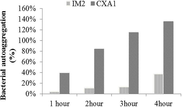
Bacterial auto aggregation response at different time intervals.
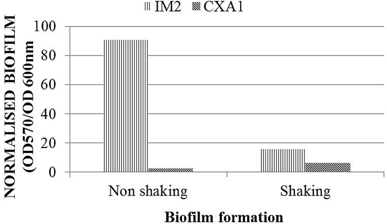
Bacterial biofilm formation at shaking and non-shaking conditions.
References
- Abdulla, A.A., Abed, T.A. and Saeed, A.M., 2014. Br. Microbiol. Res. J., 4: 2231. https://doi.org/10.9734/BMRJ/2014/6462[CrossRef]
- Ahmed, A., Minhas, K., Sahar, N.E., Aftab, O. and Sher-Khan, F., 2010. Iran. J. Publ. Hlth., 39: 109-115.
- Armas, F., Camperio, C. and Marianelli, C., 2017. PLoS One, 12: e0169543. https://doi.org/10.1371/journal.pone.0169543[CrossRef]
- Basson, A., Flemming, L.A. and Chenia, H.Y., 2008. Microb. Ecol., 55: 11-14. https://doi.org/10.1007/s00248-007-9245-y[CrossRef]
- Christensen, G.D., Simpson, W.A., Younger, J.J., Baddour, L.M., Barrett, F.F., Melton, D.M. and Beachey, E.H., 1985. J. clin. Microbiol., 22: 996-1006.
- Cray, J.A., Bell, A.N., Bhaganna, P., Mswaka, A.Y., Timson, D.J. and Hallsworth, J.E., 2013. Microb. Biotechnol., 6: 453-492. https://doi.org/10.1111/1751-7915.12027[CrossRef]
- Elyasigomari, A., Keshavarzi, D., Yusuf, M.A., Hassanzadeh, A., Marvi, N., Shahriari-Namadi, M. and Nassiri, Z., 2017. Oriental Insects, 51: 345-352. https://doi.org/10.1080/00305316.2017.1311811[CrossRef]
- Espeche, M.C., Otero, M.C., Sesma, F. and Nader-Macias, M.E., 2009. Vet. Microbiol., 135: 346-357. https://doi.org/10.1016/j.vetmic.2008.09.078[CrossRef]
- Espeche, M.C., Pellegrino, M., Frola, I., Larriestra, A., Bogni, C. and Nader-Macı´as, M.E., 2012. Anaerobe, 18: 103-109. https://doi.org/10.1016/j.anaerobe.2012.01.002[CrossRef]
- Gerhardt, G.G., Murray, R.G.E., Wood, W.A. and Kreig, N.R., 1994. Methods for general and molecular bacteriology, ASM-1325. Massachusetts Ave., N.W. Washington, D.C.
- Hall-Stoodley, L. and Stoodley, P., 2002. Curr. Opin. Biotechnol., 13: 228-233. https://doi.org/10.1016/S0958-1669(02)00318-X[CrossRef]
- Holt, J.G., Krieg, N.R., Sneath, P.H.A., Staley, J.T. and Williams, S.T., 1994. Bergey’s manual of determinative bacteriology, 7th ed. William and Wilkin Company, Baltimore, USA.
- Jankovic, T., Frece, J., Abram, M. and Gobin, I., 2012. Int. J. Sanitary Engin. Res., 6: 19-24.
- Kaplan, J.B., 2011. Int. J. Artif. Organs, 34: 737-751. https://doi.org/10.5301/ijao.5000027[CrossRef]
- Kjelleberg, S. and Molin, S., 2002. Curr. Opin. Microbiol., 5: 254-258. https://doi.org/10.1016/S1369-5274(02)00325-9[CrossRef]
- Kolenbrander, P.E., Andersen, R.N. and Holdeman, L.V., 1985. Infect. Immunol., 48: 741-746.
- Liaqat, I., Sumble, F. and Sabri, A.N., 2009. Curr. Microbiol., 59: 21-22. https://doi.org/10.1007/s00284-009-9397-8[CrossRef]
- McLaughlin, H., Farrell, A. and Quilty, B., 2006. J. environ. Sci. Health A: Tox. Hazard Subst. environ. Engin., 41: 763-777.
- Mlynek, K.D., Callahan, M.T., Shimkevitch, A.V., Farmer, J.T., Endres, J.L., Marchand, M., Bayles, K.W., Horswill, A.R. and Kaplan, J.B., 2016. Antimicrob. Agents Chemother., 60: 2639-2651. https://doi.org/10.1128/AAC.02070-15[CrossRef]
- Omar, A., Wright, J.B., Schultz, G., Burrell, R. and Nadworny, P., 2017. Microorganisms, 5: E9. https://doi.org/10.3390/microorganisms5010009[CrossRef]
- Ramalingam, B., Sekar, R., Boxall, J.B. and Biggs, C.A., 2013. Water Sci. Technol.: Water Supply, 13: 1016-1023. https://doi.org/10.2166/ws.2013.115[CrossRef]
- Rickard, A.H., McBain, A.J., Ledder, R.G., Handley, P.S and Gilbert, P., 2003. FEMS Microbiol. Lett., 220: 133-140. https://doi.org/10.1016/S0378-1097(03)00094-6[CrossRef]
- Saitou, N. and Nei., M., 1987. Mol. Biol. Evolut., 4: 406-425.
- Shahraki, G.H., Noor, H.M., Rafinejad, J., Shahard, M.K. and Bin-Ibrahim, Y., 2011. Asian Biomed., 5: 619-624.
- Simoes, L.C., Simoes, M. and Vieira, M.J., 2007. Appl. environ. Microbiol., 73: 6192-6200. https://doi.org/10.1128/AEM.00837-07[CrossRef]
- Simoes, L.C., Simoes, M. and Vieira, M.J., 2008. Appl. environ. Microbiol., 74: 1259-1263. https://doi.org/10.1128/AEM.01747-07[CrossRef]
- Tamura, K., Stecher, G., Peterson, D., Filipski, A. and Kumar, S., 2013. Mol. Biol. Evolut., 30: 2725-2729. https://doi.org/10.1093/molbev/mst197[CrossRef]
- Thurlow, L.R., Hanke, M.L., Fritz, T., Angle, A., Aldrich, A., Williams, S.H., Engebretsen, I.L., Bayles, K.W., Horswill, A.R. and Kielian, T., 2011. J. Immunol., 186: 6585-6596. https://doi.org/10.4049/jimmunol.1002794[CrossRef]
- Tittsler, R.P. and Sandholzer, L.A., 1936. J. Bact., 31: 575.
- Tshikhudo, P., Nnzeru, R., Ntushelo, K. and Mudau, F., 2013. Afr. J. Biotechnol., 12: 5975-5982. https://doi.org/10.5897/AJB2013.12057[CrossRef]
- Weiser, J., Henke, H.A., Hector, N., Both, A., Christner, M., Buttner, H., Kaplan, J.B. and Rohde, H., 2016. Int. J. med. Microbiol., 306: 471-478. https://doi.org/10.1016/j.ijmm.2016.05.015[CrossRef]
- Xu, H., Jeong, H.S., Lee, H.Y. and Ahn, J., 2009. Lett. appl. Microbiol., 49: 434-442. https://doi.org/10.1111/j.1472-765X.2009.02684.x[CrossRef]
Supplementary material
Services
Article usage statistics
Loading…