
This study evaluated the effects of NiO-NPs on oxidative stress and hematological parameters in tilapia. Fish were exposed to different NiO-NPs concentrations (0.5, 1.0, and 1.5 mg/L) for 14 days. The experiment involved collecting blood samples and various organs such as the gills, liver, kidney, and muscles of fish to evaluate activity of superoxide dismutase (SOD) and catalase (CAT). The results of the study showed that SOD activity increased in the gills, liver, kidney, and muscles of fish at 0.5 mg/L and 1.0 mg/L of NiO-NPs concentration. However, SOD activity decreased after exposure to 1.5 mg/L NiO-NPs, compared to the control group. In gills and muscles, CAT activity significantly increased at 0.5 mg/L and 1.0 mg/L NiO-NPs concentrations, while its activity decreased at 1.5 mg/L NiO-NPs, but no significant (P<0.05) variation trend was observed in the liver and kidneys compared to the control group. Exposure to NiO-NPs also had an adverse effect on hematological parameters such as red blood cells (RBCs), white blood cells (WBCs), and hemoglobin (Hb) of fish. These findings suggest that NiO-NPs can negatively impact fish and aquatic ecosystems.
Short Communication · Volume 57, Issue 3, 10 May 2025 · Open Access
Effect of Nickel Oxide Nanoparticles on Antioxidant Enzyme System and Hematology of Tilapia
· · ·
Affiliations & Notes ⌄
- 1Department of Zoology, Wildlife and Fisheries, University of Agriculture, Faisalabad 2Department of Zoology, University of Jhang, Jhang 3Department of Biosciences, COMSATS University, Islamabad * Corresponding author: sana.aziz1994@gmail.com
Article Info ⌄
- Journal:
- Pakistan Journal of Zoology
- Published:
- 10 May 2025

From the article
- Results indicated that lower concentrations (0.5 mg/L, 1.0 mg/L) of NiO-NPs increased SOD activity in all tilapia organs, while higher concentration (1.5 mg/L) led to a decrease.
- Our results showed that at lower concentrations (0.5 mg/L, 1.0 mg/L) NiO-NPs significantly increased CAT activity in the gills and muscles of O. niloticus but CAT activity is not changed significantly in the liver and kidneys of the treated group than that of the control group.
- CAT activity was significantly decreased after 1.5 mg/L exposure of NiO-NPs due to the generation of a greater amount of ROS in all organs of O. niloticus.
- Exposure to different treatments of NiO-NPs resulted in changes in SOD and CAT activity due to oxidative stress in fish.
Selected from the article's own text.
Abstract
Materials and methods
NiO-NPs were synthesized in Nanoresearch Lab, University of Agriculture, Faisalabad. Nickel chloride (NiCl2.6H2O) and sodium hydroxide were added to 50 ml of de-ionized water separately to synthesize solutions. Then NaOH solution was added dropwise in NiCl2 solutions by burette. After that, ammonia was added drop by drop for pH 10 and stirred for 2 h. After that, filtration mixture was washed twice with ethanol and distilled water and dehydrated (3 h) in an oven. The product was calcined (400 ℃) for 2 h. Nanoparticles were characterized by X-ray diffraction.
Nile tilapia, freshwater fish were collected from aquaculture ponds at Fisheries Research Farms, University of Agriculture, Faisalabad, Pakistan. Fingerlings of O. niloticus (length: 10 2.5cm, weight; 28±3 g) were chosen as test organisms after acclimation for two weeks. Commercial feed having 40% protein and 60% essential energy constituents was provided to fish twice a day.
After a period of acclimatization, the fish (n=96) were divided into twelve trial tanks, each containing eight fish including three tanks as a control group. The experimental design was triplicated. Stock solutions of NiO-NPs at different concentrations were prepared by dissolving nanoparticles in purified water using a sonicator (90 W, 30 kHz) for 5 h. Fish fingerlings were exposed to various concentrations (0.5, 1.0, and 1.5 mg/L) of NiO-NPs for 2 weeks in toxicity tests. The capillary system was utilized to provide constant airflow to the test media. Before exposing the fish to nanoparticles, the water was changed daily using a suction pump. Each tank contained 50 liters of water. Physico-chemical parameters were maintained throughout the experimental study and were as follows: DO concentration 7.00 ± 0.80 mg\L, pH 7.500 ± 0.0205 temperature 28.540 ± 0.505℃ and total hardness 90.00 ± 7.48 mg\L.
After accilimatization average activity of SOD and CAT were checked from fingerlings of same age and size and then at the end, all fishes were dissected and samples of organs such as kidneys, liver, gills, and muscle were taken out to assess the level of oxidative stress. One gram of each organ was homogenized separately in cold buffer (pH 7.4) using a homogenizer. The homogenates were then centrifuged at 10,000rpm for 15 min, and the resulting supernatants were used for determining the activity of SOD and CAT according to Weydert and Cullen (2010), with slight modifications.
Blood samples were taken from caudal vein in tubes containing potassium EDTA coagulant and stored immediately in an ice box. RBC and WBC counts were calculated using a light microscope with Neubauer haemocytometer . Hemoglobin levels were determined using a hemoglobin test kit (DIAGNOVA, Ranbaxy, India) by employing the cyanmethemoglobin method.
The water quality parameters such as temperature and dissolved oxygen were measured and noted with the help of an electronic meter (HANNA HI-9146) while pH was measured with the help of digital meters WTW in laboratories. However total ammonia, hardness and carbon dioxide were measured by following the method of APHA (2005).
All experiments were conducted in three replicates. Analysis of variance was used to check statistical differences and a comparison of means was done Tukey’s/Student Newnan-Keul tests by using Statistix 8.1 computer package.
Results and discussion
Figure 1 shows the image of the X-ray diffraction (XRD) design of NiO-NPs which indicated that powdered form of nickel oxide particles has lots of small crystals with indiscriminate alignment. XRD patterns display the 111, 200, 220 and 222 diffraction peaks that demonstrate that all the NiO-NPs were in single phase. The peaks confirmed that no other impurities were present in engineered nanoparticles. The average size (D) was 53.44nm by the Debye-Scherrer formula.
Figure 2 shows the effect of different concentrations of NiO-NPs on SOD and CAT activities in gills, liver, kidney, and muscles of tilapia. Results of the present study showed that at the 0.5 mg/L and 1.0mg/L of NiO-NPs, SOD activity increased in these organs. Exposure of 1.5 mg/L NiO-NPs exhibited a significant decreasing trend of SOD in these organs than that of control group.
The CAT activity was significantly increased in gills and muscle over 0.5 mg/L and 1.0 mg/L NiO-NPs, but no significant variation was observed in liver and kidneys of fish at these treatments as compared to the control group. After exposure to 1.5 mg/L NiO-NPs, decreased CAT activity was observed in all organs than that of the control group.
The SOD enzyme serves as the first line of protection against the toxic effects of oxygen and is usually represented as a biomarker of the synthesis of reactive oxygen species . SOD enzyme is well known for its function of catalyzing the breakdown of superoxide radical to oxygen and hydrogen peroxide. Results indicated that lower concentrations (0.5 mg/L, 1.0 mg/L) of NiO-NPs increased SOD activity in all tilapia organs, while higher concentration (1.5 mg/L) led to a decrease. Under higher pressure of nickel oxide nanoparticles reduced activity of SOD may lead to the generation of reactive oxygen species in selected organs resulting in intracellular damage ; , 2022). Jayaseelan et al. (2014) also found results similar to ours, reporting a significant decrease in SOD activity in tilapia exposed to higher concentrations of nickel nanoparticles. The interaction between nickel and the tissues causes a reduction in the activity of antioxidant enzymes, leading to oxidative stress. The CAT enzyme facilitates the conversion of hydrogen peroxide into water and oxygen at a rate of one per second . This enzyme is highly sensitive to chemical stress and plays a vital role in detecting environmental toxicity at an early stage by serving as an important oxidative stress signal . Our results showed that at lower concentrations (0.5 mg/L, 1.0 mg/L) NiO-NPs significantly increased CAT activity in the gills and muscles of O. niloticus but CAT activity is not changed significantly in the liver and kidneys of the treated group than that of the control group. CAT activity was significantly decreased after 1.5 mg/L exposure of NiO-NPs due to the generation of a greater amount of ROS in all organs of O. niloticus. Our results matched with Palermo et al. (2015) who reported that under low stress of nickel (Ni), CAT activity increased initially but after that inhibition effects were noticed. Exposure to different treatments of NiO-NPs resulted in changes in SOD and CAT activity due to oxidative stress in fish.
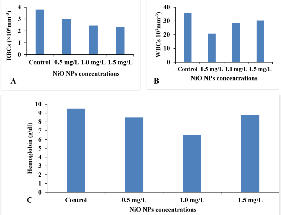
Figure 3 shows the effect of different concentration of NiO-NPs on RBC, WBC, and haemoglobin concentration of O. niloticus. The experimental results revealed that the number of red blood cells (RBCs) decreased significantly in all treatment groups of NiO-NPs, compared to the control group (P<0.05). The treatment group with 1.5 mg/L of NiO-NPs showed the lowest RBC count. Likewise, all treated groups of O. niloticus showed a decrease in the number of white blood cells (WBCs), with the least number of WBCs found in the group exposed to a concentration of 0.5 mg/L. Exposure to different NiO-NPs treatments also affected the Hb (g/dl) content of O. niloticus compared to the control group (P<0.05).
The change in hemoglobin levels indicates that NiO NPs toxicity caused a high demand for oxygen. Our findings are consistent with those of Samim and Vaseem (2021), who reported that high concentrations of NiO NPs exposure caused significant changes in all hematological parameters of fish. The significant difference in WBC count may be due to the accumulation of NiO NPs in WBCs, leading to structural deformities and changes in physiology. WBCs play a crucial role in defending the body against diseases and infections, which makes them an indicator of NiO NPs toxicity in fish.
Conclusion
Exposure to sublethal concentrations of NiO-NPs can disrupt the antioxidant systems and lead to oxidative stress in the organs of tilapia. As the dose of NiO-NPs increases, there can be more changes in tilapia blood, and higher concentrations can even lead to death.
Funding
The study received no external funding.
Ethical statement and IRB approval
The study presented here is the part of M.Phil research approved by ethical review board (ERB) at University of Agriculture, Faisalabad, Pakistan. The protocols and procedures of this study were approved by the animal use and animal care committee of the University of Veterinary and Animal Sciences, Lahore (DR/175, 05-04-2022).
Statement of conflict of interest
The authors have declared no conflict of interest.
Figures
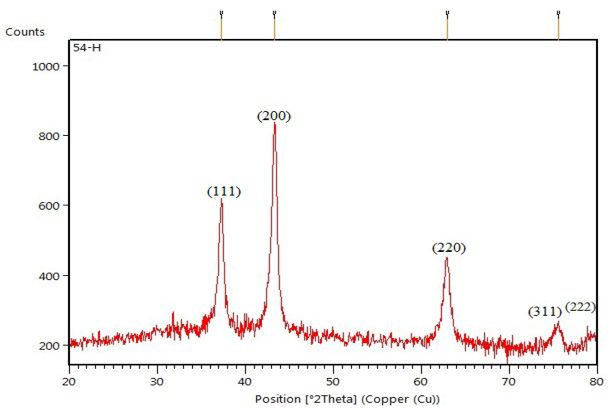
XRD-pattern of synthesized NiO-NPs.
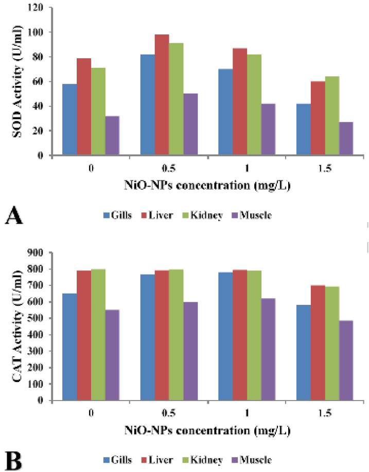
Effect of different treatments of NiO-NPs on superoxide dismutase (SOD) and catalase (CAT) activity (U/mL) in different organs of Oreochromis niloticus.
References
- APHA (American Public Health Association), 2005. Standard methods for the examination of water and waste water. 21st ed. American Public Health Association, Washington, DC. pp. 1220.
- Aziz, S., Abdulah, S., Anwar, H. and Latif, F., 2022. J. Anim. Pl. Sci., 32: 1299-1305. https://doi.org/10.36899/JAPS.2022.5.0536[CrossRef]
- Aziz, S., Abdullah, S., Anwar, H., Latif, F. and Mustfa, W., 2021. Pak. Vet. J., 41: 424-428. https://doi.org/10.29261/pakvetj/2021.044[CrossRef]
- Burgos-Aceves, M.A., Lionettiand, L. and Faggio, C., 2019. Sci. Total Environ., 670: 1170-1183. https://doi.org/10.1016/j.scitotenv.2019.03.275[CrossRef]
- Chandrasekara, H.U. and Pathiratne, A., 2005. Aquacult. Res., 36: 144-149. https://doi.org/10.1111/j.1365-2109.2004.01197.x[CrossRef]
- El-Gendy, K.S., Aly, N.M., Mahmoud, F.H., Kenawy, A., Khalek, A. and El-Sebae, H., 2010. Fd. Chem. Toxicol., 48: 215-221. https://doi.org/10.1016/j.fct.2009.10.003[CrossRef]
- Federici, G., Shaw, B.J. and Handy, R.D., 2007. Aquat. Toxicol., 84: 415-430. https://doi.org/10.1016/j.aquatox.2007.07.009[CrossRef]
- Giguere, A., Campbell, P.G.C., Hare, L. and Couture, P., 2006. Aquat. Toxicol., 77: 178-189. https://doi.org/10.1016/j.aquatox.2005.12.001[CrossRef]
- Handy, R.D., Henry, T.B., Scown, T.S., Johnston, B.D., and Tyler, C.R., 2008. Ecotoxicol.ogy, 17: 396-409.
- Hoa, L., Wang, Z. and Xing, B., 2009. J. environ. Sci., 21: 1459-1466. https://doi.org/10.1016/S1001-0742(08)62440-7[CrossRef]
- Jayaseelan, C., Abdul-Rahuman, A., Ramkumar, R., Perumal, P., Rajakumar, G., Kirthi, A.V., Santhoshkumar, T. and Marimuthu, S., 2014. Ecotoxicol. Environ. Saf., 107: 220-228. https://doi.org/10.1016/j.ecoenv.2014.06.012[CrossRef]
- Kaya, H., Akbulut, M., Celik, E.S. and Yılmaz, S., 2013. Toxicol. environ. Chem., 95: 1554-1564. https://doi.org/10.1080/02772248.2014.895363[CrossRef]
- Leonard, S.S., Harris, G.K. and Shi, X., 2004. Free Radic. Biol. Med., 37: 1921-1942. https://doi.org/10.1016/j.freeradbiomed.2004.09.010[CrossRef]
- Li, M., Zhu, L. and Lin, D., 2011. Environ. Sci. Technol., 45: 1977-1983. https://doi.org/10.1021/es102624t[CrossRef]
- Mgbenka, B.O., Oluah, N.S. and Umeike, I., 2003. Bull. environ. Contam. Toxicol., 71: 248-254. https://doi.org/10.1007/s00128-003-0157-3[CrossRef]
- Palermo, F.F., Risso, W.E., Simonato, J.D. and Martinz, C.B.R., 2015. Ecotoxol. environ. Saf., 116: 19-28. https://doi.org/10.1016/j.ecoenv.2015.02.032[CrossRef]
- Salimi, A., Sharifi, E., Noorbaksh, A. and Soltanian, S., 2007. Biophy. Chem., 125: 540-548. https://doi.org/10.1016/j.bpc.2006.11.004[CrossRef]
- Saliu, J.K. and Bawa-allah, K.A., 2012. Nat. Resour. Environ., 2: 21-26. https://doi.org/10.5923/j.re.20120201.03[CrossRef]
- Samim, A.R. and Vaseem, H., 2021. Environ. Sci. Pollut. Res., 28: 54630-54646. https://doi.org/10.1007/s11356-021-14451-y[CrossRef]
- Ward, J.E. and Kach, D.J., 2009. Mar. Environ. Res., 68: 137-142. https://doi.org/10.1016/j.marenvres.2009.05.002[CrossRef]
- Weydert, C.J. and Cullen, J.J., 2010. Nat. Prot., 5: 51-66. https://doi.org/10.1038/nprot.2009.197[CrossRef]
Services
Article usage statistics
Loading…