
The generation of reactive oxygen species appears to be responsible for gentamicin nephrotoxicity. Recent research has shown that Acacia nilotica extract (AN) retains antioxidant and anti-inflammatory activity. This study assessed AN’s inhibitory impact on rat nephrotoxicity induced by gentamicin. Twenty-four male Wister rats have sub-divided into 4 groups, the control one received saline; the second group was administered with AN extraction (5%) for fifteen days; group-3 rats were daily injected intraperitoneally with gentamicin (100 mg/kg) for fifteen days, and groups 4 include rats injected with gentamicin and with AN extraction (5%) for fifteen days as stated in groups 2 and 3. An identical normal saline volume was administered to control and gentamycin-treated rats. Serum was extracted for chemistry analysis. The tissue samples were taken for both histopathological and immunohistochemistry studies and the renal gene expression. Gentamicin significantly increased serum creatinine, urea, and uric acid. It decreased the levels SOD, catalase and GSH, with significant increases of the malondialdehyde (MDA). Co-treatment of gentamicin and AN significantly improved the aforementioned parameters through increased considerably antioxidant activity as well as up-regulation in the expression of HO-1 and Nrf2. Moreover, it showed antiapoptotic activity and down-regulation of the cox2, NF-kB, and TGF-β expression that induced by the gentamycin. Co-treatment of gentamicin and AN normalized renal injuries induced by the gentamycin administration at cellular levels. AN treatment induced ameliorative impacts against gentamycin induced nephrotoxicity at the histological, biochemical indices and the molecular levels.
Research Article · Volume 57, Issue 2, 19 February 2025 · Open Access
Effect of Acacia nilotica Against Nephrotoxicity Induced by Gentamicin in Rat: Role of Antioxidants, Anti-Inflammatory and Antiapoptotic Markers
· · · · · ·
Affiliations & Notes ⌄
- 1Department of Biology, Turabah University College, Taif University, P.O. Box 11099, Taif, Saudi Arabia. 2Department of Biology, College of Science, University of Jeddah, Saudi Arabia, P.O. Box. 80327, Jeddah 21589 3Clinical Laboratories Sciences Department, Turabah University College, Taif University, P.O. Box 11099, Taif, Saudi Arabia * Corresponding author: mmsoliman@tu.edu.sa
Article Info ⌄
- Journal:
- Pakistan Journal of Zoology
- Published:
- 19 February 2025

From the article
- Increased malondialdehyde levels are characteristic of nephrotoxicity induced by gentamicin due to high production of free radicals.
- When comparing gentamicin-only treated rats to those treated with AN and gentamicin, the latter group showed significantly less of an increase in serum creatinine, urea, and malondialdehyde.
- In this study, gentamicin treatment increased NF-κB, as indicated by the upregulation of NFkB immune-reactivity in the kidneys of the experimental animals.
- In conclusion, Acacia Nilotica extraction ameliorated nephrotoxicity induced by the gentamycin administration through its antioxidant potential and stimulation of the HO-1, Nrf2, and its antiapoptotic activity in addition to its anti-inflammatory activity through modulation of the COX2, NF-kB, and TGF-β activities.
Selected from the article's own text.
Abstract
Introduction
A common aminoglycoside antibiotic, gentamicin (GM), is reserved for the most severe infections. Nonetheless, the emergence of nephrotoxicity severely restricts its application. Estimates suggest that after receiving the antibiotic, as many as 30% of patients may develop renal impairment . Numerous studies have found multiple pathways involved in gentamicin-induced nephrotoxicity; such as ROS and reactive nitrogen species, antioxidants suppression, inflammatory processes activation, and the reduction of renal blood flow, all of which result in tubular necrosis, leukocyte infiltration, and cellular damage . Tumor necrosis factor alpha and intercellular adhesion molecule-1 are up-regulated in gentamicin-induced nephrotoxicity (ICAM-1) . Antioxidants and anti-inflammatory biomarkers have been shown to mitigate and counteract gentamicin induced kidney damage . Herbal remedies were widely used to cure several medical conditions across the globe. About 1300 species can be found in the Acacia genus , widespread in the warmest latitudes and, to a lesser extent, the tropics. The Fabaceae tree A. nilotica has several medical applications and is widely used to treat many conditions, including the common bronchitis, cold, dysentery, diarrhea, biliousness, and leukoderma . Traditional practitioners employ it to combat specific skin, mouth and bone forms. The bark and or gum can be used to treat cancers ear, eye, and testicles. The root, wood and leaves are used for tuberculosis, smallpox, and ulcers treatment, respectively . In addition to its antispasmodic and anti-hypertensive properties, A. nilotica has been linked to a wide range of other biological activities , antidiabetic , hypocholesterolemic and lowered diabetes-related liver failure risk . The current study was outlined to check the effect of AN extract against nephrotoxicity induced by gentamicin in rats at renal levels.
MATERIALS AND METHODS
Animals
A total of 24 Wistar male rats (150-155 g) were used. Animals were maintained and gained free food. Experiments done here were carried out following the guidelines of NIH and rules stated by Taif University, Saudi Arabia. Animals were handled for fifteen days before the onset of experiments.
Experimental protocol
Rats were received and allocated randomly into four groups. Group 1, rats received normal saline for fifteen days (control), Group 2 rats were treated with Acacia nilotica (AN) extraction (5%) for 15 days, Group 3, rats received gentamicin (100 mg/kg) intraperitoneally for consecutive 15 days. Group 4 injected by gentamicin in a dose of 100 mg/kg intraperitoneally and AN extract (5%) for consecutive fifteen days. An identical normal saline volume was administered to control and gentamycin-treated rats.
Sampling
After 15 days, animals were decapitated after euthanization. Blood were taken from the retero-orbital venous plexuses. After clotting, blood was centrifuged at 1000 xg for 15 min to extract serum, then kept at -30°C for future chemistry measurements. One kidney was removed from each rat, homogenized in PBS (cold). Next, centrifugation for 8 min at 2000 xp and 5°C for homogenate was done. The supernatant was kept at -20°C. The remaining part of kidney tissue was divided into 2 portions. Initial part was impeded in 10% formalin for histological and immunohistochemistry investigation , and the other part was soaked in TRIZOL reagent and kept at -70°C for future gene expression.
Biochemical assay
Biomed Diagnostics (Egypt) reagent kits were used to measure serum uric acid, urea, and creatinine levels. Superoxide dismutase (SOD) activity, reduced glutathione (GSH), catalase and malondialdehyde (MDA) were assessed following the protocols provided by the reagent kits acquired from Biodiagnostic (Egypt).
Histological and immunohistopathological screening
Kidney samples were soaked in 10% neutral formaldehyde buffer solution (Sigma-Aldrich) for in a 24 h. Tissues were fixed by a paraffin integration device . Immunohistological examination of the kidney was down as explained elsewhere . After drying kidney tissues, they were emersed in paraffin wax. Five small parts of kidney were cut, then mounted, and then incubated for 12 h. The sections were blocked for 30 min then incubated with diluted (1:1500) polyclonal anti- TGF-β, NF-kB, and COX-2 antibodies at 4° C for 12 h. Sections have rinsed by PBS 3 times then incubated with 2nd antibody (biotinylated anti-rabbit IgG, 1:500). Finally incubated with avidin-biotin-peroxidase for 30 min at room temperature. Samples were countered with hematoxylin. DAB was used to visualize the peroxidase reaction.
Gene expression-analysis (qRT-PCR)
To study gene expression in the kidney, RT-PCR was used. Various renal genes were discovered using RT-PCR. Fifty mg of renal tissue were soaked in TRIZOL reagent (Invitrogen, CA, USA) to get the total RNA. RNA concentrations were measured and RNA of ratio 1.8 was used for cDNA synthesis using the synthesis of cDNA. SYBR stain mix and primers written down in Table I were used to amplify cDNA using quantitative RT-PCR. The total reaction volume is 20 µl, which consists of 10 µl of SYBR Mix, 0.5 µl of each forward and reverse primer, and 9 µl of DNA template of each sample. The quantitative RT-PCR amplification began with 10 min of reverse transcription at 55°C. The cDNAs were amplified using 41 cycles, each consists of denaturation at 94°C for 6 sec, annealing at 55-57°C for 25 sec, and extension at 72°C for 10 sec.
During the extension process, data was collected. Analysis for curve (melting) was done to confirm specificity and authenticity of PCR products. Results were gathered using Rotor-Gene-Q (Qiagen, USA), which assessed the threshold cycle value (Ct). The amplification data was analyzed using two methodological approaches .
Table I. Primers used for RT-PCR.
| Gene name | Primer sequence 5`→ 3` | Direction | Accession number |
| CASP3 | CTGGACTGCGGTATTGAGAC | Sense | NM_012922.2 |
| CCGGGTGCGGTAGAGTAAGC | Antisense | ||
| Bax | GGCGAATTGGCGATGAACTG | Sense | NM_017059.2 |
| ATGGTTCTGATCAGCTCGGG | Antisense | ||
| Nrf2 | TGTCAGCTACTCCCAGGTTG | Sense | NM_031789.2 |
| ATCAGGGGTGGTGAAGACTG | Antisense | ||
| HO-1 | AGGATTAACTGAAGGCGAGCAT | Sense | NM_017050.1 |
| TCTACAGTTAGCAGGCCAGCAG | Antisense | ||
| Bcl-2 | GATTGTGGCCTTCTTTGAGT | Sense | NM_016993.1 |
| ATAGTTCCACAAAGGCATCC | Antisense | ||
| GAPDH | TCAAGAAGGTGGTGAAGCAG | Sense | NM_017008.4 |
| AGGTGGAAGAATGGGAGTTG | Antisense |
Statistical analysis
Data explained as means±SEM. The analysis of data was achieved using GraphPad Prism 5 program. Data was analyzed using One-way ANOVA. Tukey-Kramer post-analysis test was used to examine the means. The significance level was established at P < 0.05.
RESULTS
AN restores the biochemical and antioxidant markers in nephrotoxicity induced by gentamicin in rats
Our results showed that there were a significant elevation in renal biomarkers (uric acid, urea, and creatinine) in gentamycin-treated rats concerning the other treated rats .in addition, there were a substantial decreases in renal biomarkers levels in the AN co-treated with gentamycin treated rats concerning the gentamycin treated rats (Fig. 1). Significant decreases in the CAT, SOD, and GSH activities were accompanied by notable elevation in the levels of MDA in the gentamycin-treated rats. Conversely, AN co-treated with gentamycin-treated rats showed significant increases in the CAT, SOD, and GSH activities accompanied with prominent decline in the levels of MDA concerning the gentamycin injected rats (Fig. 2).
Effect of AN on the apoptosis and oxidative stress marked genes
Figure 3 shows significant downregulation of the HO-1, Nrf2, and Bcl2 expression with substantial upregulation of caspase-3 mRNA expression the gentamycin-treated rats compared with other groups. Otherwise, AN treated gentamycin group showed significant HO-1, Nrf2, and Bcl2 mRNA expression normalization accompanied by a marked down-regulation of the caspase-3-mRNA expression concerning the gentamycin-treated rats.
Effect of AN on renal histopathology and immunohistochemistry and renal COX-2, NF-kB, and TGF-β expression
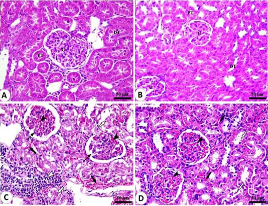
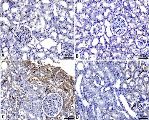
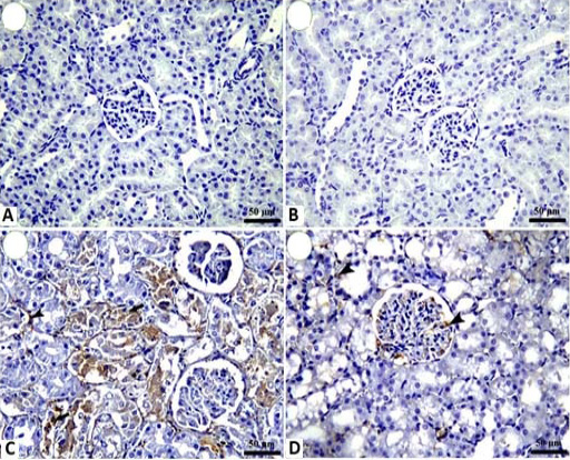
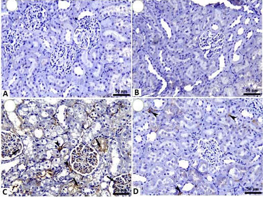
Figure 4 shows that the control group, and the AN group show intact renal corpuscles with normal glomeruli of renal parenchyma and normal proximal (PT) and distal convoluted tubules. The gentamicin-treated rats showed shrinkage of glomeruli with severe glomerular congestion and inflammatory cell infiltration. The gentamicin + AN treated group showed intact glomeruli with mild to moderate congestion in glomerular blood vessels in addition to sloughing of tubular epithelium. The COX-2 expression was mild in the control group and had an adverse expression in the control group showing weak expression of inflammatory markers (COX-2, NF-κB, TGF-β) in renal tubules. AN treated group showed negative immune reaction for COX-2, NF-κB and TGF-β in gentamicin treated group showing a significant increase of COX-2, NF-κB, and TGF-β positive renal tubular epithelium kidney of gentamicin+AN treated group showing a marked decrease in COX2, NF-κB positive renal tubular epithelium (Figs. 5, 6 and 7).
DISCUSSION
Even though gentamicin can cause kidney damage, it is still the best antibiotic for treating bacteria immune to other antibiotics. Acute kidney failure has been extensively studied in experimental settings using gentamicin-induced nephrotoxicity as an animal model . As known, oxidative stress markers can control
pathogenesis of nephrotoxicity induced by gentamicin. ROS is the major mediator in the incidence of tubular and glomerular necrosis. Initiation of the inflammatory process is greatly aided by reactive oxygen species, which do so by activating nuclear factor kappa B . One could argue that oxidative-stress and organs inflammation are the senators of pathogenesis of nephrotoxicity induced by gentamicin, creating a feedback loop that amplifies damage and linking the mechanisms responsible for tubular and glomerular alterations .
In nephrotoxicity induced by gentamicin, marked increases in creatinine and urea levels indicated a significant impairment in kidney function . At first in renal diseases, the blood creatinine level, is the most powerful indicator compared to urea. Urea concentrations rise further after parenchymal injury . Blood creatinine, BUN, MDA and uric acid, all are increased significantly after gentamicin administration, as has been previously reported ; .
Increased malondialdehyde levels are characteristic of nephrotoxicity induced by gentamicin due to high production of free radicals. When comparing gentamicin-only treated rats to those treated with AN and gentamicin, the latter group showed significantly less of an increase in serum creatinine, urea, and malondialdehyde. Our data was in harmony with as they reported that the treatment with A. nilotica was more effective than glibenclamide at reducing elevated serum urea and creatinine levels. Consumption of AN improves glomerular function, as indicated by the decrease renal biomarkers and increased creatinine clearance.
Comparing gentamicin findings, we found a decline in glutathione content and activity of antioxidants, catalase, and SOD accompanied by notable increases in the MDA. Karahan et al. (2005) showed that gentamicin did not raise renal glutathione in comparison to the control group, Antioxidant enzymes could be rendered ineffective due to the elevated ROS production seen in gentamicin-induced nephrotoxicity . The concentration of measured antioxidants were markedly increased in AN group than in gentamicin injected rats, confirming the potential antioxidant effect of AN , this action was returned to high contents tannins and polyphenols and that show antioxidant impact in addition to its scavenging ability .
Oxidative stress can stimulates NFkB which is essential for inflammation, cell proliferation, and programmed cell death . In this study, gentamicin treatment increased NF-κB, as indicated by the upregulation of NFkB immune-reactivity in the kidneys of the experimental animals. The expressions of COX-2 and TGF-1 also went up, which was associated with injury . On the other way, the co-treatment of AN+Gentamycin showed to overcome the gentamycin action on the inflammatory cytokines. This result was consistent with in which they confirmed that A. nilotica declined cyclooxygenase-2 (COX-2) stimulation in CCl4-treated rats. This effect was return to the biological activities of the AN such as anti-inflammatory due to its phenolic compounds
Apoptosis plays a key role in the initiation of inflammatory process through ROS induction. Generation of ROS activates different intrinsic and mitochondria-dependent pathways to induce apoptosis. In the present study, apoptosis induced by gentamicin occurred through the reduction in the expression of Bcl-XL and up-regulation of apoptosis associated markers as partly explained here and by others . The release of ROS can trigger apoptosis by way of an intrinsic, mitochondria-dependent pathway. Alteration in apoptosis and anti-apoptosis markers confirmed renal apoptosis induced by gentamicin. These results matched a recent study showing that gentamicin-induced renal damage was linked to the caspase-dependent apoptotic signaling pathway.
Of note, the antioxidant defensive mechanism is regulated by Nrf2 which regulates HO-1 production and expression that stop damage of cells . Substantial evidence suggests that targeting both Nrf2 and HO-1 expression may be protective against nephrotoxicity induced by gentamycin ; ; , this was supported by our finding concerning the stimulating effect of the AN on Nrf2 and HO-1 signaling pathways that were inhibited by gentamycin treatment, this effect was due to the antioxidant effect of the AN .
CONCLUSION
In conclusion, Acacia Nilotica extraction ameliorated nephrotoxicity induced by the gentamycin administration through its antioxidant potential and stimulation of the HO-1, Nrf2, and its antiapoptotic activity in addition to its anti-inflammatory activity through modulation of the COX2, NF-kB, and TGF-β activities. It restored the altered histopathological changes induced by gentamycin. Therefore, we proposed for the first time the intracellular pathway of AN against gentamycin-induced nephrotoxicity.
ACKNOWLEDGMENT
We appreciate the continuous encouragement from Taif University, Taif, Saudi Arabia.
Funding
None.
IRB approval
Not applicable.
Ethical statement
The guidelines stated by National Institutes of Health for animal use were strictly followed in this investigation.
Data availability
Data of this paper is available upon request.
Statement of conflict of interest
The authors have declared no conflict of interest.
Figures
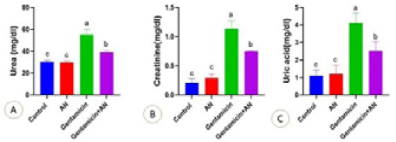
Effect of Acacia nilotica (AN) supplementation on the level of Urea (A), creatinine (B) and serum Uric acid (C) of gentamicin treated rats. Values are represented as Mean ± SEM. The column mean values with different letters superscripts were significantly (a, b, c, d) (P < 0.05) different.
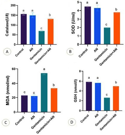
Effect of Acacia nilotica (AN) supplementation on the level of catalase (A), SOD (B), serum MDA (C) and GSH (D) of gentamicin treated rats. Values are represented as Mean ± SEM. The column mean values with different letters superscripts (a, b, c, d) were significantly (P < 0.05) different.
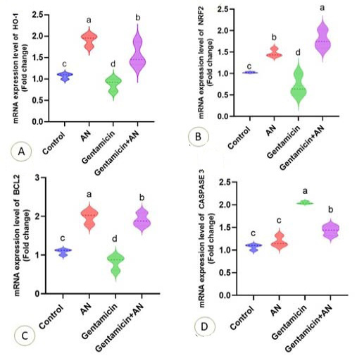
Effect of Acacia nilotica (AN) supplementation on the level mRNA expression of HO-1 (A), Nrf2 (B), BCl-2 (C) and Caspase-3 (D) of gentamicin treated rats. Values are represented as Mean ± SEM. The column mean values with different letters (a, b, c, d) superscripts were significantly P < 0.05 different.
References
- Ahmad, M., Zaman, F., Sharif, T. and Ch, M.Z., 2008. Antidiabetic and hypolipidemic effects of aqueous methanolic extract of Acacia nilotica pods in alloxan-induced diabetic rabbits. Scand. J. Lab. Anim. Sci., 35: 29.
- Ali, B.H., 1995. Gentamicin nephrotoxicity in humans and animals: Some recent research. Gen. Pharmacol., 26: 1477. https://doi.org/10.1016/0306-3623(95)00049-6[CrossRef]
- Ambasta, S.S., 1986. The useful plants of India. Publications and Information Directorate.
- Bancroft, J.D. and Layton, C., 2012. The hematoxylins and eosin. Bancroft’s theory and practice of histological techniques, pp. 173. https://doi.org/10.1016/B978-0-7020-4226-3.00010-X[CrossRef]
- Brasier, A.R., 2006. The NF-κB regulatory network. Cardiovasc. Toxicol., 6: 111. https://doi.org/10.1385/CT:6:2:111[CrossRef]
- Farombi, E. and Ekor, M., 2006. Curcumin attenuates gentamicin-induced renal oxidative damage in rats. Fd. Chem. Toxicol., 44: 1443. https://doi.org/10.1016/j.fct.2006.05.005[CrossRef]
- Galaly, S., Ahmed, O. and Mahmoud, A., 2014. Thymoquinone and curcumin prevent gentamicin-induced liver injury by attenuating oxidative stress, inflammation and apoptosis. J. Physiol. Pharmacol., 65: 823.
- Gao, X., Picchi, A. and Zhang, C., 2010. Upregulation of TNF-alpha and receptors contribute to endothelial dysfunction in zucker diabetic rats. Am. J. Biomed. Sci., 2: 1. https://doi.org/10.5099/aj100100001[CrossRef]
- Geleilete, T.J., Melo, G.C., Costa, R.S., Volpini, R.A., Soares, T.J. and Coimbra, T.M., 2002. Role of myofibroblasts, macrophages, transforming growth factor-beta endothelin, angiotensin-II, and fibronectin in the progression of tubulointerstitial nephritis induced by gentamicin. J. Nephrol., 15: 633.
- Gilani, A., Shaheen, F., Zaman, M., Janbaz, K., Shah, B. and Akhtar, M., 1999. Studies on antihypertensive and antispasmodic activities of methanol extract of Acacia nilotica pods. Phytother. Res. Int. J. Dev. Pharmacol. Toxicol. Evaluat. Nat. Prod. Deriv., 13: 665. https://doi.org/10.1002/(SICI)1099-1573(199912)13:8<665::AID-PTR563>3.0.CO;2-T[CrossRef]
- Gilbert, D., Wood, C., Kohlhepp, S., Kohnen, P., Houghton, D., Finkbeiner, H., Lindsley, J. and Bennett, W., 1989. Polyaspartic acid prevents experimental aminoglycoside nephrotoxicity. J. Infect. Dis., 159: 945. https://doi.org/10.1093/infdis/159.5.945[CrossRef]
- He, L., Peng, X., Zhu, J., Liu, G., Chen, X., Tang, C., Liu, H., Liu, F. and Peng, Y., 2015. Protective effects of curcumin on acute gentamicin-induced nephrotoxicity in rats. Can. J. Physiol. Pharmacol., 93: 275. https://doi.org/10.1139/cjpp-2014-0459[CrossRef]
- Kalaivani, T. and Mathew, L., 2010. Free radical scavenging activity from leaves of Acacia nilotica (L.) Wild. ex Delile, an Indian medicinal tree. Fd. chem. Toxicol., 48: 298. https://doi.org/10.1016/j.fct.2009.10.013[CrossRef]
- Kalayarasan, S., Prabhu, P.N., Sriram, N., Manikandan, R., Arumugam, M. and Sudhandiran, G., 2009. Diallyl sulfide enhances antioxidants and inhibits inflammation through the activation of Nrf2 against gentamicin-induced nephrotoxicity in Wistar rats. Eur. J. Pharmacol., 606: 162. https://doi.org/10.1016/j.ejphar.2008.12.055[CrossRef]
- Karahan, İ., Ateşşahin, A., Yılmaz, S., Çeribaşı, A. and Sakin, F., 2005. Protective effect of lycopene on gentamicin-induced oxidative stress and nephrotoxicity in rats. Toxicology, 215: 198. https://doi.org/10.1016/j.tox.2005.07.007[CrossRef]
- Khan, S.A., Priyamvada, S., Farooq, N., Khan, S., Khan, M.W. and Yusufi, A.N., 2009. Protective effect of green tea extract on gentamicin-induced nephrotoxicity and oxidative damage in rat kidney. Pharmacol. Res., 59: 254. https://doi.org/10.1016/j.phrs.2008.12.009[CrossRef]
- Kumar, R., 1983. Chemical and biochemical nature of fodder tree leaf tannins. J. Agric. Fd. Chem., 31: 1364. https://doi.org/10.1021/jf00120a055[CrossRef]
- Livak, K.J. and Schmittgen, T.D., 2001. Analysis of relative gene expression data using real-time quantitative PCR and the 2−ΔΔCT method. Methods, 25: 402. https://doi.org/10.1006/meth.2001.1262[CrossRef]
- Lopez-Novoa, J.M., Quiros, Y., Vicente, L., Morales, A.I. and Lopez-Hernandez, F.J., 2011. New insights into the mechanism of aminoglycoside nephrotoxicity: An integrative point of view. Kidney Int., 79: 33. https://doi.org/10.1038/ki.2010.337[CrossRef]
- Luna, L.G., 1968. Manual of histologic staining methods of the Armed Forces Institute of Pathology.
- Maciejewski, R., Ruciński, P., Burski, K. and Figura, T., 2001. Changes in glucose, cholesterol and serum lipid fraction levels in experimental diabetes. In: Annls Univ. Mar. Curie-Sklodowska. Sec. D: Med., pp. 363.
- Malkiewicz, K., Koteras, M., Folkesson, R., Brzezinski, J., Winblad, B., Szutowski, M. and Benedikz, E., 2006. Cypermethrin alters glial fibrillary acidic protein levels in the rat brain. Environ. Toxicol. Pharmacol., 21: 51. https://doi.org/10.1016/j.etap.2005.06.005[CrossRef]
- Nassan, M.A., Soliman, M.M., Aldhahrani, A., Althobaiti, F. and Alkhedaide, A.Q., 2021. Ameliorative impacts of Glycyrrhiza glabra root extract against nephrotoxicity induced by gentamicin in mice. Fd. Sci. Nutr., 9: 3405. https://doi.org/10.1002/fsn3.2183[CrossRef]
- Nitha, B. and Janardhanan, K., 2008. Aqueous-ethanolic extract of morel mushroom mycelium Morchella esculenta, protects cisplatin and gentamicin induced nephrotoxicity in mice. Fd. Chem. Toxicol., 46: 3193. https://doi.org/10.1016/j.fct.2008.07.007[CrossRef]
- Omara, E.A., Nada, S.A. and El-Toumy, S.A., 2013. Evaluation of hepatoprotective activity of the Acacia nilotica flowers on carbon tetrachloride-induced liver damage in rats. Planta Med., 79: PB36. https://doi.org/10.1055/s-0033-1351981[CrossRef]
- Omara, E.A., Nada, S.A., Farrag, A.R.H., Sharaf, W.M. and El-Toumy, S.A., 2012. Therapeutic effect of Acacia nilotica pods extract on streptozotocin induced diabetic nephropathy in rat. Phytomedicine, 19: 1059. https://doi.org/10.1016/j.phymed.2012.07.006[CrossRef]
- Seigler, D.S., 2003. Phytochemistry of Acacia sensu lato. Biochem. Syst. Ecol., 31: 845. https://doi.org/10.1016/S0305-1978(03)00082-6[CrossRef]
- Song, E., Fu, J., Xia, X., Su, C. and Song, Y., 2014. Bazhen decoction protects against acetaminophen induced acute liver injury by inhibiting oxidative stress, inflammation and apoptosis in mice. PLoS One, 9: e107405. https://doi.org/10.1371/journal.pone.0107405[CrossRef]
- Subramanian, P., Anandan, R., Jayapalan, J.J. and Hashim, O.H., 2015. Hesperidin protects gentamicin-induced nephrotoxicity via Nrf2/HO-1 signaling and inhibits inflammation mediated by NF-κB in rats. J. Funct. Fd., 13: 89. https://doi.org/10.1016/j.jff.2014.12.035[CrossRef]
- Tsuneki, H., Ishizuka, M., Terasawa, M., Wu, J.B., Sasaoka, T. and Kimura, I., 2004. Effect of green tea on blood glucose levels and serum proteomic patterns in diabetic (db/db) mice and on glucose metabolism in healthy humans. BMC Pharmacol., 4: 1. https://doi.org/10.1186/1471-2210-4-18[CrossRef]
- Yu, H., Chen, B. and Ren, Q., 2019. Baicalin relieves hypoxia-aroused H9c2 cell apoptosis by activating Nrf2/HO-1-mediated HIF1α/BNIP3 pathway. Artif. Cells, Nanomed. Biotechnol., 47: 3657. https://doi.org/10.1080/21691401.2019.1657879[CrossRef]
Services
Article usage statistics
Loading…