Prevalence of ectoparasite infestations according to the rodent species
R. norvegicus, R.r.frugivorus were the most infested individuals, they were parasitized with three groups of ectoparasites; fleas, lice, and mites. On the other hand, and R.r. alexandrinus was parasitized with only two groups of ectoparasites; fleas and mites. M. musculus didn’t show any infestation with the ectoparasites species (Table 2).
Prevalence of ectoparasite infestations according to the location.
In the present study, there is a change in the prevalence rate with different geographical locations. The highest infestation rate was localized in Rabaa although the number of rodents collected from this area was the lowest. Concerning the mites and, lice, the highest infestation rate was detected in the Qatia by 80% and 35%, respectively, while the flea highest infestation rate was detected in Rabaa by 69.57% (Table 3).
Prevalence of ectoparasite infestations according to the host sex.
The ectoparasites’ infestation rate was higher in females than males, the infestation rate of male individuals was 83.33%, while in females was 86.15% (Table 3).
Prevalence of ectoparasite infestations according to the seasons.
The highest prevalence rates were recorded in the autumn season for both Flea and Mites, meanwhile, there were no significant differences detected in lice infestations with the seasonal fluctuations (Table 3).
Morphological description of the collected ectoparasites
Morphological description of the collected fleas (Insecta: Siphonaptera)
Family: Pulicidae
Xenopsylla cheopis (Rothschild,1903)
Angulated head, wingless insects compressed laterally, dark brown, the thorax is not reduced, the thoracic segments together are wider than the first abdominal segment, absence of ctenidia, more than one post occipital bristles, and the presence of mesopleural rod Figures 1 D and E.
Echidnophaga gallinacea (Westwood, 1875)
Angulated fronts, well-developed occipital lobe, genal lobe directed backward, the thorax narrower dorsally than the tergum of the first abdominal segment, absence of ctenidia, and the presence of two posts occipital bristle Figures 1 A and B.
Family:Leptopsyllinae
Leptopsylla segnis (Schönherr, 1811)
The presence of genal and pronotal ctenidia, vertical genal ctenidia composed of four elements, and pronotal ctenidia present as presented in Figure 1C.
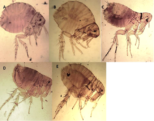
Figure 1: Light microscopy (LM) of permanent preparation of Flea spp. (X10) A&B: Echidnophaga gallinacea; C: Leptopsylla segnis; D& E: Xenopsylla cheopis
Morphological description of the collected sucking lice (Insecta: Anoplura)
Family: Hoplopleuridae (Ferris, 1951)
The Antennae were five-segmented, similar in males and females. The first pair of legs were small with slender claws, the second pair was larger with broader claws, and the paratergal plates project apically from the body with distinct tergal and sternal plates.
Hoplopleura hirsuta
Characterized by the presence of six pairs of distal abdominal setae, females presented paratergal plates by 4-6 only slightly elongated, and males with only seven tergal plates bearing a row of setae (Figures 2 A, B, C).
H. ocanthopus
Slender, 1-2 mm, characterized by large parategal plates which are four and five with two large setae on posterior margin (Figures 2 D, E, F).
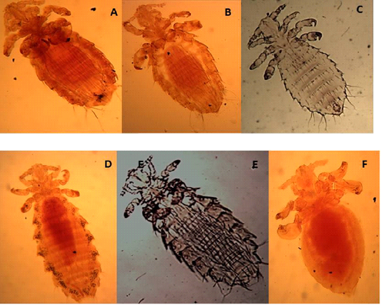
Figure 2: Light microscopy (LM) of permanent preparation of lice spp. (X40) A,B&C: Hoplopleura hirsuta; D, E& F: Hoplopleura ocanthopus
H. oenomydis
Abdomen with setae in the membrane between sternal and paratergal plates (Figures 3A and B).
Family:Polyplacidae
Polyplax spinulosa
Paratergal plates 3-5 with only dorsal apical angle produced into a point as in Figures 3C and D.
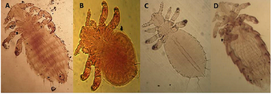
Figure 3: Light microscopy (LM) of permanent preparation of lice spp. (X40) A&B: Hoplopleura oenomydis; C& D: Polyplax spinulosa
Morphological description of the collected mites
Family: Dermanyssidae (Kolenta, 1859)
Ornithonyssus bacoti (Hirst,1931)
The dorsal plate is narrower than in the other species and tapers gradually to a blunt point Setae of the same size. There are no teeth on the chelicera (Figures 4A and B).
Dermanyssus gallinae (DeGeer, 1778)
The dorsal shield does not reach the post end and the posted margin. The setae are smaller on it than around the dorsal plate (Figures 5 A and B).
Allodermanyssus sanguine (Hirst, 1914)
Has 2 dorsal plates, the sternal plate bears all three pair of setae (Figure 4 D).
Family: Laelaptidae
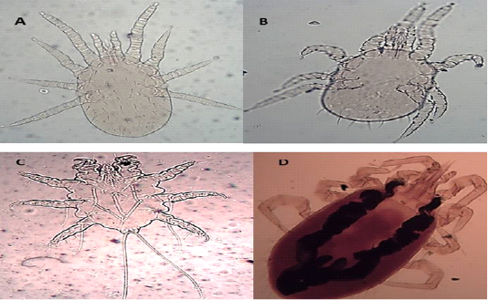
Figure 4: Light microscopy (LM) of permanent preparation of Mite spp. (X10) A&B: Ornithonyssus bacoti; C: Myobia muscula; D: Allodermanyssus sanguine
Laelaps nuttalli (Hirst, 1915)
Hypostome has a dorsal labrum of two lobes covered with minute papillae, segmented chelicerae, and ventral labium carrying two median lobes with laciniae and two lateral club-like lobes. Palpal and foreleg tarsal organs comprised ten and fifteen sensilla, respectively. Each pulvillus terminated with two medioventral claws and integumental folds beside longitudinal folds (Figures 5 C and D).
Family: Myobiidae
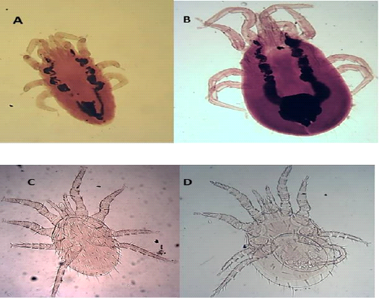
Figure 5: Light microscopy (LM) of permanent preparation of Mite spp. (X10) A&B: Dermanyssus gallinae; C& D: Laelaps nuttalli
Myobia musculi (Schranck, 1781)
has four pairs of legs, but the first pair is very short as an adaptation for grasping the hair shaft and giving the second pair of legs end in empodia (claw-like structures) (Figure 4 C).

