A three-year-old full-term Malabari doe in her third parity was referred to the outpatient unit of District Veterinary Centre, Kannur, Kerala, India for dystocia due to an abnormally large foetus. The chorioallantois had ruptured three hours back and the animal was straining. The physiological parameters of the doe were found to be normal. On per vaginal examination, after washing of perineum with 1 in 10000 potassium permanganate lotion, the vulva appeared swollen and oedematous with slightly congested vaginal mucous membrane. Oedematous limbs and scrotum of a foetus in posterior longitudinal presentation and lumbo-sacral position were found trapped and hanging from the birth canal (Figure 1A). Per-vaginal examination of
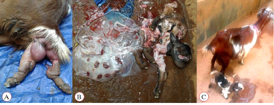
Figure 1: Malabari doe referred for dystocia due to abnormally large foetus. (A): oedematous limbs and scrotum of the foetus in dosrso-sacral position trapped at the birth canal; (B): the dead anasarcous foetus retrieved
by abdomen puncture and foetotomy along with the foetal membranes; and (C): the doe and the kid on the next day after obsterical procedure.
foetus after repulsion revealed that it was large sized and disproportionate with a severely distended abdomen that fluctuated on pressure indicating the presence of copious fluid. The vital reflexes of the foetus were absent. Based on the clinical and per-vaginal examination findings, the condition was tentatively diagnosed as dystocia due to concurrent foetal anasarca and ascites. Attempts to relieve the dystocia by gentle traction after sufficient lubrication were unsuccessful due to the distended abdomen and abnormally large size of the foetus leading to foeto-maternal disproportion. Since the foetus was confirmed as dead, a partial foetotomy of the foetus was resorted to with an aim to reduce the size of the foetus. The doe was administered epidural anaesthesia with 3 mL of 2% of lignocaine hydrochloride (Xylocaine 2%, AstraZeneca Pharma India Limited, Dundigal post, India) into the sacro-coccygeal epidural space. After lubrication with carboxy methyl cellulose, a gloved hand was inserted into the uterus and the foetal abdomen was punctured with an obstetrical hook followed by partial foetotomy using a Syme’s abscess knife to reduce the size of the foetus and retrieve the male foetus per-vaginum. Approximately six litres of ascitic fluid was drained out and the foetal parts including the eviscerated abdominal viscera were removed with gentle traction. On examination of the dead foetus, generalized oedema was observed with an enlarged and disproportionate head, swollen scrotal sac, puffed-up chin, rounded short neck and stumpy limbs, fairly developed hair coat and very thin skin. The foetal membranes along with the entrapped fluids were also removed (Figure 1B). Thereafter, one live, healthy female kid was also delivered per vaginum (Figure 1C). The doe was administered with Dextrose Normal Saline (Infutec Healthcare Ltd., Indore, India) at the rate of 10 ml/Kg body weight intravenously followed by ceftriaxone (Intacef, Intas pharmaceuticals, Ahmadabad, India) at the rate of 10 mg/Kg body weight, meloxicam (Melonex, Intas pharmaceuticals, Ahmadabad, India) at the rate of 0.2 mg/Kg body weight, oxytocin (Taj Pharmaceuticals Ltd., Mumbai, India) 5 IU (total dose) and tetanus toxoid (Serum Institute of India Pvt. Ltd., Pune, India) intramuscularly along with intrauterine administration of uterine cleanser. Antibiotic and antiinflammatory agents were continued for four more days and the animal had an uneventful recovery.

