Successful Per-Vaginal Management of Dystocia Due to Fetal Hydrocephalus in a Holstein Friesian Crossbred Cow: A Case Report
· · · · · · ·
Affiliations & Notes ⌄
1Department of Microbiology and Parasitology, Faculty of Animal Science and Veterinary Medicine, Sher-e-Bangla Agricultural University, Sher-e-Bangla Nagar, Dhaka-1207, Bangladesh
2Department of Surgery and Theriogenology, Faculty of Animal Science and Veterinary Medicine, Sher-e-Bangla Agricultural University, Sher-e-Bangla Nagar, Dhaka-1207, Bangladesh. *Correspondence | Uday Kumar Mohanta and Md. Rashedul Islam, Department of Microbiology and Parasitology, Faculty of Animal Science and Veterinary Medicine, Sher-e-Bangla Agricultural University, Sher-e-Bangla Nagar, Dhaka-1207, Bangladesh
3Department of Surgery and Theriogenology, Faculty of Animal Science and Veterinary
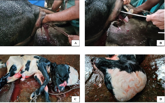
Following the palpation of the fetal head, a stab in the skin of the cranial cavity was given via a guarded knife to evacuate the fluid from the edematous head (Figure 1A).
The volume of the head was reduced in size after fluid evacuation, and with slight manipulation, the forelimbs and head of the fetus were pulled into the birth canal.
The forelimbs and obstetrical hook were pulled, and finally, the dead male fetus was successfully delivered with manual traction (Figure 1C, D).
The delivery of hydrocephalic fetus can be successfully facilitated through the evacuation of accumulated fluid after puncturing the apical soft region of the cranium, thereby reducing the head size.
Selected from the article's own text.
Abstract
A two-and-a-half-year-old Holstein Friesian crossbred dairy cow in its first parity at full term complained of straining for 11 hours with rupture of the water bag. After reaching the farm, per-vaginal examination revealed edematous and congested birth canal with completely open and relaxed cervix, and a large fluid-filled fetal head, fetus in anterior longitudinal presentation, dorsosacral position with carpal flexion, and severe straining. Based on the above clinical findings, the case was provisionally diagnosed as dystocia due to a hydrocephalic fetus. To manage the condition, an incision was made in the skin over the cranial cavity of the fetus, using a guarded embryotomy knife to drain out the excess fluid. Following the removal of fluid, the dead male fetus was successfully delivered by manual traction. After delivery, the cow was treated with antibiotics, non-steroidal anti-inflammatory drugs (NSAIDs), antihistamines, calcium, multivitamins, and multi-minerals for five days. Finally, the cow recovered uneventfully after five days.
Hydrocephalus is a dropsical condition with the accumulation of fluid as a result of an imbalance between the formation and drainage of cerebrospinal fluid (CSF), either in the ventricular system or subarachnoid space, characterized by marked enlargement of the cranium ; . It is common in animals , particularly in dairy cows. However, this condition primarily results from abnormal fetal development during pregnancy; environmental, infectious, nutritional, and genetic factors may also contribute to its occurrence ; . It is assumed that hydrocephalus might result from disturbance in the normal flow of CSF and its reabsorption . Hydrocephalus in cattle has been associated with a simple autosomal recessive gene , an autosomal dominant gene exhibiting incomplete penetrance , and hypovitaminosis A as well. It results in dystocia, and fetuses are usually delivered either by head excision followed by traction or via a cesarean section . Calves exhibiting substantial cranial enlargement typically die within 48 hours, while those with milder manifestations may survive . The present case report describes a successful per-vaginal delivery of a male hydrocephalic calf following fluid drainage from an enlarged head.
Case history and clinical observation
A two-and-a-half-years-old Holstein Friesian crossbred dairy cow, weighing 200 kg in its first parity at full term, with the manifestation of straining since the last eleven (11) hours. The water bag ruptured six (6) hours after the onset of straining, whereby a local AI technician attempted to deliver the case but failed. Then, a licensed veterinarian was called upon to treat the case. On clinical examination, the animal showed pale pink mucous membranes, a rectal temperature of 101 oF, and heart rate and respiration rate were within normal ranges. The per-vaginal examination revealed an edematous and congested birth canal with a completely open and relaxed cervix, a large fluid-filled fetal head, anterior longitudinal presentation, and dorsosacral position with carpal flexion. The examination indicated the condition of hydrocephalic fetus characterized by a significant enlargement of the cranial head. Palpation confirmed the absence of reflexes, indicating that the fetus was non-viable. Based on the above clinical findings, the case was confirmed as dystocia due to a hydrocephalus fetus.
MANAGEMENT AND DISCUSSION
At first, epidural anesthesia in the sarco-coccygeal space was performed using 5 ml 2% Lignocaine hydrochloride (G-Lidocaine, Gonoshasthaya, Bangladesh). Following the palpation of the fetal head, a stab in the skin of the cranial cavity was given via a guarded knife to evacuate the fluid from the edematous head (Figure 1A). The volume of the head was reduced in size after fluid evacuation, and with slight manipulation, the forelimbs and head of the fetus were pulled into the birth canal. Then, the forelimbs were tied with rope, and the fetus was tried to deliver, but it was unsuccessful. Then, the obstetrical hook was also applied to the inner canthus of the eye socket (Figure 1B). The forelimbs and obstetrical hook were pulled, and finally, the dead male fetus was successfully delivered with manual traction (Figure 1C, D). As manual traction is less invasive, quicker, economical, and provides fewer complications to the reproductive health of the cow compared to caesarean section (C-section), it was preferred over C-section. The fetal membranes were manually removed. The cow stood up right away after calving. Immediately after calving, Metronidazole (Diro Vet, Acme, Bangladesh) was inserted into the uterus to prevent secondary bacterial infections. The postoperative treatments included Calcium borogluconate together with magnesium, phosphorus, boric acid, and dextrose (A-Cal vet, Acme, Bangladesh) 250 ml i/v once, and Amoxicillin 2 g (Renamox vet, Renata, Bangladesh), Meloxicam 10 ml (Loxikam vet, Opsonin, Bangladesh), Diphenhydramine HCl 10 ml (Phenadryl vet, Acme, Bangladesh) each i/m for 5 days. Then, the cow recovered eventfully after five days. Fetal dropsical conditions that can lead to dystocia include hydrocephalus, ascites, hydrothorax, and anasarca , 2012). Among them, congenital hydrocephalus is morphologically characterized by a large fluid-filled sac hanging over the head. In severe cases, hydrocephalus leads to dystocia, which cannot be relieved through mutation or forced traction. Therefore, fetal delivery is typically achieved either by head incision followed by traction or through caesarean section ; . The excessive enlargement of the cranium may require a fetotomy , but in the present case, there was no excessive enlargement of the cranium except for a large volume of fluid-filled head which might facilitate per-vaginal delivery by forceful manual traction. Similar observations of per-vaginal delivery in dairy cows have been reported by several authors worldwide ; ; ; . Hydrocephalic fetus has consistently posed a significant challenge for field veterinarians. In the present case, instead of other medical interventions, an emergency veterinary surgeon was required to perform a per-vaginal delivery.
CONCLUSION
The delivery of hydrocephalic fetus can be successfully facilitated through the evacuation of accumulated fluid after puncturing the apical soft region of the cranium, thereby reducing the head size.
ACKNOWLEDGEMENTS
The authors are grateful to Upazila Livestock Officer (ULO), Upazila Livestock Office and Veterinary Hospital, Manoharganj, Cumilla, Bangladesh, for his support during the study.
Novelty Statement
Despite adopting surgical intervention like most reported cases, this case demonstrated an effective, economic, and minimally invasive alternative.
AUTHOR’S CONTRIBUTION
All the authors contributed to the research and manuscript writing.
Conflict of interest
The authors have declared no conflict of interest.
Figures
Figure 1:.
A: Incised the fetal cranial cavity. B: Applied obstetrical hook and ropes. C: Expelled the dead male fetus. D: A big hydrocephalic fetal head.
References
Arthur GH, Noakes DE, Parkinson TJ, England GCW (2001). Veterinary reproduction and obstetrics (8th ed.). Philadelphia, PA: WB Saunders. pp. 131–132.
Balasubramanian S, Ashokan SA, Seshagiri VN, Pattabisaman SR (1997). Congenital internal hydrocephalus in a calf. Indian Vet. J., 74(5): 446-447.
Bhandari RM, Kadu MS, Belorkar PM, Murudwar SS (1978). Obstetrical management of hydrocephalic fetus in buffaloes: A report of two cases. Indian Vet. J., 55(12): 1001–1002.
Bugalia NS, Chander S, Chandolia RK, Verma SK, Singh P, Sharma OK (1990). Monstrosities in cows and buffaloes. Indian Vet. J., 67(11): 1042-1043.
Dar LM, Mavi PS, Bhat GR (2012). Per vaginal delivery of a congenital hydrocephalus fetus in a crossbred primiparous heifer: A case report. Int. J. Livest. Res., 2(2): 258–260. https://www.researchgate.net/publication/272485112_Pervaginal_delivery_of_a_congenital_hydrocephalic_fetus_in_a_crossbred_primiparous_Heifer-_A_case_report
Jubb KVF, Kennedy PC, Palmer N (1993). Pathology of domestic animals. San Diego, CA: Academic Press. pp. 229–230. https://doi.org/10.1016/B978-0-12-391607-5.50007-8[CrossRef]
Jubb KVF, Kennedy PC, Palmer N (2012). Pathology of domestic animals. Harcourt Brace Jovanovich, pp. 2.
Leipold HW, Dennis SM, Huston K (1972). Congenital defects of cattle: nature, cause, and effect. Adv. Vet. Sci. Comp. Med., 16:103-50. https://pubmed.ncbi.nlm.nih.gov/4624930/
Megahed GA (2017). Extraordinary case of dystocia due to hydrocephalus foetus in dairy Egyptian cow: A case report. J. Dairy Vet. Anim. Res., 5(2): 51-51. https://doi.org/10.15406/jdvar.2017.05.00133[CrossRef]
Murali RM, Veerabramhaiah K, Praveen S, Babu A, Vardhan MS (2020). Dystocia in cattle due to fetal hydrocephalus. Indian J. Anim. Reprod., 41(1): 67–68. https://journals.acspublisher.com/index.php/ijar/article/view/3931
Noakes DE, Parkinson TJ, England GCW (2009). Veterinary reproduction and obstetrics (9th edn). London, UK: Saunders. pp. 950.
Purohit GN, Gaur M, Sharma A (2006). Dystocia in Rathi cows due to congenital hydrocephalus. Indian J. Anim. Reprod., 27(1): 98–99. https://journals.acspublisher.com/index.php/ijar/article/view/6613
Purohit GN, Kumar P, Solanki K, Shekhar C, Yadav SP (2012). Perspectives of fetal dystocia in cattle and buffalo. Vet. Sci. Dev., 2(1): 8. https://doi.org/10.4081/vsd.2012.3712[CrossRef]
Roberts SJ (1971). Veterinary obstetrics and genital diseases (Theriogenology). 2nd Edn. CBS Publishers and Distributors, New Delhi, India. pp. 69.
Roberts SJ (1986). Diagnosis and treatment of dystocia, veterinary obstetrics and genital diseases. Indian Edition, CBS Publishers, New Delhi. India. pp. 283.
Saini R, Sharma SK, Aziz PR, Mir LA, Pooniya R (2019). Congenital Hydrocephalus in a Calf and its Surgical Treatment: A Case Report. Indian J. Vet. Sci. Biotechnol., 15(1): 81–82. https://journals.acspublisher.com/index.php/ijvsbt/article/view/2563
Wasef A, Islam MR (2024). Incidence and management of dystocia of dairy cows in Manoharganj Upazila of Cumilla District, Bangladesh. Ovozoa J. Anim Reprod., 13: 178-185. https://doi.org/10.20473/ovz.v13i3.2024.178-185[CrossRef]