Animals
Inbred female Swiss albino mice (4–6 weeks old), weighing approximately 20 g, were obtained from the Laboratory Animal Resources, Indian Veterinary Research Institute, Izatnagar, India. Animal experiments were carried out after obtaining approval from the Institutional animal ethics committee. All the mice were housed under standard conditions at the animal shed facilities and provided sufficient water and food.
Bacterial Strain and Vector
The virulent Brucella abortus Strain 544 and the attenuated Brucella abortus Strain–19 was obtained from Division of Standardization, Indian Veterinary Research Institute (IVRI), Izatnagar, India. Escherichia coli DH5µ strain (Life technology, USA) was used as host for cloning of DNA vaccine construct whereas E. coli M15 strain (Qiagen, USA) was used for expression of SOD:L7/l12 fusion protein. Prokaryotic expression vector pQE–30 (Qiagen, USA) and eukaryotic expression vector pVAX1 (Invivogen, CA, USA) were used in the present study. Brucella cells were grown on Brucella broth under aerobic condition for 72 hours at 37oC and E. coli cells were grown in Luria –Bartani (LB) medium, supplemented with required antibiotics (kanamycin and ampicillin), under aerobic condition at 37oC.
Construction of pVSL DNA Vaccine:
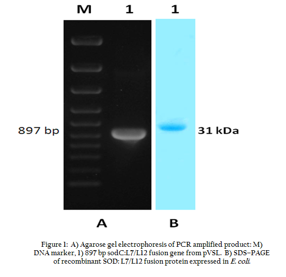
Vector pVAX1 was used to develop chimeric DNA vaccine construct encompassing Cu–Zn SOD (sodC gene) and L7/L12 ribosomal protein (l7/l12 gene) of Brucella abortus. A 522 bp DNA fragment encoding sodC gene and 375 bp l7/l12 genes were amplified from Brucella abortus strain 544 DNA by Polymerase Chain Reaction. The primer sequences of the sodC were 5'–CGGCGGATCCACCATGGGAAAGTCCTTATTTATTGCATC–3' (primer 1 with BamH I site underlined) and 5'–CGGCGTCGACTTCGATCACGCCGCAGGCAAAACG–3' (primer 2 with Sal I site underlined). The primer sequences of the l7/l12 gene were 5'–CGCCGTCGACTCGTCCGGCGGAGGCGGA–3' (primer 3 with sal I site underlined) and 5'–GCCCCTGCAGTTACTTGAGTTCAACCTTGGC–3' (primer 4 with Pst I site underlined). The PCR amplified sodC gene, without stop codon, was placed to upstream of l7/l12 gene product and ligated to pVax1 vector. Two genes were separated by a nucleotide spacer that encoded for six amino acids (2 serine and 4 glycin). The resulting plasmid was designated as pVSL. The DNA vaccine construct, pVSL, was transformed into chemically (CaCl2) competent E. coli DH5 α cells by heat shock method. Recombinant E. coli harbouring DNA vaccine construct was cultured in LB medium supplemented with kanamycin (25 μg/mL) for large scale production of DNA vaccine, pVSL. DNA vaccine constructs for individual genes, l7/l12 and sodC, were prepared in pVax1 vector and the DNA vaccine constructs were named as pVL7 /L12 and pVsod, respectively.
Expression and Purification of Sod: L7/L12 Fusion Protein
To generate recombinant SOD:L7/L12 fusion protein, the target fusion gene was cleaved from the pVSL plasmid by restriction endonucleases BamH I and Pst I. The released insert was ligated to pQE–30 expression vector and transformed into chemically competent E.coli M15 cells. Recombinant E. coli cells were grown in LB broth supplemented with kanamycin (25 μg/mL) and ampicillin (100 μg/mL) and IPTG (1mM) was used for induction of expression. Purification of SOD:L7/L12 fusion protein was made using Ni2+–NTA agarose resin (Qiagen, USA). Purified protein was identified by sodium dodecyl sulfate–polyacrylamide gel electrophoresis (SDS–PAGE). Recombinant protein for individual genes, l7/l12 and sodC was prepared using prokaryotic expression vector.
Sequencing of pVSL
The cloned L7/L12 ribosomal protein gene and sodC gene in pVSL was sequenced by dideoxy chain termination method. The nucleotide sequence was analyzed by using ‘EditSeq’ program of Lasergene software (DNASTAR, Madison, USA).
Immunization
The immunization studies were carried out in female Swiss albino mice. Each group was consisted of 15 animals. The mice were inoculated intramuscularly with 100 µg of pVSL, pVL7/L12 or pVsod in 50 µL of PBS. A booster immunization was carried out on day 21 with 50 µg of recombinant fusion protein (SOD:L7/L12), L7/L12 or Cu–Zn SOD. A group of mice was immunized with PBS as negative control. As positive control, group of mice was inoculated intraperitoneally with 1 x 10⁵ CFU of B. abortus strain 19 in 0.2 mL of PBS. Five mice from each experimental group were bled for collection of sera samples at different time intervals (0, 20, 35 and 50th day).
Analysis of Specific Antibody Responses by Elisa
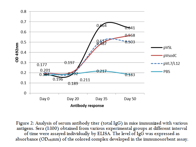
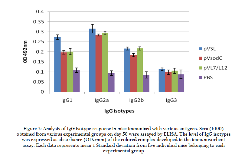
To assess the antibody response to SOD:L7/L12 fusion protein, sera samples were collected at different time points from the immunized mice. Total IgG and IgG isotype response were determined by indirect ELISA. Immunoplates (Maxisorp, Nunc) were coated with purified recombinant SOD:L7/L12 protein at a concentration of 2.5 µg/mL (100 µL/well ) diluted in carbonate–bicarbonate buffer (0.05M, pH 9.6). Goat anti–mouse IgG–HRPO conjugate (1:2500) was used as secondary antibody. ELISA plates were developed in 100 µL of freshly prepared substrate solution (200 µmol o–phenylene–diamine and 0.04% H2O2 in citrate buffer) and the reaction was stopped by addition of 50 µL of 0.18 M sulfuric acid per well. Absorbance of the developed color was measured at 492 nm. The titer of each serum was expressed as A492 value and normalized to value of control sera tested at 1:50 dilution. Similarly, titer of IgG isotypes were determined by using goat anti–mouse IgG1, IgG2a and IgG2b– HRPO conjugates (Santa Cruz, USA) at a dilution of 1:10,000. Mean values and standard deviations were calculated to depict immunoglobulin response to individual vaccine group.
Protection Study against Brucella abortus 544 Challenge
Four weeks after last vaccination, mice were challenged with a virulent culture of Brucella abortus 544 (1 X 10⁵ CFU / mice) by intraperitoneal route in 0.2 mL PBS. After 30 days of challenge, mice from each group were euthanized; spleens were aseptically removed and triturated with 2 mL of 1.5M PBS (pH7.4) with 0.1% TritonX–100 using sterile sand. Ten–fold serial dilutions of spleen cell lysate were made and 50 µL from each dilution was plated on Typtose Phosphate Broth (TPB) medium with 1.5% agar in triplicate. For S–19 vaccinated mice group, 0.1% Erythriotol was added in the plate for differentiating S–19 and Brucella abortus 544. Plates were further incubated at 37oC with 10% CO2 pressure. Colonies appeared after three days of incubation were counted and calculated as log10 colony forming unit (cfu) per spleen. Protection conferred by individual vaccine group was expressed as log10 unit of protection and it was derived by subtracting mean log10 cfu for the experimental group from the negative control group.