
Ghrelin, the endogenous ligand for the growth hormone secretagogue receptor, has been identified in the lung of many vertebrates. While ghrelin-immunopositive (ghrelin-ip) cells have been found in the cerebellum, pancreas and gastrointestinal tract of African ostrich chicks (Struthio camelus), little is known about its distribution in the lung of the African ostrich. To provide the morphological basis of ghrelin function in the African ostrich lung, the distribution and morphological characteristics of ghrelin-ip cells were investigated using immunohistochemistry. The results indicate that the lung of African ostrich was composed of interstitial tissue and parenchyma. The interstitial tissue formed a layer of serosa on the surface of the lung. The parenchyma of the lung consisted of bronchial levels, atria and respiratory capillaries. The bronchi were divided into the primary, secondary and tertiary bronchi. The wall of the tertiary bronchus had openings for the atria and respiratory capillaries. The respiratory capillaries were surrounded by a rich blood capillary bed. Ghrelin-ip cells were located among the tertiary bronchus mucosal epithelial cells, respiratory capillary epithelial cells and macrophages. The ghrelin-ip cells showed cytoplasmic staining, they were cone, oval, spindle, round or irregular in shape and tended to be restricted to a single cell. These results demonstrate the presence of ghrelin-ip cells in the lung of the African ostrich and therefore it is speculated that ghrelin may have a physiological function in the lung of the African ostrich.
Research Article · Volume 50, Issue 1, 12 January 2018 · Open Access
Distribution and Morphology of Ghrelin-Immunopositive Cells in the Lung of the African Ostrich
· · ·
Affiliations & Notes ⌄
- 1College of Animal Science, Yangtze University, Jingzhou 434103, PR China 2Ostrich Research Institute, Yangtze University, Jingzhou 434103, PR China , * Corresponding authors: wangjiaxiang1109@163.com
- 2844542539@qq.com
Article Info ⌄
- Journal:
- Pakistan Journal of Zoology
- Published:
- 12 January 2018

Abstract
Introduction
Ghrelin, a 28-amino acid gut peptide acylated at the serine-3 position with an n-octanoyl group (C8:0), has been isolated as an endogenous ligand of the growth hormone secretagogue receptor (GHSR) in the rat stomach . Among birds, chicken ghrelin is composed of 26 amino acids , while ostrich ghrelin has 28 amino acids . Ghrelin is mainly produced and secreted by A-like cells within the oxyntic glands of the stomach , and the acylation is essential for ghrelin’s activity through the GHSR . Besides its strong function to promote growth hormone secretion, ghrelin also plays roles in regulating the secretion of endocrine system, gastrointestinal motility, development of reproductive glands, feeding and energy balance ; ; , 2013; ; ; ; .
Recently, there were reports that ghrelin also participated in some physiological functions and pathological progress of lung in rats ; ; . In general, to understand or hypothesize the physiological role of newly identified peptides, it is of great significance to determine the morphological characteristics of the producing cell and its distribution. Previous studies have shown that ghrelin-ip cells were distributed in the lungs of some animals such as broiler chickens, sika deer, reindeer and rat ; ; ; . However, in the African ostrich, ghrelin-ip cells were only identified in the cerebellum, pancreas and gastrointestinal tract , 2012, 2017; , little is known about its distribution in the lung. Therefore, the distribution and morphological characteristics of ghrelin-ip cells in the lung of the African ostrich were investigated by immunohistochemistry in the present study in order to provide the morphological basis of ghrelin function in the African ostrich lung.
Materials and Methods
Animals
African ostriches (age, 5 months; weight, 35.12 ± 2.17 kg) were used for this study. African ostrich (2 females and 2 males) were obtained from the Ostrich Research Institute of Yangtze University in Hubei Province, China, where feed and water were made available ad libitum. All of the birds were maintained in a heated room with slatted plastic flooring and were fed a starter diet for postnatal days 7, which was formulated according to the specifications of the Elsenburg Ostrich Feed Database . All procedures were approved by the Animal Care and Welfare Committee of our Institute.
Tissue preparation
The ostriches were deeply anesthetized with 10% urethane (Caoyang Secondary Chemical Plant, Shanghai, China) at a dose of 1 g/kg BW, and perfused, initially with 1000 mL of 0.85% normal saline (containing 0.075% sodium citrate) and thereafter with 1500 mL of 4% paraformaldehyde phosphate-buffered solution (0.1 mol/L, pH 7.4) at 4°C. The thoracic cavity was cut open and the entire lungs were quickly removed, and gently flushed with 0.85% normal saline to remove the content, then be postfixed for more than 24 h with 4% paraformaldehyde. After immersion, the tissues were embedded in paraffin. Serial sections (5μm) were cut on a Leica microtome (Nussloch Gmbh, Germany), 2 sets of sections were prepared; one was stained by haematoxylin and eosin to observe the the morphologyy of lungs; the other was stained by immunohistochemistry to observe the distribution and morphological characteristics of ghrelin-ip cells in the lungs of the African ostrich.
Immunohistochemistry
Immunohistochemical detection of ghrelin-ip cells using rabbit anti-ghrelin was carried out by the streptavidin-biotin-peroxidase complex (SABC) method. The production and specificity of the anti-human ghrelin serum used in this study were previously reported ; it is established that this antiserum recognizes both N- and C-terminial of human ghrelin. Immunohistochemical staining was performed according to the following procedure. The sections were deparaffinized with xylene and rehydrated with decreasing concentrations of ethanol, then treated with 3% hydrogen peroxide to block endogenous peroxidase for 10 min at room temperature. After rinsing with distilled water, the sections were incubated with a citrate buffer (pH 6.0) and placed in a microwave oven until the water boiled to fully expose the antigen. After rinsing with phosphate-buffered saline (PBS), the sections were incubated with 5% normal goat serum for 20 min. After removing superfluous liquid, the sections were incubated with rabbit anti-ghrelin serum (BA1619; Boster Corporation) diluted 1:100 in PBS for 12 h in a humid chamber at 4°C. After washing with PBS for 6 min, a second incubation with biotin-conjugated anti-rabbit IgG serum (SA1022; Boster) was carried out for 20 min, and this was followed by further washing with PBS. Finally, the sections were incubated for 20 min with an SABC solution prepared according to the manufacturer’s instructions. After washing with PBS for 20 min, the sections were reacted in a diaminobenzidine-tetrachloride kit (DAB kit, AR1022, Boster Corp) for 10 min to detect immunostaining. After washing with distilled water, the sections were dehydrated with a graded ethanol series, cleared in xylene, mounted with a coverslip, and viewed under a light microscope (BH-2; Olympus, Japan). All of the incubations were carried out in a humid chamber at room temperature. Control sections were prepared using the same method, omitting the primary antibody. To examine the specificity of rabbit anti-human ghrelin antiserum, the diluted antiserum (1:100) was incubated with human ghrelin (5 μg/ml) at room temperature for 10 h, and mixtures were centrifuged at 12000 rpm for 25 min at 4°C. The supernatant was used as the primary antiserum for absorption tests.
Morphometric analysis
For each lung tissue sample, 3 cross-sections were prepared after the samples had been stained with hematoxylin and eosin and SABC stain. Further, for each lung cross-section, 10 intact, well-oriented units were selected for experiments conducted in triplicate (30 measurements for each sample). Then taking digital photographs under a light microscope with a digital camera (COOLPIX4500; Nikon, Japan).
Results
All results presented were obtained from both female and male chicks. No gender-specific effects were observed.
Histological characteristics of the lung
The lung of the African ostrich, found to be pink with no lobes, was composed of interstitial tissue and parenchyma. The interstitial tissue formed a layer of serosa on the surface of the lung, in which the connective tissue stretched into the lung parenchyma, forming a support structure for the lung, and artery could be observed in the interstitial tissue (Fig. 1A). The parenchyma consisted of the bronchial levels, atria and respiratory capillaries (Fig. 1A). The bronchi were divided into the primary, secondary and tertiary bronchus, and they formed an interconnected labyrinthine structure. The bronchus formed primary bronchi after entering the lung. The wall of the secondary bronchus mucosa had distinct longitudinal wrinkles, and the epithelium was pseudostratified ciliated columnar epithelium. The epithelium of tertiary bronchus was incomplete since it had openings for the atria and respiratory capillaries, and the epithelial cells were cone, oval or irregular in shape, the nuclei were round at the middle of the cells (Fig. 1B). There were smooth muscles around the epithelium of tertiary bronchus (Fig. 1B). The atria were small and irregular, and they also had openings for respiratory capillaries (Fig. 1C). There were abundant blood capillaries around the respiratory capillary as well (Fig. 1C). The wall of respiratory capillary was simple squamous epithelium, and the epithelial cells were oval, spindle or irregular in shape, the nuclei were oblate (Fig. 1D). In addition, macrophage and vein could be observed in interstitial tissue of lungs (Fig. 1D).
Distribution of ghrelin-immunopositive cells
Immunohistochemical streptavidin-biotin-peroxidase complex (SABC) staining showed that ghrelin-ip cells presented in the parenchyma and interstitial tissue in the lung of African ostrich, and to be varying shades of brown (Fig. 2A). Ghrelin-ip cells were located among the tertiary bronchus mucosal epithelial cells, respiratory capillary epithelial cells and macrophages, and they tended to be restricted to a single cell with different sizes (Fig. 2A). In the parenchyma, ghrelin-ip cells located among tertiary bronchus mucosal epithelial cells exhibited a stained cytoplasm, and they were cone, oval, or irregular in shape (Fig. 2B). While among respiratory capillary epithelial cells, ghrelin-ip cells were oval, spindle or irregular in shape, they also showed cytoplasmic staining (Fig. 2C). In the interstitial tissue, ghrelin-ip cells located among macrophages were irregular in shape with a stained cytoplasm (Fig. 2C). However, these staining profiles were not observed when the sections were processed with the supernatant without the primary antiserum for absorption tests as negative control (Fig. 2D).
Discussion
Structure of the African ostrich lung
The lung is an important respiratory organ in the body, and the shape, location and structure differ among animals. In the present study, we found that the lung of African ostrich had no lobes, which was similar to Falco tinnunculus (Feng et al., 2009a), Tragopan caboti , and Aguila chrysaetos (Feng et al., 2009b). However, the lung of mammals had lobes . Furthermore, the bronchi of mammals formed bronchial tree, whereas in the ostrich, they formed an interconnected labyrinthine structure, which was consistent with F. tinnunculus (Feng et al., 2009a), T. caboti and A. chrysaetos (Feng et al., 2009b). These results suggest that the lungs of avian are at a lower level of evolution than that of mammals.
In present study, the atria of the African ostrich were small and irregular, however, in the F. tinnunculus (Feng et al., 2009a), T. caboti and A. chrysaetos (Feng et al., 2009b), they were arranged radially around the tertiary bronchus. These results indicate species differences in lung tissue.
Unlike those smaller highly aerobic volant birds, ostriches have to tolerate extreme thermal stresses through sustained panting as they live on the ground . Therefore, the difference in lung tissue between the ostrich and other birds may be due to adaptation to its living environment. Further study is needed to investigate whether such differences result in different functionality of the lung.
Distribution of ghrelin immunoreactive cells in the lung of African ostrich
Recently, studies have demonstrated the presence of ghrelin-ip cells in the lungs of various animals ; ; ; . In this study, ghrelin-ip cells were found within the tertiary bronchus mucosal epithelial cells, respiratory capillary epithelial cells and macrophages. However, these distribution features of ghrelin-ip cells were different from that of broiler chicken, sika deer, reindeer and rats ; ; ; . In broiler chicken, ghrelin-ip cells were found to be located among the scattered macrophages of the lung . In sika deer, ghrelin-ip cells in the lung were mainly distributed in the epithelial cells of the bronchial wall surface and alveolar epithelial cells . Ghrelin-ip cells showed significant positive staining in the lung of reindeer, the bronchial epithelium was darker, and the cytoplasm of the alveolar cells and the nuclei were stained at high magnification . In rats, ghrelin-ip cells were expressed in the primitive epithelium of the lung at early stages of development . These results indicate that ghrelin in ostrich lung has a different distribution pattern than in other animals.
It has been demonstrated that an osmotic plate structure is distributed over the epithelium and surfaces of tertiary bronchus, atria and respiratory capillaries, whose function is similar to pulmonary surfactant of livestock, which plays significant roles in pulmonary mechanics, surfactant in airways, pulmonary vessels and blood flow ; ; ; . Additionally, Henriques-Coelho et al. (2006) found that ghrelin administration attenuated monocrotaline-induced pulmonary hypertension, pulmonary vascular remodeling, and right ventricular hypertrophy, indicating that it may modulate pulmonary hypertension. Previous studies also showed that exogenous ghrelin could prevent adverse changes in pulmonary blood flow distribution throughout the lung in rats . In present study, ghrelin-ip cells were found to be distributed in the tertiary bronchus mucosal epithelial cells and respiratory capillary epithelial cells, it suggested that ghrelin might have some effects on pulmonary physiology of African ostrich. At the same time, ghrelin-ip cells were found to be located among macrophages, so it was deduced that ghrelin is likely to play a role in immunity. Further, ghrelin has been demonstrated to have a positive effect in fetal lung development through its GHS-R1a receptor . Therefore, ghrelin may have a physiological function in the lung of the African ostrich.
Conclusion
The present study provides a morphological basis for ghrelin function in the lung of African ostrich, and it is important to perform further studies on the function and regulation mechanism of these peptides in this location for African ostrich.
Acknowledgement
The authors express appreciation for the support from the National Natural Science Foundation of China (No. 31402152).
Statement of conflict of interest
The authors report no conflicts of interest. The authors alone are responsible for the content and writing of this article.
Figures
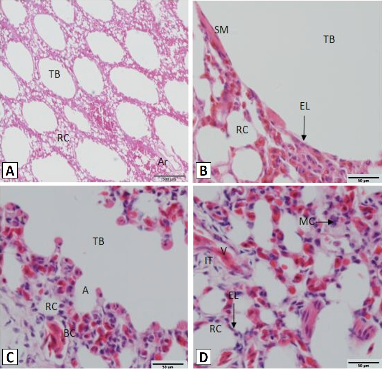
Histology of the African ostrich lungs. A, the lung parenchyma and artery; B, the tertiary bronchus; C, atria; D, respiratory capillaries. TB, tertiary bronchus; RC, respiratory capillaries; Ar, artery; SM, smooth muscle; EL, epithelial layer; A, atria; BC, blood capillary; MC, macrophage; V, vein. Scale bar: A=500 μm; B, C and D=50 μm, Stain: haematoxylin and eosin.
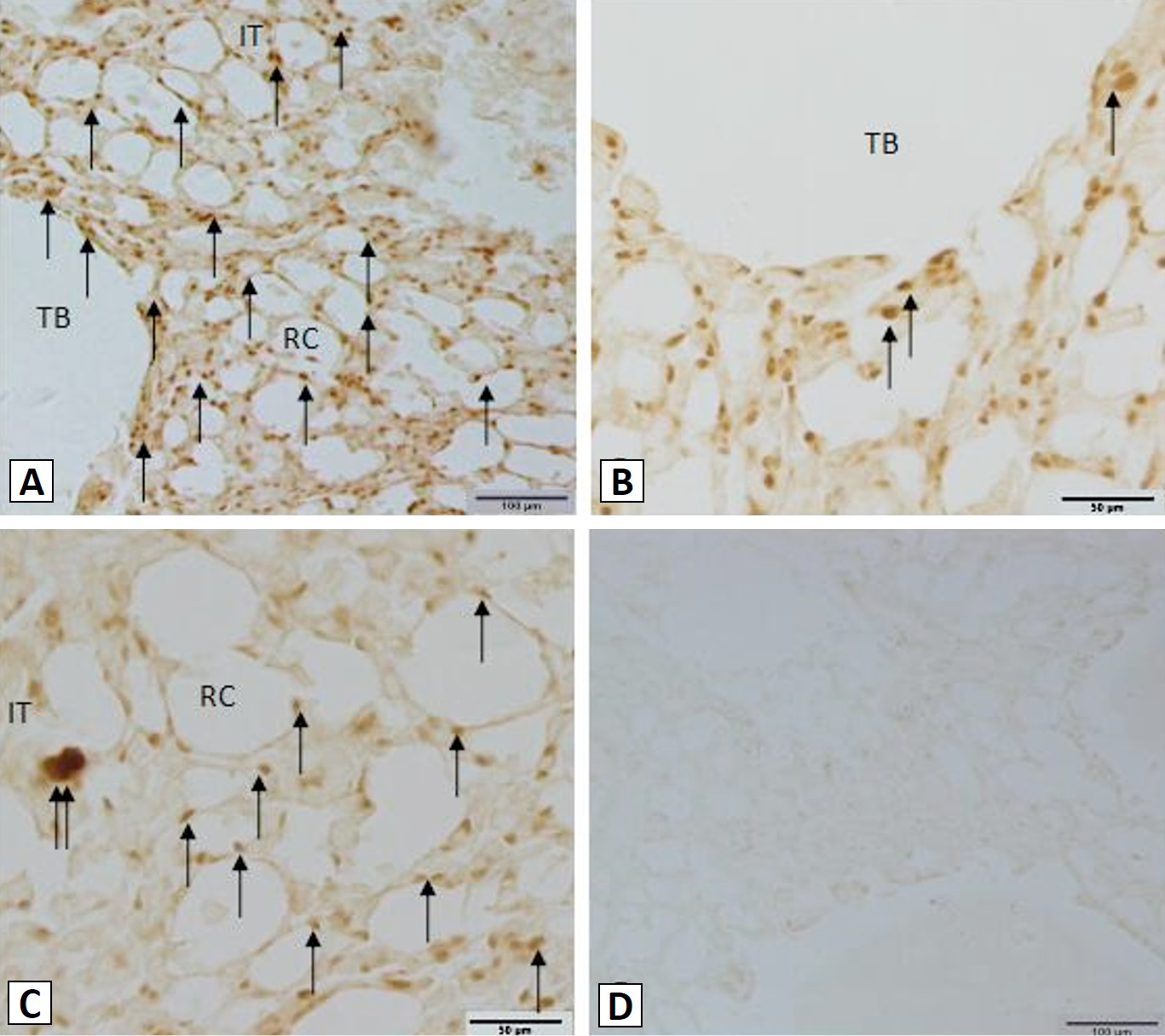
Distribution of ghrelin-ip cells in the lungs of the African ostrich. A, ghrelin-ip cells (arrows) were found within the lung; B, ghrelin-ip cells (arrows) within the tertiary bronchus mucosal epithelial cells; C, ghrelin-ip cells within the respiratory capillary epithelial cells (↑) and macrophage (↑↑); D, the control sections were negative. TB, tertiary bronchus; RC, respiratory capillary; IT, interstitial tissue. Scale bar: A and D=100 μm; B and C= 50 μm.
References
- Abizaid, A. and Horvath, T.L., 2012. Ghrelin and the central regulation of feeding and energy balance. Indian J. Endocrinol. Metab., 16(Suppl. 3): S617-S626. https://doi.org/10.4103/2230-8210.105580[CrossRef]
- Björklund, L.J., Ingimarsson, J., Curstedt, T., John, J., Robertson, B., Werner, O. and Vilstrup, C.T., 1997. Manual ventilation with a few large breaths at birth compromises the therapeutic effect of subsequent surfactant replacement in immature lambs. Pediatr. Res., 42: 348-355. https://doi.org/10.1203/00006450-199709000-00016[CrossRef]
- Brand, T.S., 2000. Elsenburg ostrich feed databases. Elsenburg Agricultural Research Centre, Elsenburg, South Africa.
- Feng, H.L., Yu, S.Y., Jia, Y.F., Deng, H.P., Wang, Y. and Niu, X.F., 2009a. Histological structure and expression of inter related poly peptides in Falco tinnunculus lung. J. Northw. Norm. Univ. (Nat. Sci.), 45: 101-106.
- Feng, H.L., Yu, S.Y., Wang, Y., Jia, Y.F. and Deng, H.P., 2009b. Histological structure and expression of KGF, c-Fos and Bax in the LUNG of Aguila chrysaetos. Chinese J. Zool., 44: 33-38.
- Healy, J.E., Bateman, J.L., Ostrom, C.E. and Florant, G.L., 2011. Peripheral ghrelin stimulates feeding behavior and positive energy balance in a sciurid hibernator. Horm. Behav., 59: 512-519. https://doi.org/10.1016/j.yhbeh.2011.01.013[CrossRef]
- Henriques-Coelho, T., Roncon-Albuquerque, Jr. R., Lourenço, A.P., Baptista, M.J., Oliveira, S.M., Brandão-Nogueira, A., Correia-Pinto, J. and Leite-Moreira, A.F., 2006. Ghrelin reverses molecular, structural and hemodynamic alterations of the right ventricle in pulmonary hypertension. Rev. Port. Cardiol., 25: 55-63.
- Im Hof, V., Gehr, P., Gerber, V., Lee, M.M. and Schürch, S., 1997. In vivo determination of surface tension in the horse trachea and in vitro model studies. Respir. Physiol., 109: 81-93. https://doi.org/10.1016/S0034-5687(97)84032-7[CrossRef]
- Kojima, M., Hosoda, H., Date, Y., Nakazato, M., Matsuo, H. and Kangawa, K., 1999. Ghrelin is a growth-hormone-releasing acylated peptide from stomach. Nature, 402: 656-660. https://doi.org/10.1038/45230[CrossRef]
- Liu, J., Zhang, M., Jin, X., Fan, Y.R., Tian, Q.Z. and Yang, Y.F., 2016. Immunohistochemical localization of ghrelin in sika deer. Anim. Husband. Vet. Med., 48: 87-91.
- Lotfi, A., Aghdam-Shahryar, H., Ghiasi-Ghalehkandi, J., Kaiya, H. and Maheri-Sis, N., 2011. Effect of in ovo ghrelin administration on subsequent serum insulin and glucose levels in newly-hatched chicks. Czech J. Anim. Sci., 8: 377-380.
- Lotfi, A., Shahryar, H.A. and Kaiya, H., 2013. Effect of in ovo, ghrelin administration on hatching results and post-hatching performance of broiler chickens. Livest. Sci., 154: 158-164. https://doi.org/10.1016/j.livsci.2013.03.020[CrossRef]
- Maina, J.N. and Nathaniel, C., 2001. A qualitative and quantitative study of the lung of an ostrich, Struthio camelus. J. exp. Biol., 204: 2313-2330.
- Nishi, Y., Yoh, J., Hiejima, H. and Kojima, M., 2011. Structures and molecular forms of the ghrelin-family peptides. Peptides, 32: 2175-2182. https://doi.org/10.1016/j.peptides.2011.07.024[CrossRef]
- Nunes, S., Nogueira-Silva, C., Dias, E., Moura, R.S. and Correia-Pinto, J., 2008. Ghrelin and obestatin: Different role in fetal lung development? Peptides, 29: 2150-2158. https://doi.org/10.1016/j.peptides.2008.08.012[CrossRef]
- Ohno, T., Mochiki, E. and Kuwano, H., 2010. The roles of motilin and ghrelin in gastrointestinal motility. Int. J. Pept., 2010: 466-469. https://doi.org/10.1155/2010/820794[CrossRef]
- Santos, M., Bastos, P., Gonzaga, S., Roriz, J.M., Baptista, M.J., Nogueira-Silva, C., Melo-Rocha, G., Henriques-Coelho, T., Roncon-Albuquerque, R. Jr., Leite-Moreira, A.F., De Krijger, R.R., Tibboel, D., Rottier, R. and Correia-Pinto, J., 2006. Ghrelin expression in human and rat fetal lungs and the effect of ghrelin administration in nitrofen-induced congenital diaphragmatic hernia. Pediatr. Res., 59: 531-537. https://doi.org/10.1203/01.pdr.0000202748.66359.a9[CrossRef]
- Sato, T., Nakamura, Y., Shiimura, Y., Ohgusu, H., Kangawa, K. and Kojima, M., 2012. Structure, regulation and function of ghrelin. J. Biochem., 151: 119-128. https://doi.org/10.1093/jb/mvr134[CrossRef]
- Schwenke, D.O., Gray, E.A., Pearson, J.T., Sonobe, T., Ishibashi-Ueda, H., Campillo, I., Kangawa, K., Umetani, K. and Shirai, M., 2011. Exogenous ghrelin improves blood flow distribution in pulmonary hypertension-assessed using synchrotron radiation microangiography. Pflugers Arch., 462: 397-406. https://doi.org/10.1007/s00424-011-0992-8[CrossRef]
- Shi, L., Du, X., Jiang, H. and Xie, J., 2017. Ghrelin and neurodegenerative disorders-A review. Mol. Neurobiol., 54: 1144-1155. https://doi.org/10.1007/s12035-016-9729-1[CrossRef]
- Sirotkin, A.V., Pavlova, S., Tena-Sempere, M., Grossmann, R., Jiménez, M.R., Rodriguez, J.M. and Valenzuela, F., 2013. Food restriction, ghrelin, its antagonist and obestatin control expression of ghrelin and its receptor in chicken hypothalamus and ovary. Comp. Biochem. Physiol. A: Mol. Integr. Physiol., 164: 141-153. https://doi.org/10.1016/j.cbpa.2012.07.010[CrossRef]
- Vizcarra, J.A., Wright, H. and Vizcarra, A., 2012. The effect of passive immunization against ghrelin on feed and water intake in turkeys. Poult. Sci., 91: 2305-2309. https://doi.org/10.3382/ps.2012-02206[CrossRef]
- Wada, K., Jobe, A.H. and Ikegami, M., 1997. Tidal volume effects on surfactant treatment responses with the initiation of ventilation in preterm lambs. J. appl. Physiol., 83: 1054-1061.
- Wagner, M.H., Segerer, H., Koch, H., Scheid, A. and Obladen, M., 1996. Circulatory changes after surfactant bolus instillation in lung-lavaged adult rabbits. Exp. Lung Res., 22: 667-676. https://doi.org/10.3109/01902149609070036[CrossRef]
- Wang, J.X., Li, P., Peng, K.M. and Jin, S.H., 2011. cDNA cloning of ghrelin and ontogeny of ghrelin mRNA expression in the gastrointestinal tract of African ostrich chicks. Regul. Pept., 167: 50-55. https://doi.org/10.1016/j.regpep.2010.11.009[CrossRef]
- Wang, J.X. and Li, P., 2017. Distribution and morphology of ghrelin-immunopositive cells in the pancreas of the African ostrich. Pakistan J. Zool., 49: 1391-1396
- Wang, J.X., Li, P., Zhang, X.T. and Ye, L.X., 2017. Distribution and developmental changes of ghrelin-immunopositive cells in the pancreas of African ostrich chicks (Struthio camelus). Poult. Sci., 96: 3445-3451. https://doi.org/10.3382/ps/pex145[CrossRef]
- Wang, J.X., Li, P. and Zhou, Y., 2012. Distribution and morphology of ghrelin-immunopositive cells in the cerebellum of the African ostrich. Tissue Cell, 44: 373-377. https://doi.org/10.1016/j.tice.2012.06.004[CrossRef]
- Wang, J.X., Peng, K.M., Liu, H.Z., Song, H., Chen, X. and Liu, M., 2009. Distribution and developmental changes in ghrelin-immunopositive cells in the gastrointestinal tract of African ostrich chicks. Regul. Pept., 154: 97-101. https://doi.org/10.1016/j.regpep.2009.02.005[CrossRef]
- Wang, S.L., Liu, J.H., Chen, X.J., Cao, Y.C., Li, L. and Jiang, W.X., 2006. The observation of anatomy and morphology of Tragopan caboti lung. Progr. Vet. Med., 27: 82-85.
- Wei, FM., Li, Y.G., Ye, Y.L., Zhang, Y., Ma, Y.J. and Jiang, Q.Y., 2010. Localization and development of ghrelin-immunopositive cells in periphery organs of broiler chickens. Acta Vet. Zootech. Sin., 41: 341-346.
- Yarto-Jaramillo, E., 2011. Respiratory system anatomy, physiology, and disease: Guinea pigs and chinchillas. Vet. Clin. N. Am. Exot. Anim. Pract., 14: 339-355. https://doi.org/10.1016/j.cvex.2011.03.008[CrossRef]
- Zhang, M., Jin, X., Liu, J. and Yang, Y.F., 2016. The examination of expression and location of ghrelin in the reindeer organs. Acta Vet. Zootech. Sin., 47: 2510-2519.
Services
Article usage statistics
Loading…