
Reindeer is the only deer species in both sexes grow antlers. Many apparent differences exist in antlers of female and male reindeer and sika deer. Insulin like growth factor 1 (IGF1), Keratinocyte growth factor (KGF) and Nerve Growth Factor (NGF) are essential for antler growth. To investigate whether epigenetic regulation of the growth factors is important in growth of sika deer, female and male reindeer antlers, methylation status were evaluated using bisulfite sequencing PCR (BSP). The 5´ flanking regions of IGF1, KGF and NGF were cloned from reindeer and sika deer antlers, and the lengths were 2089bp/2107bp, 1474bp/1474bp and 865bp/865bp, respectively. Based on bioinformatics analysis, the regions of IGF1 (-50bp ~ +615bp), KGF (-285bp ~ +207bp) and NGF (-388bp ~ +109bp) were selected for studying the methylation status. The antler mesenchyme of male reindeer was the only methylation sample in IGF1 (2.23 ± 0.92%), significantly higher in methylation level than that of female reindeer and sika deer (0.01<P<0.05). Methylation levels of KGF in antler mesenchyme were highly significantly higher in male reindeer and sika deer (45.53 ± 3.87% and 42.20 ± 1.91%, respectively), compared to female reindeer (0.00 ± 0.00%, P<0.01). Methylation level of NGF in antler mesenchyme was highly significantly higher in female reindeer (33.90 ± 3.57%), compared to male reindeer and sika deer (17.20 ± 3.57% and 21.57 ± 1.21%, respectively. P<0.01). We concluded that different methylation patterns of IGF1, KGF and NGF existed among antler mesenchyme of sika deer, female and male reindeer, and KGF might be an important candidate for regulating the unique growth of female reindeer antler.
Research Article · Volume 55, Issue 3, 15 March 2023 · Open Access
Differential DNA Methylation of Growth Factors in Antlers of Sika Deer and Reindeer
· · · · · ·
Affiliations & Notes ⌄
- 1College of Wildlife and Protected Area, Northeast Forestry University, Harbin 150040, China 2Nature Reserve Planning and Research Institute, East China University of Technology, Nanchang 330013, China 3Key Laboratory of National Forestry and Grassland Administration on Forest Ecosystem Protection and Restoration of Poyang Lake Watershed, Jiangxi Agricultural University, Nanchang 330045, China 4College of Bioengineering, Daqing Normal University, Daqing 163712, China Jian-Cheng Zhai and Sheng-Nan
Article Info ⌄
- Journal:
- Pakistan Journal of Zoology
- Published:
- 15 March 2023

From the article
- Therefore, DNA methylation of candidate genes may be the prerequisite for studying the apparent difference in antlers among female, male reindeer and sika deer.
- Therefore, we obtained the 5´ flanking region of IGF1, KGF and NGF from the antler mesenchyme of reindeer and sika deer in the current study, and the length of IGF1, KGF and NGF in reindeer and sika deer were 2089bp/ 2107bp, 1474bp/ 1474bp and 865bp/ 865bp, respectively.
- Based on bioinformatics analysis, we selected the regions of IGF1 (-50bp ~ +615bp), KGF (-285bp ~ +207bp) and NGF (-388bp ~ +109bp) for the study of methylation patterns in female, male reindeer and sika deer antler mesenchyme.
Selected from the article's own text.
Abstract
INTRODUCTION
Antlers are the unique mammalian appendages that regenerate completely every year . Reindeer (Rangifer tarandus) is the only extant species of deer in which females as well as males normally develop antlers . However, many apparent differences exist in antlers between sika deer, and female and male reindeer, such as branch size, complexity and regeneration time. Genes involved in regulating the growth and development of antler are being gradually discovered, and the regulatory mechanisms are also being revealed, laying the foundation for further studies on the regulation difference of these genes in antlers of sika deer, female and male reindeer. Therefore, in order to explore the molecular mechanisms underlying antlers, more knowledge about the interplay of genes involved in the antler growth of sika deer, female and male reindeer is required.
The whole cycle of antler growth is regulated by environmental and hormonal factors, including testosterone and growth factors ; ; ; ; . Locally produced growth factors are required to control and stimulate antler growth. Insulin like growth factor 1 (IGF1), a polypeptide of about 7.5 kDa, has a role in the development of cartilage matrix . Numerous in vitro studies have demonstrated that IGF1 plays a crucial role in the regulation of antler growth especially from the tip region ; ; . Earlier studies of IGF1 have proved be essential for somatic growth and promotes bone cell replication and differentiation in antler. Keratinocyte growth factor (KGF), a member of the rapidly growing fibroblast growth factor (FGF) family of mitogens, has been found differentially expressed in the antler tip . It has been demonstrated that KGF stimulates proliferation and migration of antler cells, but it also affects differentiation processes ; . Nerve growth factor (NGF) is involved in many aspects of nerve growth. Very little is known about nerve regeneration and antler innervation during antler renewal. A previous study revealed that NGF appears to be a likely candidate in attracting nerve fibers towards their target field to fulfill the role of attracting nerves in antlers ; .
Gene expression changes are crucial for the progression of cell differentiation in sika deer, female and male reindeer antler. On the other hand, epigenetic changes are important in the heritability and control of cellular gene expression pattern during antler cell differentiation. DNA methylation is one of the major epigenetic mechanisms, can hinder binding of transcription factors to the promoter to inhibit gene transcription ; Wang and Xu, 2014). The dynamic nature of DNA methylation changes has been shown in a recent study on different tissues regeneration, including antler, Xenopus laevis tail, rodent spinal cord and zebra fish retina ; ; . And it might participate in the regulation of the apparent difference in antlers of sika deer, female and male reindeer. In this study, we have evaluated the role of epigenetic regulation of IGF1, KGF and NGF in reserve mesenchyme of sika deer, and female and male reindeer antler tip. Moreover, the different methylation patterns of these genes in antler tip reserve mesenchyme was explored among sika deer, and female and male reindeer. These results may offer a deeper understanding of the molecular mechanisms underlying antler and provide important clues for the apparent differences exist in antlers between the sika deer, and female and male reindeer.
MATERIALS AND METHODS
Sample collection
Antlers were collected from three male, three female healthy reindeer and three male healthy sika deer 60 days after casting of the previous year’s hard antler at Liaoyang Qianshan Chenglong Technology Co., Ltd. in Liaoning province, and the reindeer were introduced from the Ewenki reindeer herding of Aoluguya, Inner Mongolia, China. They were all 5-year-old. The distal 5 cm from each antler was collected for this study. Following the established methods , reserve mesenchyme was rapidly separated from five morphologically defined layers (dermis, reserve mesenchyme, precartilage, transition zone, and cartilage, Fig. 1) under a dissecting microscope, snap frozen in liquid nitrogen until further processing. All procedures used in this study were approved by the Institutional Animal Care and Use Committee of Northeast Forestry University (Harbin, China) (UT-31; 90 20 June 2014).
DNA preparation
Total genomic DNA was isolated from the collected reserve mesenchyme samples of female and male reindeer antler tip using the genomic DNA extraction kit (DP304-02, Tiangen, Beijing, China), according to the manufacturer’s instructions. The content and purity of the extracted DNA was detected by the UV-visible spectrophotometer (NanoDrop 2000c, Thermo Scientific, USA). Extracts were frozen at -20°C.
Normal DNA PCR products sequencing
The promoter of reindeer IGF1, KGF and NGF genes were cloned using the genomic DNA extracted from reserve mesenchyme of reindeer antler tip. Primers were designed from 5´ flanking conserved region of IGF1, KGF and NGF genes of closely related species, such as Odocoileus virginianus texanus, Bos taurus, Capra hircus and Ovis aries (Table I). The DNA fragment was amplified in 10 μL reaction mixture containing 0.1 μL rTaq dNA polymerase (2.5 U/μL, TaKaRa, Dalian, China), 1 μL 10* buffer (Mg2+ plus), 0.8 μL dNTP mixture (2.5 mM each), 0.5 μL of each primer (10 μmol/L), 1 μL of DNA template (<1 μg) and sterilized deionized water up to 10 μL. The PCR protocol was 5 min initial denaturation at 95°C, followed by 35 cycles of 30 s at 95°C, 30 s at 55°C, 1 min at 72°C, and a final extension step of 10 min at 72°C. The amplification product was examined by electrophoresis through a 1% agarose gel and extracted from the gel using the Quick Gel Extraction Kit (BioTeke, Beijing, China). The purified product was ligated into the pMD 18-T vector (TaKaRa, Dalian, China) and used for the transformation of Escherichia coli DH5α, which were grown on LB plates containing 100 μg/mL ampicillin, Isopropyl β-D-1-thiogalactopyranoside (IPTG) and 5-bromo-4-chloro-3-indolyl-β-D-galactopyranoside (X-gal). Through PCR confirmation, the positive recombinant clones were sequenced.
Methylation analysis
For each sample, 400 ng of DNA was subjected to bisulfite conversion using the EZ DNA Methylation-GoldTM Kit (Zymo Research, CA, USA). Through this process, the unmethylated cytosine was converted to uracil, whereas methylated cytosine remained unchanged. Primers for bisulfite sequencing PCR (BSP) were designed and synthesized to amplify bisulfite-treated DNA utilizing the online MethPrimer 2.0 software (http://www.urogene.org/methprimer2). The primer sequences are listed in Table I. PCR was performed in 10 μL of reaction mixture containing 1 μL bisulfite-treated DNA, 1 μL 10* Buffer, 0.8 μL dNTP Mixture (2.5 mM each), 0.5 μL of each primer (10 μmol/L), 0.05 μL HS Taq DNA Polymerase (2.5 U/μL, TaKaRa, Dalian, China) and 6.15 μL sterilized deionized water. The PCR protocol was 5 min initial denaturation at 95°C, followed by 40 cycles of 30 s at 95°C, 30 s at 55°C, 1 min at 72°C, and a final extension step of 10 min at 72°C. Following separation by 1% agarose gel electrophoresis, the PCR product was excised, purified and inserted into the pMD 18-T vector (TaKaRa, Dalian, China). The recombinant clones were used to transform Escherichia coli DH5α cells. Positive recombinant clones were selected on LB agar plates, and confirmed by PCR and DNA sequencing (10-20 positive recombinant clones were selected from each sample).
Data processing and analysis
Methylation sequencing results were analyzed using Bisulfite Sequencing DNA Methylation Analysis software (BISMA, http://services.ibc.uni-stuttgart.de/BDPC/BISMA/) . Statistical analyses were carried out using SPSS 19.0 software (SPSS, Chicago, IL, USA). The differences between and within multiple groups were analyzed by one-way ANOVA or Student t test. P<0.05 was considered to be statistically significant.
RESULTS
Cloning of 5´ flanking regions of IGF1, KGF and NGF
The total genomic DNA extracted from reserve mesenchyme of reindeer and sika deer antler showed high quality, and the OD260/OD280 value were both between 1.8 and 2.0 (Fig. 2A). We obtained the 5´ flanking regions of reindeer and sika deer IGF1, KGF and NGF genes from antler genomic DNA (Fig. 2B, C, D). The amplification length of IGF1 in reindeer and sika deer were 2089bp and 2107bp, respectively. The amplification length of KGF and NGF in reindeer and sika deer were both 1474bp and 865bp, respectively. An NCBI BLAST search was performed using the obtained sequences of IGF1, KGF and NGF and showed high sequence identity with white-tailed deer, cattle and goat. Residues shown to be basic for the biological activity of IGF1, KGF and NGF in other species were strictly conserved in the reindeer and sika deer sequence. Based on the high level of homology between this sequence and the nucleotide sequence of IGF1, KGF and NGF in other species, we concluded that we had isolated the reindeer and sika deer IGF1, KGF and NGF 5´ flanking regions.
Table I. Primers for promoter amplification PCR and bisulfite sequencing PCR analysis.
| Primer | Sequence of primer (5’-3’) | Annealing (°C) | Size (bp) |
| IGF1 normal primer | F: CCCMGCCTACSTCTTGATGAT R: AAATAACTCCCAGTGCCGAAA | 55 | 2089 |
| KGF normal primer | F: CCTGGYGGTAAAGTAGTG R: ACGGCTCAAAAGTCTRGT | 55 | 1474 |
| NGF normal primer | F: CYCCTGGGTGCKCTTTTT R: GGAACTCCACGACCTGATAG | 57 | 865 |
| IGF1 BSP primer | F: TGAGGGGAGTTAATTATAAAGTTG R: AATATTAAAAACAATATCACATTTCAA | 53 | 666 |
| KGF BSP primer | F: TAGTTGTTATGAGAAAAGTTAAAAA R: AAAAACAAATAACAATCACTTACTTATTC | 51 | 493 |
| NGF BSP primer | F: TAGGAGGAGTAGAAGTTTAGGGTAGG R: AAACTTCTTAAATTCTTAACCCC | 53 | 468 |
Bioinformatics analysis of 5´ flanking regions of IGF1, KGF and NGF
The 5´ flanking regions of IGF1, KGF and NGF contained multiple transcription factor binding sites as predicted by bioinformatics analysis. Bioinformatics software analysis revealed that the 5´ flanking region of IGF1 of reindeer contained two TATA boxes (-444bp and +159bp) and four CAAT boxes (-798bp, -41bp, +659bp and +719bp), and the 5´ flanking region of IGF1 of sika deer contained three TATA boxes (-962bp, -447bp and +159bp) and four CAAT boxes (-801bp, -42bp, +659bp and +719bp). The IGF1 5´ flanking region of reindeer and sika deer were not detected the CpG island, but contained 12 CpG sites. The results of Proscan and Promoter 2.0 Prediction Server showed that the reindeer and sika deer IGF1 gene core promoter region was located in the region from-113 bp to +191bp. The 5´ flanking regions of KGF of reindeer contained three TATA boxes (-779bp, -512bp and -352bp) and one CAAT boxes (-70bp), and the 5´ flanking region of KGF of sika deer contained one TATA boxes (-962bp) and one CAAT boxes (-70bp). The KGF 5´ flanking regions of reindeer and sika deer were not detected the CpG island, and contained 3 CpG sites. Proscan and Promoter 2.0 Prediction Server did not detected the reindeer and sika deer KGF gene core promoter region. In the 5´ flanking regions of NGF of reindeer and sika deer, we did not detect the TATA boxes, CAAT boxes and core promoter region. The CpG island prediction software were used to predict two CpG islands with a relatively high score and the islands were located from −295bp to +47bp and +114bp to +401bp, with a length of 249bp and 288bp.
Methylation status of IGF1, KGF and NGF 5´ flanking regions in sika deer, and female and male reindeer antlers
To investigate the methylation difference of IGF1, KGF and NGF genes in female, male reindeer and sika deer antler, we performed BSP using genomic DNA from reserve mesenchyme of them. The size of the amplified fragments corresponded with the expected product sizes and each primer pair amplified a single specific product, which was cloned and sequenced (Figs. 3B, 4B, 5B). Prior to analysis, strict quality control was performed to remove potentially unreliable measurements. The CpG units that failed to produce data from more than 30% of samples (unreliable CpG units) and samples missing more than 30% of the data points (unreliable samples) were discarded . A total of 94 correct clones of CpG island-containing fragments were obtained and confirmed by sequence.
The methylation statuses of IGF1 (-50bp ~ +615bp, Fig. 3A), KGF (-285bp ~ +207bp, Fig. 4A) and NGF (-388bp ~ +109bp, Fig. 5A) were analyzed from antler mesenchyme of 3 heads of female, male reindeer and sika deer. The methylation levels of IGF1 in antler mesenchyme of female reindeer and sika deer were both 0.00 ± 0.00%, significantly lower than that in male reindeer (2.23 ± 0.92%, 0.01<P<0.05. Fig. 3C). The methylation levels of KGF were highly significantly higher in male reindeer and sika deer antler mesenchyme (45.53 ± 3.87% and 42.20 ± 1.91%, respectively), compared to a level of 0.00 ± 0.00% in female reindeer antler mesenchyme (P<0.01). Methylation levels in KGF were significantly different between male reindeer and sika deer antler mesenchyme (0.01<P<0.05, Fig. 4C). The methylation level of NGF was highly significantly higher in female reindeer antler mesenchyme (33.90 ± 3.57%), compared to male reindeer and sika deer antler mesenchyme (17.20 ± 3.57% and 21.57 ± 1.21%, respectively. P<0.01). Methylation levels in NGF were significantly different between male reindeer and sika deer antler mesenchyme (0.01<P<0.05, Fig. 5C).
To investigate whether altered methylation patterns exist in IGF1, KGF and NGF of female, male reindeer and sika deer antler mesenchyme, we analyzed the CpG sites of IGF1, KGF and NGF in them. Sites analysis revealed that CpG1 and CpG2 of IGF1 in female reindeer antler mesenchyme were the only two methylation CpG sites (16.67 ± 5.77% and 10.00 ± 10.00%, respectively), which was supported by consistent methylation at the sites in the female reindeer antler mesenchyme. The methylation levels of all CpG sites of KGF were highly significantly higher, with a median level in the male reindeer and sika deer antler mesenchyme (33.33 ± 5.77% ~50.00 ± 0.00%), compared to the female reindeer antler mesenchyme (0.00 ± 0.00%, P<0.01). The results of sites analysis of NGF revealed that the CpG sites of hypermethylation were mainly concentrated from CpG1 to CpG9 in female, male reindeer and sika deer antler mesenchyme. 17, 15 and 4 of 30 CpG sites of NGF were significantly different in the methylation levels of antler mesenchyme of the female and male reindeer, female reindeer and sika deer, male reindeer and sika deer, respectively (P<0.05).
DISCUSSION
Epigenetic modifications are involved in heritable gene expression patterns. In normal mammalian somatic cells, most CpG sites are methylated and methylation is also thought to prevent chromatin instability . DNA methylation and demethylation in regulatory regions represents an epigenetic change that profoundly affects gene expression, which depends on the genomic CpG context: promoter methylation is associated with gene silencing, gene body methylation has variable effects on gene transcription, and intergenic methylation may affect gene expression through enhancer regulation ; . It’s reported that the demethylation of Xenopus ef1-α in fin regeneration compared with the adult zebrafish caudal fin tissue was considered that methylation acted as a potential means of transgene silencing . DNA methylation has been found to be involved in the process of pedicle periosteum potentiation of antler . Therefore, DNA methylation of candidate genes may be the prerequisite for studying the apparent difference in antlers among female, male reindeer and sika deer.
The eukaryotic promoter is relatively complex and lies upstream of the structural gene 5´-terminus. The promoter can instruct the assembly of RNA polymerase holoenzyme on template DNA to initiate transcription . Therefore, we obtained the 5´ flanking region of IGF1, KGF and NGF from the antler mesenchyme of reindeer and sika deer in the current study, and the length of IGF1, KGF and NGF in reindeer and sika deer were 2089bp/ 2107bp, 1474bp/ 1474bp and 865bp/ 865bp, respectively. The typical eukaryotic promoter mainly includes the TATA box, initiator, GC box, and a CAAT box . Based on bioinformatics analysis, we selected the regions of IGF1 (-50bp ~ +615bp), KGF (-285bp ~ +207bp) and NGF (-388bp ~ +109bp) for the study of methylation patterns in female, male reindeer and sika deer antler mesenchyme.
We performed quantitative methylation analysis of IGF1, KGF and NGF genes in antler mesenchyme of female, male reindeer and sika deer using bisulfite sequencing PCR (BSP). A significant difference was observed in the methylation status of IGF1 in antler mesenchyme when comparing the female reindeer and sika deer to the male reindeer (P<0.05). And the methylation level of male reindeer antler mesenchyme, the only methylation sample, was very low (2.23 ± 0.92%). The results were consistent with the high expression in the antler mesenchyme of IGF1 gene ; . The hypomethylation of IGF1 gene might significantly promote the proliferation of antler stem cells and had a significant influence on the speed of antler growth. We speculated that the IGF1 gene was the key factor to control the growth rate of antler of female, male reindeer and sika deer.
Though the antler mesenchyme of male reindeer and sika deer both had medium methylation levels in KGF gene (45.53 ± 3.87% and 42.20 ± 1.91%, respectively), there is no methylation in the antler mesenchyme of female reindeer. The highly significant differences were observed in the methylation levels of antler mesenchyme of KGF when comparing separately the male reindeer and sika deer to the female reindeer (P<0.01). Antlers are usually the male character of the deer family, while reindeer is the only deer species in both male and female grow antlers. Relative to the antlers of male reindeer and sika deer, and the female reindeer antler has occurred the aberrant methylation. Therefore, the non-methylation of KGF gene in female reindeer antler mesenchyme could be considered to provide a new research direction in the research of female reindeer antler growth. However, additional functional studies are needed to clarify whether the aberrant DNA methylation of KGF gene in the female reindeer antler mesenchyme are biologically relevant.
We investigated the methylation status of NGF gene in female, male reindeer and sika deer antler mesenchyme and observed that the NGF promoter was hypermethylated in female reindeer antler mesenchyme and hypomethylated in male reindeer and sika deer antler mesenchyme. Meanwhile, the methylation level of sika deer antler mesenchyme was significantly higher than that of male reindeer antler mesenchyme. In general, a different methylation profile of NGF had been shown to have an impact on apparent difference in antlers, suggesting the involvement of epigenetic mechanisms in the regulation of NGF gene expression in female, male reindeer and sika deer antler mesenchyme. These studies need to be confirmed and extended in a larger group, where multivariate analysis will be possible
In summary, our results demonstrated that the different methylation of IGF1, KGF and NGF genes existed in the antlers of female, male reindeer and sika deer, and the methylation of KGF gene might play an important role in controlling the antler growth of female reindeer. This knowledge could have implications in revealing the underlying molecular mechanisms of antler and may help elucidate epigenetic variations in the apparent difference in antlers among female, male reindeer and sika deer.
ACKNOWLEDGEMENTS
This study was financially supported by the Fundamental Research Funds for the Central Universities (2572016AA42), Natural Science Foundation of Heilongjiang Province (C2017012), Scientific Research Fund (DHBK2019064) and the Project for Field Rescue and Captive Breeding of Endangered Species of the State Forestry Administration of China (2012).
Statement of conflict of interest
The authors have declared no conflict of interest.
Figures
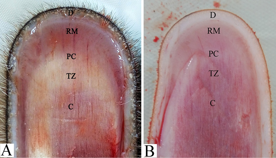
A schematic diagram of the apical structure of reindeer antler (A) and sika deer antler (B). D, dermis; RM, reserve mesenchyme; P, precartilage; TZ, transition zone; C, cartilage.
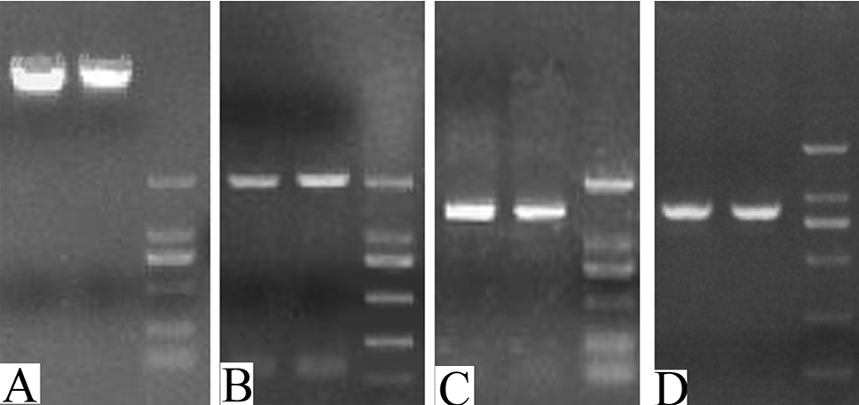
Electrophoresis of genomic DNA and IGF1, KGF and NGF gene promoters’ amplification. A: Genomic DNA of reindeer and sika deer antler reserve mesenchyme. B, C, and D, represent the amplification of reindeer and sika deer IGF1, KGF and NGF gene promoters, respectively.
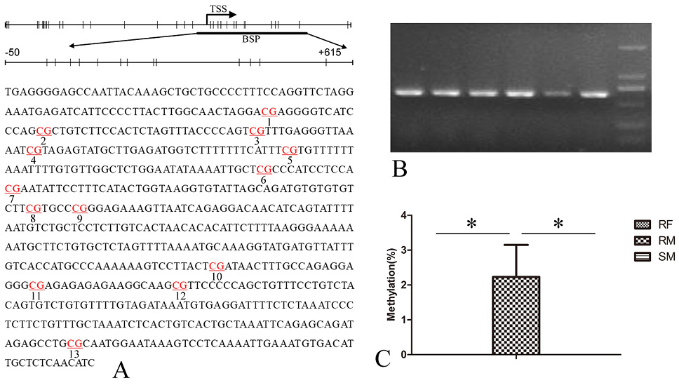
Methylation status of IGF1 gene in antler mesenchyme of female, male reindeer and sika deer. A: A schematic represents the distribution of the CpG site in the IGF1 gene and the analyzed sequence represents a 666 base pair fragment (positions −50 ~ +615) in the promoter region of IGF1 gene. B: Electrophoresis of BSP products of IGF1. C: IGF1 methylation levels in antler mesenchyme of female, male reindeer and sika deer. TSS, transcription start sites; vertical line, CpG sites; RF, female reindeer antler mesenchyme; RM, male reindeer antler mesenchyme; SM, male sika deer antler mesenchyme; *0.01<P<0.05, **P<0.01.
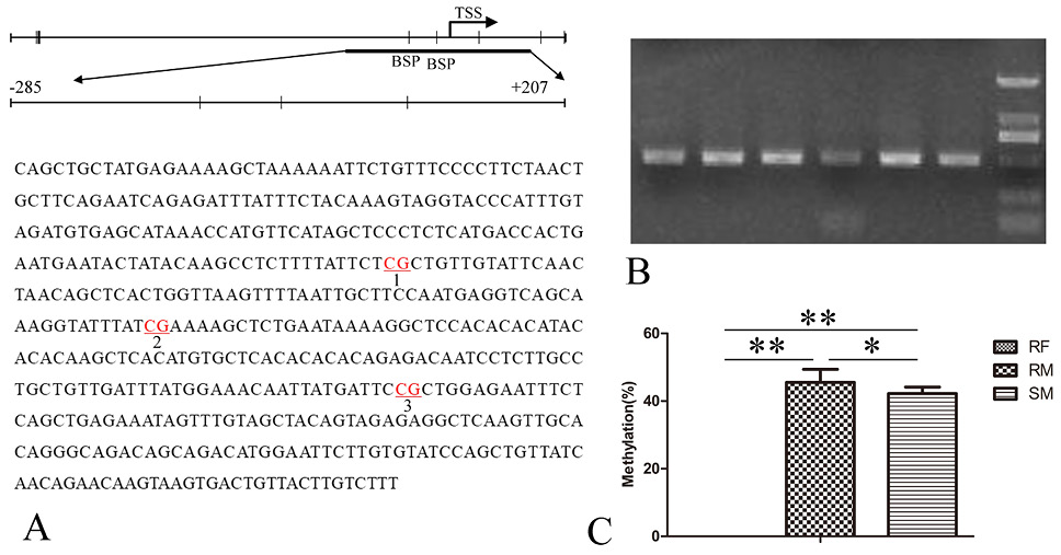
Methylation status of KGF gene in antler mesenchyme of female, male reindeer and sika deer. A: A schematic represents the distribution of the CpG site in the KGF gene and the analyzed sequence represents a 493 base pair fragment (positions −285 ~ +207) in the promoter region of KGF gene. B: Electrophoresis of BSP products of KGF. C: KGF methylation levels in antler mesenchyme of female, male reindeer and sika deer. For abbreviations and statistical detail, see Figure 3.
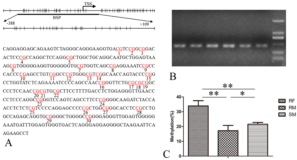
Methylation status of NGF gene in antler mesenchyme of female, male reindeer and sika deer. A: A schematic represents the distribution of the CpG site in the NGF gene and the analyzed sequence represents a 468 base pair fragment (positions −388 ~ +109) in the promoter region of NGF gene. B: Electrophoresis of BSP products of NGF. C: NGF methylation levels in antler mesenchyme of female, male reindeer and sika deer. For abbreviations and statistical detail, see Figure 3.
References
- Alpdogan, O., Hubbard, V. M., Muriglan, S.J., Goldberg, G.L., Kochman, A.A., Eng, J.M., Smith, O.M., Boyd, R.L., and Brink, M.R., 2006. Keratinocyte growth factor (KGF) is required for post-natal thymic regeneration. Blood, 107: 2453-2460. https://doi.org/10.1182/blood-2005-07-2831[CrossRef]
- Elliott, J.L., Oldham, J.M., Ambler, G.R., Bass, J.J., Spencer, G.S., Hodgkinson, S.C., Breier, B.H., Gluckman, P.D., and Suttie, J.M., 1992. Presence of insulin-like growth factor-I receptors and absence of growth hormone receptors in the antler tip. Endocrinology, 130: 2513-2520. https://doi.org/10.1210/endo.130.5.1315246[CrossRef]
- Elliott, J.L., Oldham, J.M., Ambler, G.R., Molan, P.C., Spencer, G.S., Hodgkinson, S.C., Breier, B.H., Gluckman, P.D., Suttie, J.M., and Bass, J.J., 1993. Receptors for insulin-like growth factor-II in the growing tip of the deer antler. J. Endocrinol., 138: 233-242. https://doi.org/10.1677/joe.0.1380233[CrossRef]
- Feng, H.H., Bi, X.M., Zhao, L.H., Song, Y., Yue, Z.P., Zhang, X.M., Li, C.Y., and Deng, X.M., 2007. Effects of insulin-like growth factor 1 on the in vitro proliferation of antler organic center cells at different growing periods. J. Clin. Rehabil. Tissue Eng. Res., 11: 7373-7376.
- Gelfman, S., Cohen, N., Yearim, A., and Ast, G., 2013. DNA-methylation effect on cotranscriptional splicing is dependent on GC architecture of the exon–intron structure. Genome Res., 23: 789-799. https://doi.org/10.1101/gr.143503.112[CrossRef]
- Grunstein, M., 1997. Histone acetylation in chromatin structure and transcription. Nature, 389: 349-352. https://doi.org/10.1038/38664[CrossRef]
- Huo, Y., Schirf, V.R., and Winters, W.D. 1997. The differential expression of NGFS-like substance from fresh pilose antler of Cervus nippon Temminck. Biomed. Sci. Instrum., 33: 541.
- Jeltsch, A., 2002. Beyond Watson and Crick: DNA methylation and molecular enzymology of DNA methyltransferases. Chembiochem, 3: 275-293. https://doi.org/10.1002/1439-7633(20020402)3:4<274::AID-CBIC274>3.0.CO;2-S[CrossRef]
- Kim, T.H., Barrera, L.O., Zheng, M., Qu, C., Singer, M.A., Richmond, T.A., Wu, Y., Green, R.D., and Ren, B., 2005. A high-resolution map of active promoters in the human genome. Nature, 436: 876. https://doi.org/10.1038/nature03877[CrossRef]
- Leader-Williams, N., 1979. Age-related changes in the testicular and antler cycles of reindeer, Rangifer tarandus. J. Reprod. Fertil., 57: 117-126. https://doi.org/10.1530/jrf.0.0570117[CrossRef]
- Li, C., and Suttie, J.M., 2003. Tissue collection methods for antler research. Eur. J. Morphol., 41: 23-30. https://doi.org/10.1076/ejom.41.1.23.28106[CrossRef]
- Li, C., Clark, D.E., Lord, E.A., Stanton, J.A., and Suttie, J.M., 2002. Sampling technique to discriminate the different tissue layers of growing antler tips for gene discovery. Anat. Rec., 268: 125. https://doi.org/10.1002/ar.10120[CrossRef]
- Li, C., Stanton, J.A., Robertson, T.M., Suttie, J.M., Sheard, P.W., Harris, A.J., and Clark, D.E., 2007. Nerve Growth Factor mRNA Expression in the Regenerating Antler Tip of Red Deer (Cervus elaphus). PLoS One, 2: e148. https://doi.org/10.1371/journal.pone.0000148[CrossRef]
- Liu, M., Yao, B., Zhang, H., Guo, H., Hu, D., Wang, Q., and Zhao, Y. 2014. Identification of novel reference genes using sika deer antler transcriptome expression data and their validation for quantitative gene expression analysis. Genes Genom., 36: 573-582. https://doi.org/10.1007/s13258-014-0193-x[CrossRef]
- Momparler, R.L., and Bovenzi, V., 2000. DNA methylation and cancer. J. Cell. Physiol., 183: 145. https://doi.org/10.1002/(SICI)1097-4652(200005)183:2<145::AID-JCP1>3.0.CO;2-V[CrossRef]
- Ollikainen, M., Smith, K.R., Joo, J.H., Hong, K.N., Andronikos, R., Novakovic, B., Aziz, N.K.A., Carlin, J.B., Morley, R., and Saffery, R., 2010. DNA methylation analysis of multiple tissues from newborn twins reveals both genetic and intrauterine components to variation in the human neonatal epigenome. Hum. Mol. Genet., 19: 4176-4188. https://doi.org/10.1093/hmg/ddq336[CrossRef]
- Powell, C., Elsaeidi, F., and Goldman, D., 2012. Injury-dependent Müller glia and ganglion cell reprogramming during tissue regeneration requires Apobec2a and Apobec2b. J. Neurosci., 32: 1096-1109. https://doi.org/10.1523/JNEUROSCI.5603-11.2012[CrossRef]
- Price, J., Faucheux, C., and Allen, S., 2005. Deer antlers as a model of Mammalian regeneration. Curr. Top. Dev. Biol., 67: 1-48. https://doi.org/10.1016/S0070-2153(05)67001-9[CrossRef]
- Price, J.S., Oyajobi, B.O., Oreffo, R.O., and Russell, R.G., 1994. Cells cultured from the growing tip of red deer antler express alkaline phosphatase and proliferate in response to insulin-like growth factor-I. J. Endocrinol., 143: R9. https://doi.org/10.1677/joe.0.143R009[CrossRef]
- Rohde, C., Zhang, Y.Y., Reinhardt, R., and Jeltsch, A., 2010. BISMA fast and accurate bisulfite sequencing data analysis of individual clones from unique and repetitive sequences. BMC Bioinf., 11: 12. https://doi.org/10.1186/1471-2105-11-230[CrossRef]
- Sadighi, M., Li, C., Littlejohn, R.P., and Suttie, J.M., 2001. Effects of testosterone either alone or with IGF-I on growth of cells derived from the proliferation zone of regenerating antlers in vitro. Growth Horm. Igf Res., 11: 240. https://doi.org/10.1054/ghir.2001.0232[CrossRef]
- Stadler, M.B., Murr, R., Burger, L., Ivanek, R., Lienert, F., Schöler, A., Van, N.E., Wirbelauer, C., Oakeley, E.J., and Gaidatzis, D., 2011. DNA-binding factors shape the mouse methylome at distal regulatory regions. Nature, 480: 490-495. https://doi.org/10.1038/nature10716[CrossRef]
- Suttie, J.M., Fennessy, P.F., Corson, I.D., Laas, F.J., Crosbie, S.F., Butler, J.H., and Gluckman, P.D., 1989. Pulsatile growth hormone, insulin-like growth factors and antler development in red deer (Cervus elaphus scoticus) stags. J. Endocrinol., 121: 351. https://doi.org/10.1677/joe.0.1210351[CrossRef]
- Suttie, J.M., White, R.G., Breier, B.H., and Gluckman, P.D., 1991. Photoperiod associated changes in insulin-like growth factor-I in reindeer. Endocrinology, 129: 679-682. https://doi.org/10.1210/endo-129-2-679[CrossRef]
- Thummel, R., Burket, C.T., and Hyde, D.R., 2015. Two different transgenes to study gene silencing and re-expression during zebrafish caudal fin and retinal regeneration. Sci. World J., 6: 65-81. https://doi.org/10.1100/tsw.2006.328[CrossRef]
- Wang, R., and Xu, J., 2014. Genomic DNA methylation and histone methylation. Hereditas, 36: 191.
- Werner, S., 1998. Keratinocyte growth factor: A unique player in epithelial repair processes. Cytokine Growth Factor, 9: 153-165. https://doi.org/10.1016/S1359-6101(98)00010-0[CrossRef]
- Yakushiji, N., Suzuki, M., Satoh, A., Sagai, T., Shiroishi, T., Kobayashi, H., Sasaki, H.H., and Tamura, K., 2007. Correlation between Shh expression and DNA methylation status of the limb-specific Shh enhancer region during limb regeneration in amphibians. Dev. Biol., 312: 171-182. https://doi.org/10.1016/j.ydbio.2007.09.022[CrossRef]
- Yang, C., Chu, W., Xiao, L.U., and Chunyi, L.I., 2016. Comparative study on methods of genome-wide DNA methylation assay in antler stem cells. J. Jilin Agric. Univ., 37: 463-468.
- Yang, C., Lu, X., Sun, H., Chu, W.H., and Li, C., 2016. Analysis of genomewide DNA methylation reveals differences in DNA methylation levels between dormant and naturally as well as artificially potentiated pedicle periosteum of sika deer (Cervus nippon). J. Exp. Zool. B Mol. Dev. Evol., 326: 375-383. https://doi.org/10.1002/jez.b.22695[CrossRef]
- Yao, B., Zhao, Y., Wang, Q., Zhang, M., Liu, M., Liu, H., and Li, J., 2011. De novo characterization of the antler tip of Chinese Sika deer transcriptome and analysis of gene expression related to rapid growth. Mol. Cell. Biochem., 364: 93-100. https://doi.org/10.1007/s11010-011-1209-3[CrossRef]
Cited by 1 article
In journals published by ResearchersLinks.
Services
Article usage statistics
Loading…