Research Article · Volume 14, Issue 3, 11 March 2026 · Open Access
Diagnosis of Feline Panleukopenia and Endoparasite Coinfection in Cats in Bandar Lampung, Indonesia
· · · · · · · · ·
Affiliations & Notes ⌄
1Graduate student of Magister Sains Veteriner, Faculty of Veterinary Medicine, Universitas Gadjah Mada, Yogyakarta 55281, Indonesia
2Graduate student of Doctor Sains Veteriner, Faculty of Veterinary Medicine, Universitas Gadjah Mada, Yogyakarta 55281, Indonesia
3Department of Parasitology, Faculty of Veterinary Medicine, Universitas Gadjah Mada, Yogyakarta 55281, Indonesia
4Department of Pathology, Faculty of Veterinary Medicine, Universitas Gadjah Mada, Yogyakarta 55281, Indonesia
5Department of Internal Medicine, Faculty of Veterinary Medicine, Universitas Gadjah Mada, Yogyakarta 55281, Indonesia. *Correspondence | Soedarmanto Indarjulianto, Department of Internal Medicine, Faculty of Veterinary Medicine, Gadjah Mada University, Yogyakarta 55281, Indonesia
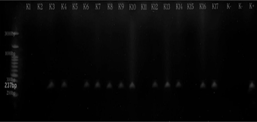
Of the 17 cats with clinical suspicion, 12 (70.6%) were PCR-positive for FPV and 5 (29.4%) were PCR-negative (Figure 2).
Endoparasite coinfection was detected in 7/12 (58.3%) FPV-positive cats, while 5/12 (41.7%) showed no parasites.
This study confirms that FPL in Bandar Lampung primarily affects young, unvaccinated cats, commonly presenting with leukopenia and severe gastrointestinal signs.
PCR demonstrated higher diagnostic sensitivity than antigen rapid testing.
Selected from the article's own text.
Abstract
Feline panleukopenia (FPL) is a highly contagious and immunosuppressive viral disease in cats that is often complicated by secondary infections, such as gastrointestinal endoparasites. This study describes the clinical, hematological, parasitological, and molecular characteristics of PCR-confirmed FPL cases in Bandar Lampung, Indonesia, and examines the potential influence of endoparasite coinfection on clinical severity. Seventeen cats with suspected FPL underwent clinical and hematological assessment, FPV rapid antigen testing, microscopic fecal examination, PCR, and DNA sequencing. Of these, twelve cats (70.6%, 12/17) were confirmed positive for FPL by PCR. Leukopenia was observed in 88.2% (15/17) of clinically suspected cats. Gastrointestinal endoparasite coinfection was identified in 58.3% (7/12) of PCR-confirmed cases, predominantly Toxocara sp. and Dipylidium caninum. Descriptive assessment indicated that coinfected cats experienced more severe gastrointestinal symptoms, particularly diarrhea and dehydration. Sequencing of two representative samples revealed ≥94% nucleotide identity with FPV strains circulating in East Asia. These findings highlight the limitations of relying on rapid antigen testing alone. The frequent occurrence of gastrointestinal parasitic coinfection and its presence with more severe disease supports the integration of molecular diagnostics and fecal examination, particularly in unvaccinated cats presenting with severe gastrointestinal signs.
Keywords | Coinfection, Endoparasite, Feline panleukopenia, PCR, Sequencing DNA
Introduction
Feline Panleukopenia (FPL) is an acute, highly contagious viral disease of cats with substantial morbidity and mortality worldwide. It predominantly affects kittens under one year of age, with mortality rates ranging from 25–90% and infection rates nearing 100% in susceptible populations ; ; ; . This disease is caused by the Feline Panleukopenia Virus (FPV), a single-stranded, non-enveloped DNA virus that is highly resistant to environmental conditions and capable of surviving for prolong period outside the host . FPV has a global distribution, with high prevalence in regions of dense cat populations and low vaccination coverage b; ; ; . Transmission of FPV occurs primarily through direct contact with infected cats or their body fluids, including feces, vomit, saliva, and urine. Transmission transmission can also occur through the transplacental route from the mother to the fetus . In addition, indirect transmission through mechanical vectors can occur via humans, fleas, and flies, play a significant rol due to the virus’s environmental persistance ; ;
Feline Panleukopenia Virus exhibits a strong tropism for actively dividing cells, especially those in the intestinal epithelium, lymphoid tissue, and bone marrow, causing severe enteritis, leukopenia, immunosuppression, and intestinal mucosal damage ; . The initial clinical symptoms include fever, lethargy, anorexia, leukopenia, and dehydration, which then manifest as leukopenia, vomiting, diarrhea, and severe depression and can end in sudden death ; ; .
Laboratory tests that can be performed to confirm the diagnosis of FPL include hematological examination, antigen and antibody detection, direct red blood cell agglutination, hemagglutination-inhibition test to detect the presence of antibodies in the sample, immunofluorescence antibodies, virus isolation, and viral DNA detection using polymerase chain reaction (PCR) ; ; . Molecular diagnosis via PCR and real-time PCR enables high-sensitivity, high-specificity differentiation of FPV from canine parvovirus (CPV), often using minor groove binder (MGB) probes (Decaro et al., 2010a). FPV is characterized by a subacute-to-peracute infection, often accompanied by sudden death .
The immunosuppressive condition caused by FPV infection makes cat patients susceptible to secondary infections, including parasitic coinfections. One of the secondary infections that often occurs is coinfection with gastrointestinal endoparasites such as Toxocara sp., Ancylostoma sp., Giardia sp., Cystoisospora sp., and Dipylidium caninum (Miro et al., 2013). These endoparasites are associated with gastrointestinal signs such as diarrhea, vomiting, anorexia, and dehydration, which overlap with FPL clinical manifestation, complicating clinical diagnosis and exacerbating disease severity . Similar clinical presentation may also be observed in other gastroenteritis disease, further emphasizing the diagnostic challenge ; .
Several studies indicate a high prevalence of endoparasites in shelter or unvaccinated cats. For example, a study by Gates and Nolan (2009) found that up to 45% of shelter cats were infected with at least one type of endoparasite. This is particularly relevant for FPL patients, most of whom come from environments with poor sanitation and a history of not being vaccinated, which are also high-risk factors for parasitic infestations. Coinfection of FPV with endoparasites can worsen the prognosis and hinder the effectiveness of supportive care therapy. Parasitic infections can exacerbate fluid and nutrient loss, worsen mucosal damage, and impair drug absorption .
The prognosis generally depends on the severity of the disease, timeliness of diagnosis, and adequacy of the supportive care . Cats with a poor prognosis often experience complications from secondary infections and coinfections . Therefore, it is important to conduct a thorough diagnosis, not only to detect the presence of gastrointestinal endoparasites but also to identify the possibility of coinfection.
Bandar Lampung is a region with a rapidly expanding small-animal veterinary sector and a high pet cat density. Based on data from the Lampung Provincial Animal Health and Veterinary Services Office (Disnakkeswan), the cat population has increased year by year over the past five years, mirroring the rise in reported FPL cases. Routine FPL diagnosis in Bandar Lampung has historically relied on clinical signs, hematology, and rapid FPV-Ag tests, with little use of molecular confirmation or systematic screening for gastrointestinal endoparasite coinfection. This diagnostic gap may contribute to misclassification, underestimation of disease severity, and suboptimal treatment strategies. Therefore, integrating molecular FPV detection with systematic parasitological evaluation is essential for accurately characterizing FPL cases in this region.
Materials and Methods
Sample collection
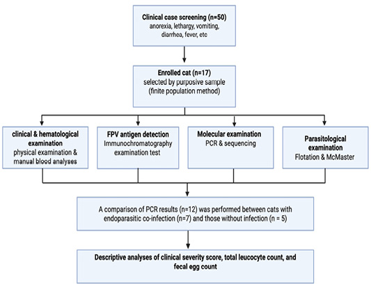
Cats were enrolled via purposive sampling, selecting individuals presenting with one or more clinical signs suggestive of FPV infection, including anorexia, lethargy, dehydration, fever (>39.3 °C), vomiting, diarrhea, or halitosis. From an initial pool of 50 cats meeting the clinical case definition, 17 were enrolled based on a sample size calculation for a finite population (Figure 1). Ethical clearance was obtained from the Animal Research and Ethics Committee, Faculty of Veterinary Medicine, Universitas Gadjah Mada, No: 46/EC-FKH/int./2025.
Clinical and hematological examination
All cats received a standardized clinical assessment comprising history-taking (anamnesis), physical examination, and laboratory tests ; . The anamnesis included information on vaccination status, deworming history, and environmental conditions (indoor/outdoor). All findings were recorded and analyzed. For hematological evaluation, blood samples were collected from the cephalic or saphenous vein into EDTA tubes and analyzed manually. Hematological analysis was performed manually: total leukocyte counts were obtained using a hemocytometer, and differential counts were evaluated from Wright-Giemsa-stained blood smears. Quality control was ensured through duplicate counts and slide re-evaluation by a second examiner. The results were compared with reference ranges .
Antigen immunochromatography examination/ antigen rapid test
FPV antigen detection was performed using a commercial immunochromatographic kit (PetX JandG Biotech Ltd., UK). Fresh fecal samples were collected using the sterile applicator provided in the kit and mixed thoroughly with the supplied extraction buffer until a homogenous suspension was obtained. Approximately 3–4 drops of the extracted sample were then dispensed into the sample well of the cassette. The cassette was left undisturbed at room temperature for 5–10 minutes.
The appearance of two distinct colored bands on the control line (C) and test line (T) was interpreted as a positive result, while the presence of only the control line indicated a negative outcome. Tests lacking a visible control line were considered invalid and repeated with a new cassette. All results were recorded immediately after the recommended reading time window to avoid misinterpretation.
Molecular examination
To prevent contamination, separate work areas were maintained for DNA extraction, reagent preparation, amplification, and electrophoresis. Aerosol-resistant tips were used, and each PCR run included positive and negative controls. DNA extraction from blood samples was performed using the gSYNCTM DNA Extraction Kit (Geneaid Biotech Ltd., Taiwan) following the manufacturer’s protocol. Amplification of the FPV VP2 gene was conducted using FPV-specific primers designed by Zhang et al. (2019), consisting of FPV-F (5′-CATACATGGCAAACAAATAGAGCA-3′) and FPV-R (5′-TGTTTTAAATGGCCCTTGTGTAGA-3′), targeting a 237 bp amplicon. PCR reactions were prepared using KOD Fx Neo polymerase, and amplification was carried out for 30 cycles in a thermal cycler. The cycling conditions were as follows: Initial denaturation at 95 °C for 3 min, followed by 35 cycles of denaturation at 95 °C for 30 s, annealing at 55 °C for 30 s, and extension at 72 °C for 30 s, with a final extension at 72 °C for 5 min. PCR amplicons were separated by electrophoresis on a 2% agarose gel stained with RedSafe, with a DNA ladder and controls included on each gel. Bands were visualized under a UV transilluminator and documented. The two best samples testing positive by PCR were subsequently subjected to bidirectional DNA sequencing at the Integrated Research and Testing Laboratory (LPPT), Universitas Gadjah Mada, Yogyakarta. Sequence identity was confirmed through BLAST analysis using the NCBI database.
Parasite examination
Fresh fecal samples were obtained via rectal swabs using sterile cotton-tipped applicators. Fecal samples were examined microscopically using standard flotation methods, and parasite eggs or proglottids were identified based on morphological keys ; . Quantitative fecal egg counts (EPG) Toxocara sp. were determined using the McMaster technique, while Dipylidium sp. determined based on presence/absence of proglottid. Fecal samples were homogenized, mixed with flotation solution, and loaded into McMaster counting chambers. Eggs were counted under 100x magnification and multiplied by the standard McMaster factor to obtain EPG values. Fecal egg counts (EPG) were categorized semi-quantitatively as mild (<500 EPG), moderate (500–2000 EPG), or severe (>2000 EPG), based on standard parasitological methods.
Results
Of the 17 cats with clinical suspicion, 12 (70.6%) were PCR-positive for FPV and 5 (29.4%) were PCR-negative (Figure 2). FPV antigen rapid testing detected infection in 10/12 PCR-positive cats (83.3%). Endoparasite coinfection was detected in 7/12 (58.3%) FPV-positive cats, while 5/12 (41.7%) showed no parasites. All PCR-confirmed cats were unvaccinated and had not been dewormed. Clinical severity was classified descriptively based on dehydration status, diarrhea characteristics, and lethargy (Table 1).
Table 1: Characteristics of cats with PCR-confirmed Feline Panleukopenia (n=12).
Parameter
Number of infected FPL
Percentage (%)
Age (months)
<6
8
66.7
6-12
4
33.3
Sex
Male
9
75.0
Female
3
25.0
Leukocyte (cell/mm3)
<5.500
12
100.0
Antigen rapid test
Positive
10/12
83.3
Negative
2/12
16.7
Note*: normal leukocyte counts 5,500-19,500 cells/mm3 (Tilley and Smith, 2011).
Clinical signs observed in the study population are summarized in Table 2. The most prevalent signs were anorexia (100%), lethargy (100%), diarrhea (100%), fever (100%), vomiting (100%), and dehydration (91.7%). Less common findings included halitosis (16.7%), hypersalivation (16.7%), stomatitis (8.3%), nasal discharge (8.3%), and otitis (8.3%). Hematological evaluation revealed leukopenia (<5,500 cells/μL) in all FPV-positive cats.
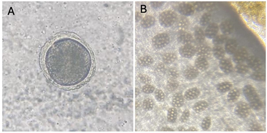
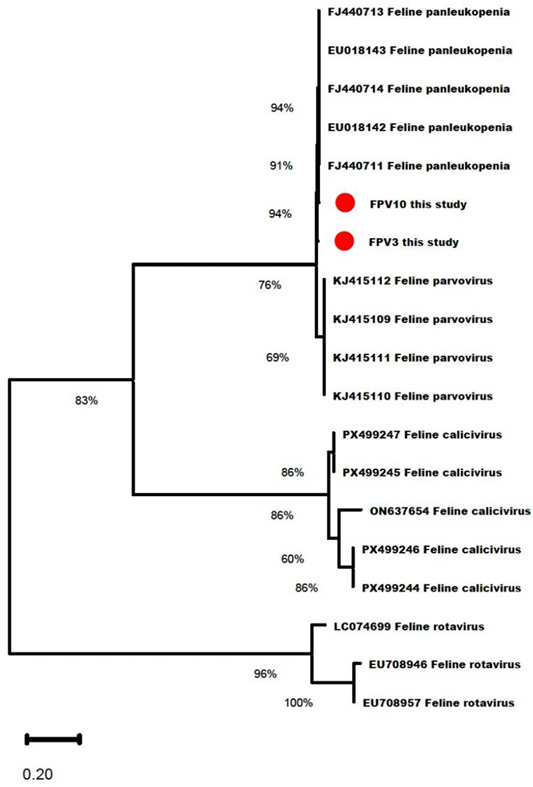
Gastrointestinal parasite screening was performed in 12 FPV-confirmed cats. Seven cats (58.3%) were positive for endoparasites and five (41.7%) were negative. The parasites detected were Toxocara sp. (n = 2), Dipylidium caninum (n = 4), and mixed Toxocara sp. + Dipylidium caninum infection (n= 1). Quantitative McMaster analysis of Toxocara sp. showed EPG values ranging from 2600 to 3200 in infected cats. Cats with parasitic coinfection displayed more severe gastrointestinal signs descriptively, including mucoid or hemorrhagic diarrhea, moderate to severe dehydration, anorexia, and profound lethargy (Table 3). The most common parasites identified were Toxocara sp. eggs and Dipylidium caninum proglottids (Figure 3). Sequencing of two PCR-positive samples produced nucleotide fragments of approximately 180–200 bp. BLAST analysis revealed ≥94% identity with FPV sequences to strains from East Asia, especially strain with accession number MN400978 (China) (Figure 4).
Table 2: Clinical symptoms of cats infected with Feline Panleukopenia Virus (FPV) (n=12).
No
Symptoms
Number of infected
Percentage (%)
1
Anorexia
12/12
100
2
Lethargy
12/12
100
3
Fever
12/12
100
4
Vomit
12/12
100
5
Diarrhea
12/12
100
6
Dehydration
11/12
91.7
7
Halitosis
2/12
16.7
8
Hypersalivation
2/12
16.7
9
Stomatitis
1/12
8.3
10
Nasal discharge
1/12
8.3
11
Otitis
1/12
8.3
Table 3: Gastrointestinal endoparasites, Fecal Egg Count (EPG), and Clinical Severity Score in FPV-positive cats (n=12) (EPG terminology clarified; Clinical Severity Score defined as descriptive assessment).
No
Parasite species
n
Mean EPG
Range EPG
% Clinical severity
1
Toxocara sp.
2
3025
2850-3200
100
2
Dipylidium caninum
4
-
-
100
3
Mixed infection
1
3100
-
100
4
No parasites
5
0
0
0
Note: Clinical severity score was classified based on dehydration (moderate-severe), diarrhea (mucoid or hemorrhagic), and lethargy recorded during physical examination.
Discussion
Feline panleukopenia is a highly contagious and often fatal viral disease characterized by acute systemic illness, severe gastrointestinal involvement, and profound immunosuppression, particularly in young and immunologically naïve cats. In this study, most FPV-positive
cats were under six months old, consistent with reports identifying kittens as the most susceptible group ; ; ; ; . This vulnerability stems from FPV’s tropism for rapidly dividing cells ; ; and the waning of maternally derived antibodies (MDA), creating a period where kittens are neither fully protected nor responsive to vaccination ; ; ; ; . This immunological gap contributes to high morbidity and mortality, with most FPV-related deaths occurring in kittens . Additionally, higher total body water in younger animals predisposes them to rapid dehydration and poorer outcomes .
Although males were slightly more affected than females, the difference was minimal, supporting the notion that sex is not a strong predisposing factor ; ; ; . Contradictory findings in other studies ; ; may reflect behavioral differences rather than biological susceptibility. Male cats, especially feral individuals, may be exposed more frequently due to larger roaming ranges and polygynous reproductive behavior ; . Regardless of sex, the durable environmental persistence of FPV facilitates indirect transmission ; ; , leaving all cats equally vulnerable.
The disease course in this study was characterized by acute illness with prominent gastrointestinal signs, which align with previous reports ; ; ; . The pattern reflects the underlying pathogenesis of FPV, which is characterized by extensive destruction of intestinal crypts, resulting in villus atrophy, malabsorption, increased permeability, and diarrhea ; ; ; ; . However, diarrhea could also result from endoparasite infections, including Giardia sp., Toxocara sp., Ancylostoma sp., Cystoisospora sp., and Dipylidium caninum, all of which can independently impair intestinal function .
Cats with coinfection appear to exhibit more severe gastrointestinal signs that may be caused by immunosuppression, leading the host to reduce resistance to parasitic infections (Miro et al., 2013). Parasites such as Toxocara sp. and Dipylidium caninum can intensify the mucosal inflammation, nutrient malabsorption, and systemic immune dysregulation (Bowman et al., 2002; ; Traversa et al., 2010). Although statistical comparison was not feasible due to the limited sample size, these findings align with earlier reports indicating that viral–parasitic coinfections worsen clinical outcomes .
Leukopenia is the classical hematological profile of FPV infection, presented and mainly as a result of the viral destruction of bone marrow precursor cells ; ; ; . The findings align with earlier studies reporting leukopenia in the majority of FPV-positive cats ; ; ; ; that also could be the prognostic indicator since some findings show that it was associated with increased mortality ; ; ; . Parasitic coinfection may also influence leukocyte differentials, as helminth infections are known to induce eosinophilia or reactive neutrophilic responses, potentially modifying the typical hematological profile of FPV infection ; Traversa et al., 2010).
Rapid antigen testing demonstrated lower sensitivity than PCR, aligning with previous studies ; ; . Sensitivity is affected by shedding dynamics and peaks before clinical signs appear ; . Low viral load, degraded antigen, improper storage, fecal consistency, and subjective interpretation of faint bands all contribute to false negatives ; b; . In contrast, PCR detection using FPV-specific primers confirmed infection in a greater number of samples and remains the reference diagnostic method in this case due to the sensitivity ; ; ; .
Sequencing of selected PCR-positive samples further validated the assay’s specificity. Although phylogenetic analysis was not the primary aim, the high similarity to a Chinese FPV strain suggests genetic relatedness of strains circulating in Bandar Lampung. The global mobility of domestic animals may influence the spread of similar FPV strains across countries . Furthermore, the fact that all cats in this study were never dewormed or vaccinated also facilitated the virus’s spread, highlighting the urgent need for improved preventive measures. Vaccination programs for owned and community cats, routine deworming protocols integrated into primary veterinary care, structured client education on preventive medicine, and standardized vaccination-deworming policies for individual owners, shelters, and rescue facilities should be more widely promoted and regulated by the government. Establishing these measures could curb the spread of the virus and improve overall feline health in the region.
Conclusion
This study confirms that FPL in Bandar Lampung primarily affects young, unvaccinated cats, commonly presenting with leukopenia and severe gastrointestinal signs. PCR demonstrated higher diagnostic sensitivity than antigen rapid testing. Coinfection with gastrointestinal endoparasites was common and descriptively linked to more severe clinical disease. For clinicians, combining PCR with fecal examination should be considered in severe or non-responsive cases, while acknowledging the cost constraints of routine practice.
Acknowledgments
The authors thank the Directorate of Research and Community Service, Directorate General for Research and Development, Ministry of Higher Education, Science, and Technology, Indonesia, for the Penelitian Fundamental grant based on Decree Number 0419/C3/DT.05.00/2025 and Agreement/ Contract Number 067/C3/DT.05.00/PL/2025 and 2391/UN1/DITLIT/PT.01.03/2025.
Novelty Statement
This study provides the first integrated molecular (PCR and sequencing) and parasitological assessment of Feline Panleukopenia in Bandar Lampung, Indonesia, demonstrating a high prevalence of gastrointestinal endoparasite coinfection associated with increased clinical severity. These findings support the integration of molecular diagnostics with routine fecal examination to enhance diagnostic accuracy and patient management.
Authors’ contribution
SI and YRN: Conceptualization and supervision. AP, YN, AN, and ADP: Clinical examination. AP, SW, and YRN: Microscopic and molecular analysis. MAA and SW: Hematological analysis. AP and FN: Data analysis and manuscript preparation. All authors reviewed and approved the final manuscript.
Generative AI and AI-assisted technology statement
Artificial intelligence was used solely to improve language and clarity. It was not involved in the study design, data analysis, or interpretation. All scientific content and conclusions are the responsibility of the authors.
Conflict of interest
The authors have declared no conflict of interest.
Figures
Figure 1:.
Flowchart of the study design and diagnostic workflow of feline panleukopenia and gastrointestinal endoparasite coinfection.
Figure 2:.
Agarose gel electrophoresis of FPV VP2 gene amplification (237 bp). M: DNA ladder; PC: positive control; NC: negative control; lanes 1–12: clinical samples.
Figure 3:.
Gastrointestinal endoparasites identified in FPL-positive cats. A) Toxocara sp. egg (1,000x magnification). (B) Dipylidium sp. (100x magnification).
Figure 4:.
Phylogenetic tree of FPV VP2 gene sequences obtained in this study compared with reference FPV strains from GenBank. The study isolates a cluster closely related to East Asian FPV strains, particularly accession number MN400978 (China). The tree was constructed using the Neighbor-Joining method with 1000 bootstrap replications.
References
Abd-Eldaim M, Beall MJ, Kennedy MA (2009). Detection of feline panleukopenia virus using a commerical elisa for canine parvovirus. Vet. Ther, 10(4): E1-6.
Al-Bayati HAM (2016). Detection of feline parvovirus (fpv) from cats infected with enteritis using rapid test and polymerase chain reaction in Iraq. Kufa J. Vet. Med. Sci., 7(2).https://doi.org/10.36326/kjvs/2016/v7i24346[CrossRef]
Altschul SF, Gish W, Miller W, Myers EW, Lipman DJ (1990). Basic local alignment search tool. J. Mol. Biol., 215(3): 403–410. https://doi.org/10.1016/S0022-2836(05)80360-2[CrossRef]
Awad RA, Khalil WK, Attallah AG (2018). Epidemiology and diagnosis of feline panleukopenia virus in Egypt: Clinical and molecular diagnosis in cats. Vet. World, 11(5): 578.https://doi.org/10.14202/vetworld.2018.578-584[CrossRef]
Baratelli M, Molist-Badiola J, Puigredon-Fontanet A, Pascual M, Boix O, Mora-Igual FX, Woodward M, Lavazza A, Capucci L (2020). Characterization of the maternally derived antibody immunity against rhdv-2 after administration in breeding does of an inactivated vaccine. Vaccines, 8(3): 484.https://doi.org/10.3390/vaccines8030484[CrossRef]
Barrs VR (2019). Feline panleukopenia: a re-emergent disease. Vet. Clin. Small Anim. Pract., 49(4): 651-670.https://doi.org/10.1016/j.cvsm.2019.02.006[CrossRef]
Bengsen AJ, Algar D, Ballard G, Buckmaster T, Comer S, Fleming PJ, Friend JA, Johnston M, McGregor G, Moseby K, Zewe F (2016). Feral cat home-range size varies predictably with landscape productivity and population density. J. Zool., 298(2): 112-120. https://doi.org/10.1111/jzo.12290[CrossRef]
Bowman DD (2014). Georgis’ Parasitology for Veterinarians 10th Ed. Elsevier Health Sciences.
Bowman DD, Hendrix CM, Lindsay DS, Barr SC (2002). Feline Clinical Parasitology, 1st Ed. Ames: Iowa State University Press.
Bowman DD, Montgomery SP, Zajac AM, Eberhard ML, Kazacos KR (2010). Hookworms of dog and cats as agents of cutaneous larva migrans. Trends. Parasitol, 26(2): 162-167. https://doi.org/10.1016/j.pt.2010.01.005[CrossRef]
Claus MA, Levy JK, MacDonald K, Tucker SJ, Crawford PC (2006). Immunoglobulin concentrations in feline colostrum and milk, and the requirement of colostrum for passive transfer of immunity to neonatal kittens. J. Feline Med. Surg., 8(3): 184-191.https://doi.org/10.1016/j.jfms.2006.01.001[CrossRef]
Cooper PJ, Walker AW, Reyes JL (2012). The roles of helminths in host immune regulation: Implications for autoimmune and inflammatory diseases. Curr. Opin. Immunol., 24(6): 658–665.
Day MJ, Horzinek MC, Schultz RD, Squires RA (2016). Guidelines for the vaccination of dogs and cats. J. Small Anim. Pract., 57(1): 4-8. https://doi.org/10.1111/jsap.2_12431[CrossRef]
Decaro N, Buonavoglia C (2012). Canine parvovirus. A review of epidemiological and diagnostic aspects, with emphasis on type 2c. Vet. Microbiol., 155(1): 1–12. https://doi.org/10.1016/j.vetmic.2011.09.007[CrossRef]
Decaro N, Buonavoglia D, Desario C, Amorisco F, Colaianni ML, Parisi A, Terio V, Elia G, Lucente MS, Cavalli A, Martella V, Buonavoglia C (2010a). Characterisation of canine parvovirus strains isolated from cats with feline panleukopenia. Res Vet Sci. 89(2): 275-278. https://doi.org/10.1016/j.rvsc.2010.03.001[CrossRef]
Decaro N, Desario C, Beall MJ, Cavalli A, Campolo M, DiMarco AA, Amorisco F, Colaianni ML, Buonavoglia C (2010b). detection of canine parvovirus type 2c by a commercially available in-house rapid test. Vet. J., 184 (3): 373-375.https://doi.org/10.1016/j.tvjl.2009.04.006[CrossRef]
Duarte AM, Jenkins TP, Latrofa MS, Giannelli A, Papadopoulos E, de Carvalho LM, Nolan MJ, Otranto D, Cantacessi C (2016). Helminth infections and gut microbiota - a feline perspective. Parasit Vectors. 9(1):625. https://doi.org/10.1186/s13071-016-1908-4[CrossRef]
Englar RE (2017). Performing the small animal physical examination. John Wiley and Sons.
Esfandiari J, Klingeborn B (2000). A comparative study of a new rapid and one-step test for the detection of parvovirus in faeces from dogs, cats and mink. J. Vet. Med. Ser. B, 47(2): 145-153.https://doi.org/10.1046/j.1439-0450.2000.00328.x[CrossRef]
Figueiredo CA, Barreto ML, Rodrigues LC, Cooper PJ, Silva NB, Amorim LD, Alcantara-Neves NM (2010). Chronic intestinal helminth infections are associated with immune hyporesponsiveness and induction of a regulatory network. Infect. Immun. 78(7):3160-3167. https://doi.org/10.1128/iai.01228-09[CrossRef]
Gates MC, Nolan TJ (2009). Endoparasite prevalence and recurrence across different age groups of dogs and cats. Vet. Parasitol., 166(1-2): 153-158. https://doi.org/10.1016/j.vetpar.2009.07.041[CrossRef]
Greene CE (2012). Infectious diseases of the dog and cat 4th Edition. Saunders Elsevier. St. Louis, Missouri, USA.
Hermawan IP, Darantika G, Tage RA, Desiandura K, Wardhani HCP (2023). Studi kasus: Kesembuhan kasus feline panleukopenia pada kucing mocca di Surabaya. J. Kajian Vet., 11(1): 10-18.https://doi.org/10.35508/jkv.v11i1.7627[CrossRef]
Imoto Y, Yoshida Y, Yagisawa F, Kuroiwa H, Kuroiwa T (2011). The cell cycle, including the mitotic cycle and organelle division cycles, as revealed by cytological observations. J. Electron Microsc., 60 (Supp. 1): S117-S136. https://doi.org/10.1093/jmicro/dfr034[CrossRef]
Indarjulianto S, Widiyono I, Raharjo S, Nururrozi A (2025). Pemeriksaan klinis hewan kesayangan (clinical examination of pet animal). Yogyakarta: Gadjah Mada University Press.
Isaya, R., Isaya R, Ciccarelli S, Enache D, Specchi S, Pesaresi M, Ferri F, Porporato F, Auriemma E, Contiero B, Coppola LM, Zini E (2021). Gastrointestinal ultrasonographic findings in cats with Feline panleukopenia: a case series. BMC Vet. Res., 17(20):1-8. https://doi.org/10.1186/s12917-020-02720-w[CrossRef]
Islam M, Rahmad MS, Rony SA, Uddin MJ, Rahman AKMA (2010). Antigenic detection of feline panleukopenia virus in local breed cats at tangail district in Bangladesh. Int. J. Biores., 2(11): 25-28.
Jacobson LS, Janke KJ, Giacinti J, Weese JS (2021). Diagnostic testing for feline panleukopenia in a shelter setting: A prospective, observational study. J. Feline Med. Surg., 23(12): 1192-1199. https://doi.org/10.1177/1098612X211005301[CrossRef]
Jakel V, Cussler K, Hanschmann KM, Truyen U, Konig M, Kamphuis E, Duchow K (2012). Vaccination against feline panleukopenia: implications from a field study in kittens. BMC Veterinary Research, 8(1): 1-8.https://doi.org/10.1186/1746-6148-8-62[CrossRef]
Kruse BD, Unterer S, Horlacher K, Sauter-Louis C, Hartmann K (2010). Prognostic factors in cats with feline panleukopenia. J. Vet. Intern. Med., 24(6): 1271-1276. https://doi.org/10.1111/j.1939-1676.2010.0604.x[CrossRef]
Kusumawardhani SW, Chandra AH, Yehuda LA, Viska M, Widyastuti (2019). Catatan dokter hewan: Penyakit Infeksius pada Kucing. IPB press. Bogor, Indonesia.
Lamm CG, Rezabek GB (2008). Parvovirus infection in domestic companion animals. Small Anim. Pract., 38(4): 837-850. https://doi.org/10.1016/j.cvsm.2008.03.008[CrossRef]
Lee JA, Cohn LA (2017). Fluid therapy for pediatric patients. Vet. Clin. Small Anim., 47: 373-382. https://doi.org/10.1016/j.cvsm.2016.09.010[CrossRef]
Lian ATY, Chircop M (2016). Mitosis in animal cells. Encycl. Cell Biol., 3: 479-493.https://doi.org/10.1016/B978-0-12-394447-4.30064-5[CrossRef]
Mahendra YN, Yuliani MGA, Agus W, Driyanto Miyayu, S Sofyan (2020). A Case Study of Feline Panleukopenia in Cats at The Educational Animal Hospital of Universitas Airlangga. J. Appl. Vet. Sci. Technol., 1(1): 6-11.https://doi.org/10.20473/javest.V1.I1.2020.6-10[CrossRef]
Mende K, Stuetzer B, Truyen U, Hartmann K (2014). Evaluation of an in-house dot enzyme-linked immunosorbent assay to detect antibodies against feline panleukopenia virus. J. Feline Med. Surg., 16(10): 805-811.https://doi.org/10.1177/1098612X14520812[CrossRef]
Miller L, Janeczko S, Hurley KF (2021). Infectious disease management in animal shelters, second edition. Ames, Iowa: Wiley-Blackwell. pp. 337-366.
Mircean V, Györke A, Cozma V (2012). Prevalence and risk factors of gastrointestinal parasites in cats from Romania. Parasite, 19(1): 61–66. https://doi.org/10.1016/j.vetpar.2011.08.022[CrossRef]
Miro G, Montoya A, Roura X, Galvez R, Sainz A (2013). Seropositivity rates for agents of canine vector-borne diseases in Spain: A multicentre study. Parasites Vectors, 6(117). https://doi.org/10.1186/1756-3305-6-117[CrossRef]
Mosallanejad B, Avizeh R, Najafabadi MG (2009). Antigenic detection of felinepanleukopenia virus (FPV) in diarrhoeic companion cats in Ahvaz Area. Iran. J. Vet. Res., 10(3): 289-293.
Moschidou P, Martella V, Lorusso E, Desario C, Pinto P, Losurdo M, Catella C, Parisi A, Banyai K, Buonavoglia C (2011). Mixed Infection by feline astrovirus and feline panleukopenia virus in a domestic cat with gastroenteritis and panleukopenia. J. Vet. Diagn. Invest., 23(3): 581-584.https://doi.org/10.1177/1040638711404149[CrossRef]
NCBI. National Center for Biotechnology Information (NCBI). https://blast.ncbi.nlm.nih.gov
Pandey S (2022). Feline panleukopenia infections: Treatment and control in Nepal. Eur. J. Vet. Med., 2(1): 10-14. https://doi.org/10.24018/ejvetmed.2022.2.1.19[CrossRef]
Pfankuche VM, Jo WK, Van Der Vries E, Jungwirth N, Lorenzen S, Osterhaus ADME, Baumgartner W, Puff C (2017). Neuronal vacuolization in feline panleukopenia virus infection. Vet. Pathol., 5(2): 294-297.https://doi.org/10.1177/0300985817738096[CrossRef]
Porporato F, Horzinek MC, Hofmann-Lehmann R, Ferri F, Gerardi G, Contiero B, Vezzosi T, Rocchi P, Auriemma E, Lutz H, Zini E (2018). Survival Estimates and outcome predictors for shelter cats with feline panleukopenia virus infection. J. Am. Vet. Med. Assoc., 253(2): 188-195.https://doi.org/10.2460/javma.253.2.188[CrossRef]
Purnamaningsih H, Indarjulianto S, Yanuartono Y, Nururrozi A, Widiyono I, Rahardjo S, Hartati S, Rusmihayati (2020). Gambaran leukosit kucing penderita feline panleukopenia. J. Sains Vet., 38(2): 121-125.https://doi.org/10.22146/jsv.50202[CrossRef]
Purnamaningsih H, Indarjulianto S, Yanuartono, Nururrozi A, Widiyono I, Rahardjo S, Hartati S, Rusmihayati (2022). Diagnosis Feline panleukopenia berdasar total leukosit dan uji feline parvovirus-antigen pada kucing-kucing diare. J. Vet., 23(1): 36-41.https://doi.org/10.19087/jveteriner.2022.23.1.36[CrossRef]
Putri R, Sumiarto B, Mulyani GT (2020). Faktor-faktor risiko feline panleukopenia pada kucing di daerah istimewa yogyakarta. J. Sain Vet., 38(3): 206-213.https://doi.org/10.22146/jsv.48922[CrossRef]
Putri R, Wahyuwardani S (2022). Koinfeksi pada Kejadian Panleukopenia Kucing: Suatu Kajian Pustaka. J. Vet., 23(1): 121-129.https://doi.org/10.19087/jveteriner.2022.23.1.121[CrossRef]
Raj VP, Haryanto A (2020). Clinical Study and rapid detection of feline parvovirus in suspected cats by polymerase chain reaction method. Indones. J. Vet. Sci., 1(1): 15-23.https://doi.org/10.22146/ijvs.v1i1.44835[CrossRef]
Rehme T, Hartmann K, Truyen U, Zablotski Y, Bergmann M (2022). Feline panleukopenia outbreaks and risk factors in cats in animal shelters. Viruses, pp. 1-14. https://doi.org/10.3390/v14061248[CrossRef]
Rice JK (2017). Successful treatment of feline panleukopenia: A guideline for rescuers and veterinarians, Part I. J. Vet. Sci. Med. Diagn., 6(2): 1-5. https://doi.org/10.4172/2325-9590.1000223[CrossRef]
Robertson ID, Irwin PJ, Lymbery AJ, Thompson RCA (2000). The Role of companion animals in the emergence of parasite zoonoses. Int. J. Parasitol., 30: 12-13. https://doi.org/10.1016/S0020-7519(00)00134-X[CrossRef]
Stuetzer B, Hartmann K (2014). Feline parpovirus infection and associated diseases. Vet. J., 201(2): 150-155.https://doi.org/10.1016/j.tvjl.2014.05.027[CrossRef]
Sykes JE (2014). Canine and feline infectious diseases. Elsevier Health Science. pp. 187-194.https://doi.org/10.1016/B978-1-4377-0795-3.00019-3[CrossRef]
Tilley LP, Smith FWK (2011). Blackwell’s five-minute veterinary consult: Canine and feline. 5th ed. Wiley-Blackwell, Ames, IA, USA
Tinky SS, Ambily R, Nair SR, Mini M (2015). Utility of a rapid immunochromatographic strip test in detecting canine parvovirus infection compared with polymerase chain reaction. Vet. World, 8(4): 523-526. https://doi.org/10.14202/vetworld.2015.523-526[CrossRef]
Traversa D, Di Cesare A, Conboy G (2010). Canine and feline cardiopulmonary parasitic nematodes in Europe: Emerging and underestimated. Parasit. Vectors 3: 62. https://doi.org/10.1186/1756-3305-3-62[CrossRef]
Trotman TK (2015). Gastroenteritis. Small Animal Critical Care Medicine, pp. 622- 626. https://doi.org/10.1016/B978-1-4557-0306-7.00117-3[CrossRef]
Truyen U, Addie D, Belak S, Boucraut-Baralon C, Egberink H, Frymus T, Gruffydd-Jones T, Hartmann K, Hosie MJ, Lloret A, Lutz H, Marsilio F, Pennisi MG, Radford AD, Thiry E, Horzinek MC (2009). Feline panleukopenia ABCD guidelines on prevention and management. J. Feline Med. Surg., 11(7): 538-546. https://doi.org/10.1016/j.jfms.2009.05.002[CrossRef]
Weese JS, Evason MD (2019). Infectious diseases of the dog and cat. 4th ed. Elsevier, St. Louis, MO, USA. https://doi.org/10.1201/9780429186646[CrossRef]
Zajac AM, Conboy GA (2012). Veterinary clinical parasitology. 8th ed. Wiley-Blackwell, Chichester, West Sussex, UK.
Zenad MM, Radhy AM (2020). Clinical, serological and antigenic study of feline panleukopenia virus in vats in Baghdad, Iraq. Iraqi J. Vet. Sci., 34(2): 435-439. https://doi.org/10.33899/ijvs.2019.125960.1201[CrossRef]
Zhang Q, Niu J, Yi S, Dong G, Yu D, Guo Y, Huang H, Hu G (2019). Development and application of a multiplex pcr method for the simultaneous detection and differentiation of feline panleukopenia virus, feline bocavirus, and feline astrovirus. Arch. Virol., 164(11): 2761-2768.https://doi.org/10.1007/s00705-019-04394-8[CrossRef]
Zhang Z, Li Y, Ullah S, Chen L, Ning S, Lu L, Lin W, Li Z (2022). Home range and activity patterns of free-ranging cats: A case study from a Chinese university campus. Animals, 12(9): 1141. https://doi.org/10.3390/ani12091141[CrossRef]