
A significant clinical challenge is posed by wound infections in disease development, therefore, nowadays multifunctional hydrogel dressings have revolutionized the ways of infection prevention. They provide effective treatment and easy monitoring for wound recovery. In connection to this, our study developed and characterized a novel topical hydrogel based on Chitosan and Gelatin, incorporated with Matricaria chamomilla (chamomile) extract, for its combined anti-inflammatory, antioxidant, and antimicrobial properties. The optimal formulation was achieved using chitosan (as polymer matrix), gelatin (as gelatinizing agent) and cross-linking them with NaOH as PH adjuster Chamomile extract served as additive to impart antimicrobial and antioxidant properties to prepared hydrogel. Physicochemical characterization confirmed the hydrogels’ suitability for wound management. Gel fraction determination yielded a value of 42 %, indicating effective cross-linking and structural integrity. The DPPH radical scavenging assay revealed 58.6 % DPPH free radical scavenging indicating potential to combat oxidative damage. Antimicrobial evaluation demonstrated the hydrogels’ efficacy against common wound pathogens i.e. Escherichia coli (Gram-negative) and Streptococcus spp. (Gram-positive) with 13 and 14 mm zone of inhibition. An acetic-acid dissolved hydrogel variant showed enhanced activity against E. coli (16 mm zone) comparable to Ciprofloxacin. The successful integration of bioactive chamomile extract into a structurally stable, absorbent, and bioactive chitosan-gelatin matrix positions this hydrogel as a promising, naturally derived advanced wound dressing.
Research Article · Volume 39, Issue 3, 13 September 2026 · Open Access
Development of Biopolymeric Chamomile Hydrogel for Potential Wound Healing Applications
· · · · · ·
Affiliations & Notes ⌄
- 1Institute of Biochemistry, University of Sindh, Jamshoro, Pakistan. *Correspondence | Beenish Khanzada, Institute of Biochemistry, University of Sindh, Jamshoro, Pakistan
- 2Email: beenish@usindh.edu.pk
Article Info ⌄
- Journal:
- Pakistan Journal of Agricultural Research
- Published:
- 13 September 2026
From the article
- A novel naturally derived topical hydrogel was successfully formulated using a combination of chitosan gelatin and chamomile flower extract.
- The determination of a 42 % gel fraction indicates a successfully cross-linked structure with substantial insoluble polymer content, ensuring the hydrogel’s mechanical integrity and durability during use.
- Significant bioactive compounds from chamomile extract imparted around 60 % antioxidant activities to the hydrogel and the fabricated gel was found to inhibit common wound bacterial pathogens i.e. E. coli and S. aureus effectively.
- This hydrogel combination created a stable three-dimensional polymeric network suitable for wound healing applications.
Selected from the article's own text.
Abstract
Introduction
A prominent crucial role is played by human skin in combating various types of wound infections. Our skin not only provides physical protection and barrier to a variety of invaded microorganisms but also assists in respiration, excretion and body temperature regulation. Normally, the restoration of skin integrity after injury is made possible with a minimal scar via combination of coagulation, hemostasis, inflammation, proliferation and remodeling . Various local (e.g. oxygenation, local wound area and depth etc.) and systemic factors (i.e. age, gender, hormonal imbalance, ischemia, alcoholism, obesity etc.) can interrupt the healing process .
Hydrogels can be regarded as polymer chains cross-linked in a three-dimensional networking, that have the tendency to absorb fluid. Due to their high-water content, porosity and softened structure of hydrogels, they show a close resemblance with native extracellular matrix. These characteristics make them compatible with biological systems and applicable in wound healing . The porous structure of hydrogels makes them able for controlled release of loaded pharmaceutical agents thus can contribute to delivery of phenols, flavonoids or even medicine or growth factors indispensable for tissue regeneration. Literature shows the extensive use of natural and synthetic polymers for formulating hydrogels with their wound care related advantages i.e. biodegradability, biocompatibility and non-toxicity .
Chitosan, a natural cationic polysaccharide with desirable and tunable biomedical properties is considered ideal for use in wound care formulations. Chitosan has hemostatic, anti-bacterial and anti-inflammatory properties and is also found to boost endogenous hyaluronan (HA) at the wound position to stimulate wound healing. Biodegradability, high biocompatibility, non-toxicity and muco-adhesiveness are additional plus points of chitosan for use in wound dressings .
Gelatin is another biodegradable and biocompatible biopolymer, obtained from the skin, connective tissues and bones collagen via Partial hydrolysis or thermal denaturation. Chemical cross linking or modifications in gelatin is facilitated by its molecular architecture . The part of gelatin in wound healing, tissue regeneration is well acknowledged as it concentrates the large volumes of RBC’s and platelets via quick blood absorption and increase blood clot formation, plugging wounds and hemostasis .
Chamomile is a member of Compositae family, having two varieties: German and Roman chamomile i.e. Matric aria chamomilla and Chamaemelum nobile respectively. German chamomile is particularly the most common kind used for medicinal applications . It contains essential oil, polyphenols, flavonoids, Terpenoids, bisabol and chamazulene that have been shown to impart anti-inflammatory, antibacterial, anti-allergic, and antifungal properties. These phytochemicals from chamomile extract have the potential to aid in wound healing process by reducing inflammation and oxidative stress .
Several limitations such as low biodegradation, high toxicity, low stability of already reported hydrogels led us to develop new research combinations for wound care applications. To the best of our knowledge, Chamomile flowers extract has not been explored yet for hydrogel synthesis along with Chitosan Gelatin Combinations that displays novelty of our research. In this work, we have developed herbal hydrogel formulation using chitosan, gelatin and chamomile flower with antioxidant and antibacterial properties to serve as therapeutic wound dressing.
Materials and Methods
Materials
Low molecular weight Chitosan (chemsavors) and gelatin (Rossmoor), chamomile flowers, Acetic acid, NAOH, Distilled water, Methanol.
Preparation of chamomile extract
Chamomile (Matri Caria Chamomilla) flowers were first collected and dried under shade at room temperature to preserve their active constituents. Once dried, the flowers were finely ground using a grinder to obtain a coarse powder. Following a previously reported extraction method , Chamomile powder was soaked in methanol. The mixture was kept in a closed container and allowed to macerate for 48 hours at room temperature with occasional shaking to enhance extraction. The obtained solution was filtered via Whatman filter paper, then filtrate was stored at 4 oC for use in subsequent hydrogel synthesis.
Synthesis of CS/GEL/CE hydrogels
For the fabrication of hydrogels ; , a 2 % chitosan solution was prepared by dissolving 1 g of chitosan in 1% acetic acid solution allowing it to stir at 700 rpm for 24 h at room temperature. A 15% Gelatin aqueous solution was prepared by dissolving Gelatin (7.5 g in 50 ml warm water) at 40 oC at 900 rpm for 2 h. Both chitosan and gelatin solution were mixed and homogenized by stirring at 700 rpm for at least 4 h. Different volume ratios of CS, GEL, and CE solutions were utilized for hydrogel fabrication such as 3:1, 2:5 and 3:10. Next, 4 ml of chamomile flower extract was added to all ratios of chitosan gelatin homogenized solution and allowed to stir at 800 RPM for 2 h. The pH of the solution was adjusted by adding 3 M NAOH to obtained final gelation. Consequently, hydrogel solution was poured in a beaker finally, the and dried at room temperature for 24 hours.
Characterization of hydrogel
The physical appearance, homogeneity and color of the CS/GEL/CE Hydrogels were assessed using images captured with a Redmi Note-11 Pro mobile camera.
FTIR spectroscopy
CS/GEL/CE Hydrogels were directly positioned on the FTIR spectrometer. FTIR spectra were documented over 4000–400 cm⁻¹ to examine functional groups and interactions among different hydrogel molecules. The peaks acquired were plotted as wavenumber (cm−1) in X axis and as percentage transmittance in Y-axis.
Scanning electron microscopy
The shape of CS/GEL/CE Hydrogels were evaluated by SEM using scanning electron microscope (JEOL JSM-6490 LV, Tokyo, Japan). Thin films of the hydrogel were prepared (by oven drying at 50˚C) and then placed on a carbon coated copper grid for microscopic analysis.
Gel fraction determination
Following a previous method with some modifications , hydrogel was cut into small pieces. Initial weight of hydrogel was noted (W1) before drying them in the oven at 60 °C for alternating hours (1, 2, 3 or 4 hours); after each hour. Then, the hydrogel was immersed in distilled water at room temperature for 24 hours. Then hydrogel was re-dried in the oven at 60 °C until it reached a constant weight (Wo).
Gel fraction determination = (W1 - Wo) / Wo × 100%
DPPH free radical scavenging
For DPPH free radical scavenging analysis of CS/GEL/CE Hydrogels , 0.05 g of hydrogel was dissolved in 20 mL of 1 % (v/v) acetic acid and stirred for 20 minutes until a homogeneous solution was obtained. A freshly prepared DPPH solution (0.1 mM) was used as the radical source and was protected from light to prevent degradation. For the assay, 2 mL of DPPH solution was added to 2 mL of the hydrogel sample solution in test tubes, and the reaction mixtures were gently shaken and incubated at room temperature in the dark for 30 minutes to allow interaction between DPPH radicals and the antioxidant components of the hydrogel. The negative control consisted of DPPH solution without gel sample, while methanol without DPPH was used as the blank, ascorbic acid was used as positive control. After incubation, the absorbance of all samples was measured at 517 nm using a UV–Visible spectrophotometer.

Antibacterial assay
Antimicrobial activity of CS/GEL/CE Hydrogels was tested against gram positive Staphylococcus aureus and gram negative Escherichia coli via well diffusion assay . Two type of hydrogel samples were used i.e. acetic acid dissolved hydrogel solution (0.05 g hydrogel in 20 ml acetic acid solution) and pure hydrogel (0.05 g). Primarily, all the bacterial cultures were grown on nutrient agar media for 24 hrs at 37oC. Media (i.e., Nutrient agar and Muller Hinton (MH)) were prepared as per manufacturer’s instructions. All the Media were autoclaved at 121 oC for 15-20 min at 15 lbs. Antimicrobial activity was achieved using fresh culture on Muller Hinton agar media by well diffusion method. Wells were punched using a sterile borer (6 mm in diameter) in media plates with bacterial inoculums. Approximately, 60µl of the hydrogel solution were added to the respective wells with proper labelling. Pure acetic acid was used as control. All the plates were incubated at 24 hrs at 37 oC. Zone of inhibition was observed and measured in millimeters. Moreover, the minimum inhibition concentration (MIC) was observed using 96-well plates via well diffusion method.
Results and Discussion
The visual images of prepared hydrogels are shown in Figure 1, indicating the homogenous appearance and uniform texture of chamomile hydrogels. Sample 1 (chitosan and gelatin gel without CE) are clear and white in color, while sample 2 show yellow and light yellowish colors, respectively, due to CE incorporation.
FTIR analysis
FTIR was carried out to assess the intermolecular interactions between specific functional groups of different components of hydrogel i.e. chitosan, gelatin and chamomile extract. Figure 2 shows the FTIR spectra of chamomile extract compared to fabricated chamomile-based hydrogel. A broad absorption band of approximately around 3334.32 cm-1 was observed in both spectra, which represents the O-H stretching vibration (hydroxyl groups) which are present in the polysaccharide structure of chitosan, the protein chains of gelatin and the polyphenolic compounds in chamomile . This peak indicates hydrogen bonding between chamomile extract and hydrogel matrix, as evidenced in previous studies which the characteristic broad peak from 3000 cm−1 to 3600 cm−1, centered at approximately 3350 cm−1, which indicated the presence of hydroxyl, amino and hydrogen-bonded (-OH) groups . It was also observed that N-H/O-H stretching vibration peaks lost their sharpness in fabricated hydrogels as compared to CE spectra. This might be due to the addition of CE and the presence polyphenolic hydroxyl groups in CE .
SEM analysis
SEM surface images of CS/GEL/CE Hydrogels in Figure 3 show that prepared hydrogel possesses a highly fibrous and porous structure, that is good for cell migration and adhesion and is important for tissue regeneration in the healing process. The porous hydrogels actually provide a continuous moist surface at the wound site and are known to release active ingredients incorporated in them, for prolonged periods to inhibit bacterial invasion and aids in healing process. These results are in agreement with literature studies, where CS/PVA hydrogels are reported to enhance their porous network by increasing Chitosan ratio ; . Literature supports that porous hydrogel structure is most appropriate for acute wounds treatment .
Gel fraction determination
The fabricated hydrogel weight was seen to be decreased from 2.8 g to 1.4 g within 4 hours, after which the weight remained constant. This indicates the formation of a stable cross-linked gel network. Figure 4A clearly shows that the gel weight from 0 to 4 hours’ decreases. indicating that soluble and non-cross-linked polymer fractions are lost alternatively
during the drying process. Using the final constant weight, the gel fraction of the hydrogel was 42% (Figure 4B), indicating the proportion of insoluble and cross-linked polymer content within the hydrogel matrix .
Antibacterial assay
Distinct antibacterial behavior was observed in antibacterial assay where wells were labeled as: AA → Acetic acid + Hydrogel, Gel → Hydrogel, C → Control (acetic acid).
Table 1: Zone of inhibition (mm) of prepared hydrogel.
|
Sample |
Zone of inhibition (mm) |
|
|
E. coli |
S. aureus |
|
|
Hydrogel |
13 |
14 |
|
Acetic-acid gel |
16 |
11 |
|
Control (Pure acetic acid) |
--- |
--- |
|
Ciprofloxacin (+ve Control) |
21 |
19 |
The chitosan-based hydrogel incorporated with chamomile (Matri Caria chamomilla) dry leaf extract exhibited clear antibacterial activity against Escherichia coli (Gram-negative) and Streptococcus spp. (Gram-positive), with 13 mm and 14 mm zones Control (Acetic acid) showed negligible zones whereas in comparison Ciprofloxacin (20 µg/ml) depicted highest zone of inhibition (Table 1).
This antimicrobial activity is mainly attributed to the cationic nature of chitosan which interacts electrostatically with negatively charged bacterial cell walls, leading to membrane disruption and leakage of intracellular contents. The higher inhibition observed against Streptococcus spp. (Figure 5) can be explained by the thick peptidoglycan layer of Gram-positive bacteria, which contains teichoic acids that enhance interaction with positively charged chitosan . The presence of chamomile within the hydrogel further enhances this antibacterial effect. Chamomile contains bioactive phytochemicals such as flavonoids and phenolic compounds, which are gradually released from the hydrogel matrix and contribute to bacterial growth inhibition . A hydrogels’ mechanical and physical properties are largely governed by a network of bonding interactions (Hydrogen bonding, vander wall forces or hydrophobic interactions etc.) formed in between chitosan, gelatin and plant extract (chamomile as cross linker). A blend of chitosan and gelatin in hydrogel assist in amino and hydroxyl groups of chitosan connection with flexible peptide chains of gelatin to make a more enhanced network with improved biological properties .
DPPH radical scavenging assay
This method is widely accepted for determining the hydrogen- or electron-donating capacity of antioxidant systems and provides a reliable indication of free radical scavenging potential. DPPH assay results demonstrated that acetic acid dissolved chamomile hydrogel showed around 60% DPPH free radical scavenging activity and this property was quite visible as the color of solution was changed to light yellow (Figure 6). Comparatively, Ascorbic acid assisted as positive control depicting an IC50 of 0.01 mg/ml. Chitosan is reported to have an antioxidant activity because of its ability of hydroxyl and amine units to make bonds with free radicals. Due to this reason, antioxidant activity will be governed by the functional groups on the active surface of the chitosan gels and flexibility of the chitosan chains . The antioxidant activity of the gel can be attributed primarily to the presence of chamomile along with chitosan and their interaction within the gel network. The results obtained in this study are in good agreement with the findings reported by a previous study where hydrogels containing reversible imine linkages showed improved free radical scavenging ability compared to unmodified chitosan. The fabricated CS/GEL/CE Hydrogel with strong DPPH scavenging indicates its ability to struggle with oxidative damage, which is advantageous for chronic wounds management .
Conclusion
A novel naturally derived topical hydrogel was successfully formulated using a combination of chitosan gelatin and chamomile flower extract. The determination of a 42 % gel fraction indicates a successfully cross-linked structure with substantial insoluble polymer content, ensuring the hydrogel’s mechanical integrity and durability during use. Significant bioactive compounds from chamomile extract imparted around 60 % antioxidant activities to the hydrogel and the fabricated gel was found to inhibit common wound bacterial pathogens i.e. E. coli and S. aureus effectively. This hydrogel combination created a stable three-dimensional polymeric network suitable for wound healing applications.
Acknowledgement
We would like to acknowledge Prof. Dr. Naseem Aslam Channa, Institute of Biochemistry and Dr. Abdul Nabi Mirjat, Institute of Microbiology, University of Sindh Jamshoro for their help in FTIR and Antimicrobial assays.
Novelty Statement
Chamomile flowers extract has not been explored yet to synthesize Chitosan gelatin based hydrogel with enhanced antioxidant and antimicrobial properties that demonstrate the novelty of our research.
Author’s Contribution
Beenish Khanzada: Supervision, writing, review and editing.
Zainab Saleem-Uddin: Methodology, writing original draft.
Fatima Khan: Conceptualization, investigation.
Aliza Zehra: Project administration, formal analysis.
Faryal Sharif: Validation, Visualization.
Raheen Wasim: Data curation, resources.
Zainab Kashif: Software, validation.
Data availability
Data requested will be provided on demand.
Funding statement
This study received no funding.
Generative AI and AI assisted technology statement
The authors declare that no generative AI and AI assisted technology was used in the creation of this manuscript.
Conflict of interest
The authors have declared no conflict of interest.
Figures
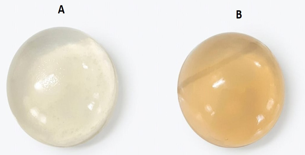
(a) Hydrogel without CE, (b) Hydrogel with CE.
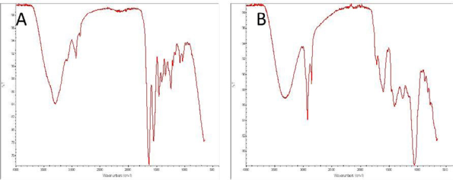
FTIR spectra of (a) hydrogel (b) chamomile extract
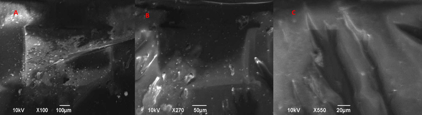
SEM analysis of CS/GEL/CE Hydrogels (a) at 100 µm (b). at 50 µm (c). at 20 µm
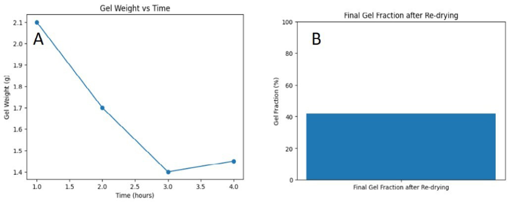
(a) Gel weight as function of time (b) Final gel fraction percentage.
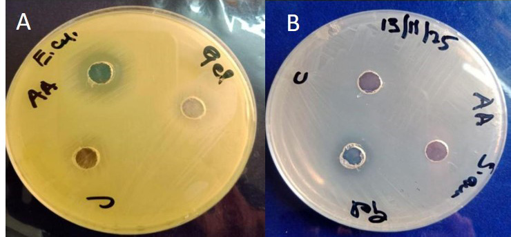
Zone of inhibition of hydrogel Against (a). E. coli (b). S. aureus.
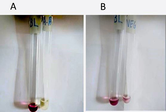
Visual color changes in DPPH Assay of (a) Acetic acid dissolved hydrogel; (b) Negative control.
References
- Amin, M.A. and Abdel-Raheem, I.T., 2014. Accelerated wound healing and anti-inflammatory effects of physically cross linked polyvinyl alcohol–chitosan hydrogel containing honey bee venom in diabetic rats. Arch. Pharma. Res., 37(8): 1016-1031. https://doi.org/10.1007/s12272-013-0308-y[CrossRef]
- Ardean, C., Davidescu, C.M., Nemeş, N.S., Negrea, A., Ciopec, M., Duteanu, N., Negrea, P., Duda-Seiman, D. and Musta, V., 2021. Factors influencing the antibacterial activity of chitosan and chitosan modified by functionalization. Int. J. Mol. Sci., 22(14): 7449. https://doi.org/10.3390/ijms22147449[CrossRef]
- Călina, I.C., Scărișoreanu, A., Demeter, M., Mănăilă, E. and Crăciun, G., 2025. Strategies for incorporating natural therapeutic agents into hydrogel dressings: Innovations in wound healing. Polymers, 17(23): 3105. https://doi.org/10.3390/polym17233105[CrossRef]
- Chauhan, R., Singh, S., Kumar, V., Kumar, A., Kumari, A., Rathore, S., Kumar, R. and Singh, S., 2021. A comprehensive review on biology, genetic improvement, agro and process technology of German chamomile (Matricaria chamomilla L.). Plants, 11(1): 29. https://doi.org/10.3390/plants11010029[CrossRef]
- Farasati Far, B., Naimi-Jamal, M.R., Jahanbakhshi, M., Hadizadeh, A., Dehghan, S. and Hadizadeh, S., 2024. Enhanced antibacterial activity of porous chitosan-based hydrogels crosslinked with gelatin and metal ions. Sci. Rep., 14(1): 7505. https://doi.org/10.1038/s41598-024-58174-9[CrossRef]
- Ferreira, L.M.d.M.C., Modesto, Y.Y., Souza, P.D.Q.D., Nascimento, F.C.D.A., Pereira, R.R., Converti, A., Lynch, D.G., Brasil, D.D.S.B., Da Silva, E.O. and Silva-Júnior, J.O., C., 2024. Characterization, biocompatibility and antioxidant activity of hydrogels containing propolis extract as an alternative treatment in wound healing. Pharmaceuticals, 17(5): 575. https://doi.org/10.3390/ph17050575[CrossRef]
- Fukuda, K., Ito, Y. and Amagai, M., 2025. Barrier integrity and immunity: Exploring the cutaneous front line in health and disease. Ann. Rev. Immunol., 43(1): 219-252. https://doi.org/10.1146/annurev-immunol-082323-030832[CrossRef]
- Hromiš, N.M., Lazić, V.L., Popović, S.Z., Šuput, D.Z. and Bulut, S.N., 2017. Antioxidative activity of chitosan and chitosan based biopolymer film. Food Feed Res., 44(2): 91-100-191-100. https://doi.org/10.5937/FFR1702091H[CrossRef]
- Iftime, M.M., Ailiesei, G.L. and Ailincai, D., 2024. Tuning antioxidant function through dynamic design of chitosan-based hydrogels. Gels, 10(10): 655. https://doi.org/10.3390/gels10100655[CrossRef]
- Kaçoğlu, H.S., Ceylan, Ö. and Çelebi, M., 2024. Comparative study of the effect of cross-linking degree on chitosan hydrogels synthesized with low and medium molecular weight chitosan. Polymer Eng. Sci., 64(3): 1326-1339. https://doi.org/10.1002/pen.26619[CrossRef]
- Kosimaningrum, W.E., Barleany, D.R., Sako, V.N. and Ristiyanti, R., 2020. Preparation of gelatin-chitosan-honey-based hydrogel for potential active material of wound care dressing application. Mater. Sci. Forum, https://doi.org/10.4028/www.scientific.net/MSF.988.162[CrossRef]
- Li, L., Chen, D., Chen, J., Yang, C., Zeng, Y., Jin, T., Zhang, Y., Sun, X., Mao, H. and Mu, Z., 2023. Gelatin and catechol-modified quaternary chitosan cotton dressings with rapid hemostasis and high-efficiency antimicrobial capacity to manage severe bleeding wounds. Mater. Design, 229: 111927. https://doi.org/10.1016/j.matdes.2023.111927[CrossRef]
- Maita, K.C., Avila, F.R., Torres-Guzman, R.A., Garcia, J.P., Eldaly, A.S., Palmieri, L., Emam, O.S., Ho, O. and Forte, A.J., 2022. Local anti-inflammatory effect and immunomodulatory activity of chitosan-based dressing in skin wound healing: A systematic review. J. Clin. Transl. Res., 8(6): 488. https://doi.org/10.18053/jctres.08.202206.007[CrossRef]
- Melnyk, N., Nyczka, A., Piwowarski, J.P. and Granica, S., 2024. Traditional use of chamomile flowers (Matricariae flos) in inflammatory-associated skin disorders. Prosp. Pharma. Sci., 22(4): 59-73. https://doi.org/10.56782/pps.215[CrossRef]
- Mujahid, M., Zubair, M., Yaqoob, A., Shahzad, S. and Ullah, A., 2025. Formulation and evaluation of licorice-extract-enhanced chitosan, PVA, and gelatin-derived hydrogels for wound dressing. Bioengineering, 12(5): 439. https://doi.org/10.3390/bioengineering12050439[CrossRef]
- Naranđa, J., Bračič, M., Maver, U. and Trojner, T., 2025. Recent advancements in smart hydrogel-based materials in cartilage tissue engineering. Materials, 18(11): 2576. https://doi.org/10.3390/ma18112576[CrossRef]
- Razavi, Z.S., Aliniay, S.S., Yousefi, M.H., Javaheri, F., Rahimi, B.M.R., Afkhami, H. and Heidari, F., 2025. Application of novel strategies in chronic wound management with focusing on pressure ulcers: New perspective. Arch. Dermatol. Res., 317(1): 320. https://doi.org/10.1007/s00403-024-03790-8[CrossRef]
- Razif, R., Zainuddin, A.N.Z., Zawawi, N.A., Maarof, M., Ahmad, H., Mohd Nor, F. and Fauzi, M.B., 2026. Preliminary genipin-crosslinked gelatin–asiaticoside injectable hydrogel for future biomaterials ink: Physicochemical properties and cytocompatibility for wound repair. Artif. Cells, Nanomed. Biotechnol., 54(1): 159-191. https://doi.org/10.1080/21691401.2026.2627111[CrossRef]
- Saputra, A.S., Prihatiningsih, T. and Santoso, O., 2021. Antibacterial properties of hydrogel membranes infused with liquid smoke on growth inhibition of Staphylococcus aureus. Malays. J. Med. Health Sci., 17(3): 2636-9346.
- Sepp, J., Koshovyi, O., Jakstas, V., Žvikas, V., Botsula, I., Kireyev, I., Tsemenko, K., Kukhtenko, O., Kogermann, K. and Heinämäki, J., 2024. Phytochemical, technological, and pharmacological study on the galenic dry extracts prepared from German chamomile (Matricaria chamomilla L.) flowers. Plants, 13(3): 350. https://doi.org/10.3390/plants13030350[CrossRef]
- Singh, B., Singh, J., Sharma, V., Kumari, A. and Sharma, D., 2023. Exploring bioactive aloe-vera and sterculia gum to develop hydrogel wound dressings by graft-copolymerization. Food Hydrocoll. Health, 4: 100168. https://doi.org/10.1016/j.fhfh.2023.100168[CrossRef]
- Verdolino, D.V., Thomason, H.A., Fotticchia, A. and Cartmell, S., 2021. Wound dressings: Curbing inflammation in chronic wound healing. Emerg. Topics Life Sci., 5(4): 523-537. https://doi.org/10.1042/ETLS20200346[CrossRef]
- Zhou, T., Chen, S., Ding, X., Hu, Z., Cen, L. and Zhang, X., 2021. Fabrication and characterization of collagen/PVA dual-layer membranes for periodontal bone regeneration. Front. Bioeng. Biotechnol., 9: 630977. https://doi.org/10.3389/fbioe.2021.630977[CrossRef]
- Zhou, Y., Zhang, C., Liang, K., Li, J., Yang, H., Liu, X., Yin, X., Chen, D. and Xu, W., 2018. Photopolymerized water-soluble maleilated chitosan/methacrylated poly (vinyl alcohol) hydrogels as potential tissue engineering scaffolds. Int. J. Biol. Macromol., 106: 227-233. https://doi.org/10.1016/j.ijbiomac.2017.08.002[CrossRef]
Services
Article usage statistics
Loading…