
Mycoplasma bovis is the second most important mycoplasmas causing a variety of diseases in cattle worldwide. This study was aimed to report the PCR identification of M. bovis from slaughtered cattle in northeastern Nigeria. Four hundred and eighty (480) samples of lung tissues (180), nasal swab (180), ear swab (60) and pleural fluid (60) were collected from 190 heads of cattle at slaughter in Yola and Jalingo abattoirs in Adamawa and Taraba States, respectively Samples were processed based on standard laboratory protocols. An overall Mycoplasma bovis isolation rate of 0.83% (4/480) was obtained. Based on the states studied, 1 (0.35%) and 3 (1.53%) M. bovis were isolated from Adamawa and Taraba States, respectively with an insignificant association between M. bovis infection and the states sampled (P>0.05). Based on organs/site sampled, 2 (5.40%) isolates of M. bovis were from lung tissues and 1 (2.70%) were from both pleural fluid and ear swab samples. All the 4 isolates were confirmed as M. bovis by the presence of one band of 734 bp. The study had established first isolation of M. bovis from the ear canal of cattle at slaughter in the study area. Therefore, large scale epidemiological studies that will reveal the true prevalence and distribution of M. bovis infection in cattle population in Nigeria are recommended.
Research Article · Volume 6, Issue 2, 22 August 2020 · Open Access
Detection of Mycoplasma bovis from Cattle Presented for Slaughter in Adamawa and Taraba States, Northeastern Nigeria
· · · · · · · ·
Affiliations & Notes ⌄
- 1Faculty of Veterinary Medicine, Ahmadu Bello University, Zaria, Nigeria
- 2Bacteria Vaccine Production Division, National Veterinary Research Institute, Vom, Nigeria
- 3Faculty of Veterinary Medicine, University of Ilorin, Ilorin, Nigeria
- 4Faculty of Veterinary Medicine, University of Abuja, Abuja, Nigeria
- 5Istituto Zooprofilattico Sperimentale dell’ Abruzzo e del Molise “G. Caporale”, Teramo, Italy. *Correspondence | Markus I. Francis, Faculty of Veterinary Medicine, Ahmadu Bello University, Zaria, Nigeria
- 6Email: markfrancis4u@gmail.com
Article Info ⌄
- Journal:
- Veterinary Sciences: Research and Reviews
- Published:
- 22 August 2020

From the article
- Based on the two States sampled, 1 (0.35%) and 3 (1.53%) isolates of M. bovis were isolated from Adamawa and Taraba States respectively with no statistically significant difference observed between M. bovis infection and the states sampled (P>0.05).
- Mycoplasma bovis infection was identified to be higher in Taraba State than in Adamawa State even though there was an insignificant statistical difference (P>0.05).
- Mycoplasma bovis was isolated and identified from the ear canal of cattle in Taraba State with a 1.67% isolation rate.
- The present study has demonstrated M. bovis from lung tissues, pleural fluid and ear canal of cattle and thus, the first report of isolation of M. bovis from an ear canal of cattle at slaughter in the study area.
Selected from the article's own text.
Abstract
Introduction
Mycoplasma bovis (M. bovis) is the second most important mycoplasmas of cattle causing respiratory disease, mastitis, arthritis, otitis media, keratoconjunctivitis and a variety of other diseases in cattle worldwide . The organism was first isolated from a cow with mastitis in 1961 in the USA . It lacks cell wall, had a complex nutritional requirement and a low G+C content of 23.8-32.9 mol % . It does ferment glucose nor hydrolyses arginine but instead uses organic acids such as lactate and pyruvate as energy source for growth ; . Mycoplasma bovis has been reported to be transmitted in respiratory secretions through aerosols, nose-to-nose contact, or indirectly via feed, water, housing, or other formite ; .
Diagnosis of M. bovis infection can be performed by serology, isolation and polymerase chain reaction (PCR) on lung tissues ; . Diagnosis by culture requires complex media, specialized equipment and technical skills with growth apparent by 48 hours . The PCR has been used to detect M. bovis from clinical materials with sensitivity higher than that of culture method . Many PCR systems have been routinely described for specific identification of M. bovis which target 16S rRNA gene , uvrC gene , oppD/F gene , and Vsp gene . These had resulted in improved specificity for M. bovis detection in samples.
Nigeria is endowed with abundant livestock resources with 19.7 million estimated livestock population and diseases have been reported as a setback in actualization of animal production . Mycoplasma bovis infection is an important emerging disease infecting cattle in Nigeria resulting in high economic losses . The disease is poorly understood among livestock owners and animal health handlers, therefore needed to establish the distribution of the disease in the study area. The present study was aimed to detect M. bovis from cattle presented for slaughter in Adamawa and Taraba States, northeastern Nigeria.
Materials and Methods
Study area
This study was carried out in two northeastern states of Nigeria, namely; Yola Abattoir in Adamawa State and Jalingo Abattoir in Taraba State (Figure 1). Yola Abattoir based on 3D Global Positioning System (GPS) is located on the Latitude 9°13’33.3”N and Longitude 12°27’10.1”E with an average of 70 - 80 cattle being slaughtered daily and higher during festivals period. Jalingo Abattoir based on 3D Global Positioning System (GPS) is located on the Lattitude 8°54’12.1”N and Longitude 11°21’14.3”E with an average of 40-50 cattle slaughtered daily.
Collection and processing of samples
The samples used for this study were obtained from cattle slaughtered at abattoirs within the study States. The period of collection was from February 2016 to February 2017. A total of four hundred and eighty (480) samples of lung tissues suspected of pneumonia (180), nasal swab (180), ear swab (60) and pleural fluid (60) were collected from 190 heads of cattle at slaughter with cases of suspected pneumonia indicative of M. bovis infections (120 from Adamawa State while 70 from Taraba State). Two hundred and eighty-four (284) and one hundred and ninety-six (196) samples (tissues and swabs/pleural fluids) from cattle were collected from the abattoirs in Adamawa and Taraba States, respectively. The samples were transported to the National Veterinary Research Institute Zonal Offices in Yola and Jalingo, respectively where they are processed in 2 ml sterile Nalgene® cryo vials containing 1.5 ml of pleuropneumonia-like organism (PPLO) broth and were later transported in refrigerated Coleman box to the Mycoplasma Laboratory, National Veterinary Research Institute (NVRI) Vom, Plateau State and then stored at -20°C prior to further processing.
Microbiological culture of mycoplasmas
The samples were processed according to the method described . The transport media (PPLO broth) containing various samples were filtered by passing through 0.45µm millipore antibacterial filters which is permeable to organisms smaller than 0.45 µm in sizes such as Mycoplasma and viruses. The filtrate was then incubated at 37oC in screw-capped bottles for three (3) days under CO2 and later cultured onto PPLO agar. These were incubated at 37oC in a 5% CO2 atmosphere with maximum humidity and examined daily for evidence of growth for 1-2 weeks using a stereomicroscope. Isolated colonies having the classical ‘fried eggs’ appearance with dense raised centres (nipples) were triple-cloned by aseptically transferring a small agar block containing an isolated Mycoplasma colony into 10 ml PPLO broth and incubated for 72 hours. Both broth and agar media were considered negative if no comet or Mycoplasma-like colony was observed after 14 days of incubation. Cloned isolates were stored in triplets at -4°C, -20ºC and -80°C. These isolates were later transported under cold chain by FedEx® Red Star Express Courier to OIE Reference Laboratory for Contagious Bovine Pleuropneumonia (CBPP) in Istituto Zooprofilattico Sperimentale dell’ Abruzzo e del Molise (IZSAM) “G. Caporale”, Teramo, Italy. On receiving the isolates, they were stored at -20°C, the isolates were then removed, verified and then registered under exotic disease unit before being processed.
Detection of Mycoplasma bovis based on 16S rRNA genes
Mycoplasma bovis DNA from the presumptive culture was extracted from 500 µl of broth cultures using a commercially available automated nucleic acid extraction system the Maxwell® 16 Tissue/cell DNA Purification kits (Promega, USA) with Maxwell® 16 instrument (Promega, USA) following manufacturer’s instructions. The extracted DNA was resuspended in 500 µl TE buffer (10 mM Tris- HCl, 1 mM EDTA, pH 7.6) and was stored at -20°C until analyzed.
The following PCR mix was used for each sample; 25 µl of master mix (Taq Polymerase, dNTPs, MgCl2, Taq Buffer), 18 µl nuclease free water, 1 µl forward primer, 1 µl reverse primer and 5 µl of DNA sample as described by .
Primers designed for 16S rRNA were used to amplify 734 bp of 16S rRNA gene for Mycoplasma bovis. The forward primer was MBOF2: 5’-GAA GCG AAA GTA GCA TAG GAA ATG AT-3’ and the reverse primer was MBOR2: 5’-CGT CGT CCC CAC CTT CCT CCC G-3’. The samples were then subjected to amplification reactions consisting of one cycle of initial denaturation at 94°C for 5 minutes, followed by 40 cycles of denaturation at 94°C for 30 seconds, annealing at 59°C for 30 seconds, extension at 72°C for 30 seconds, a final extension step at 72°C for 5 minutes and standby temperature at 4°C. The amplified products were detected by electrophoresis in 1% agarose gel electrophoresis. The resultant bands were stained with ethidium bromide and visualized under ultraviolet light. Mycoplasma bovis reference strain PG45 was used as a positive control, while negative control contained only the master mix.
Data analysis
Data collected were analyzed using simple percentages, while Fisher’s Exact test was used to test the level of significance between the variables and the value of P<0.05 is considered statistically significant.
Results and Discussion
Thirty-seven (37/480) field isolates of Mycoplasma species were identified on culture. Out of which 4 (4/37) yielded positive growth exhibiting fried egg colonies (Figure 2) with a slight presence of film typical of Mycoplasma bovis on PPLO agar, giving an overall isolation rate of 0.83% (4/480) (Table 1) while the rest of the isolates do not exhibit typical mycoplasma colonies.
Table 1: Isolation rates of Mycoplasma bovis from slaughtered cattle in Adamawa and Taraba States, northeastern Nigeria.
| Source of examined isolates | No. of samples | No. of M. bovis identified (%) | P-value |
| States | |||
| Adamawa | 284 | 1 (0.35) | |
| Taraba | 196 | 3 (1.53) | 0.3095 |
| Organs | |||
| Lungs | 180 | 2 (1.11) | |
| Pleural fluid | 180 | 1 (0.56) | |
| Nasal swab | 60 | 0 (0.00) | |
| Ear swab | 60 | 1 (1.67) | 0.8409 |
| Total | 480 | 4 (0.83) |
Based on the two States sampled, 1 (0.35%) and 3 (1.53%) isolates of M. bovis were isolated from Adamawa and Taraba States respectively with no statistically significant difference observed between M. bovis infection and the states sampled (P>0.05). Based on organs/site sampled, 2 (5.40%) isolates of M. bovis were isolated from lung tissues and 1 (2.70%) was isolated from both pleural fluid and ear swab samples while no M. bovis isolate was identified from nasal swab with an insignificant statistical difference (P>0.05). Mycoplasma bovis was recovered from lung tissues, pleural fluid and ear swab samples collected (Table 1).
The four (4) isolates were confirmed by PCR based on 16S rRNA gene as Mycoplasma bovis by the presence of one band of 734 bp at the same distance with positive control that was used (Figure 3).
Mycoplasma species are pathogenic Mollicutes affecting animals, humans and plants and therefore of great concern in human and veterinary medicine . The isolation and identification rate of 0.83 % (4/480) of M. bovis reported in this study is lower than 2.0 % isolation by from pneumonic lungs of cattle from Adamawa State and 8.9 % isolation from pneumonic lungs of cattle from two northwestern States of Nigeria. The difference in the isolation rates may be attributed to the delayed analysis of the samples after collection. The isolation of M. bovis from pneumonic lungs of slaughtered cattle shows that M. bovis and its interrelated infection may be occurring in the study area. This finding is of clinical significance because these few infected cattle could likely be a threat and basis of infection to several vulnerable and in-contact herds and farms in the study area and hence leading to increase in prevalence. Similar observations were made ; ; ; .
Mycoplasma bovis infection was identified to be higher in Taraba State than in Adamawa State even though there was an insignificant statistical difference (P>0.05). This signifies a higher risk of susceptibility to infection with M. bovis for cattle in Taraba State than their counterpart in Adamawa State, even though there was a strong link in the movement of cattle between the two states in search of forage and favorable market. Similar findings have been reported by previous authors ; .
Based on organs/site sampled, M. bovis was identified higher in lung tissues than pleural fluid and ear swab. This might be attributed to the fact that, the predilection site for most Mycoplasma species is the respiratory system , where these organisms multiply and cause pathological lesions . Dissemination to other organs and parts of the body occurs as the infection progresses to a chronic stage leading to the recovery of these pathogens in the pleural sac, nasal and ear canals .
Mycoplasma bovis was isolated and identified from the ear canal of cattle in Taraba State with a 1.67% isolation rate. This is of scientific significance as the organism has been previously reported to cause otitis media in cattle . To the best of our knowledge, this is the first report of isolation of M. bovis from the bovine ear canal in Nigeria. The presence of the M. bovis in these sites is not surprising in view of the fact that they are widely distributed in nature. Considering the ability of other ruminant mycoplasmas to colonize the ear canal, ear involvement by M. bovis may not be doubtful. The assumptions on the occurrence of M. bovis in the ear canal include the anatomical connection between the pharynx and middle ear via Eustachian tube for the pathogenesis of otitis media due to M. bovis infection in cattle ; ; . Also the mycoplasmaemia developed for the period of acute infection may involve dissemination of mycoplasmas from primary site of infection (the respiratory system) to other organs and systems including the auditory organs where the organisms were isolated . The report was consistent with previous report who also for the first time reported M. bovis and a crowd of other mycoplasmas from the ear canal of cattle in Brazil.
Molecular identification of isolates from broth culture, identified 16S rRNA gene of M. bovis isolates TL43, TL46, TES57 and AP106, and was consistent with previous reports ; , 2005).
Conclusion
The present study has demonstrated M. bovis from lung tissues, pleural fluid and ear canal of cattle and thus, the first report of isolation of M. bovis from an ear canal of cattle at slaughter in the study area. We therefore, recommend large scale epidemiological studies that will reveal the prevalence and distribution of M. bovis infection in cattle populations in Nigeria.
Acknowledgments
The authors are most grateful to the management of National Veterinary Research Institute Vom, Plateau State Nigeria for primary isolation, and OIE Reference Laboratory for CBPP at Istituto Zooprofilattico Sperimentale dell Abruzzo e Molise ‘G Caporale’, Campo Boario Teramo, Italy for free bench space and technical support for this work.
Authors’ Contribution
CNK, JA, LA, MAR, GOE and MIF conceived and designed the study. PIA and MIF collected and analyzed samples. FS, MS and MIF performed molecular identification. MIF, PIA, MAR and FS drafted the manuscript. All authors read and approved the final manuscript for submission.
Conflict of interest
The authors have declared no conflict of interest.
Figures
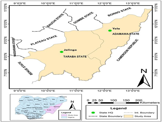
Map of Adamawa and Taraba States, North-eastern Nigeria (MLS, 2010).
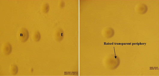
Colonies of Mycoplasma bovis from lung sample on PPLO agar showing ‘fried egg’ colonies with nipples ‘n’ and raised transparent periphery ‘t’ observed under Stereomicroscope (X40).
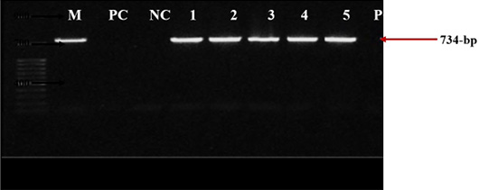
PCR amplification of Mycoplasma bovis 16S rRNA gene on 1% agarose gel with primers MBOF2 and MBOR2. M: molecular marker of 50-bp; PC: positive control reference strain PG45 (734-bp); NC: negative control; Lanes 1-5: represents samples TL41, TL43, TL46, TES57 and AP106.
References
- Ajuwape, A.T.P., Ikheloa, J.O., Ojo, M.O., Alaka, O.O. and Adatosoye, A.J., 2003. Biochemical and serological identification of mycoplasmas isolated from pneumonic bovine lungs in Nigeria. Isr. J. Vet. Med., Microbiol. Parasitol., 58: 2-8.
- Alberti, A., Addis, M.F., Chessa, B., Cubeddu, T., Profiti, M., Rosati, S., Ruiu, A. and Pittau, M., 2006. Molecular and antigenic characterization of Mycoplasma bovis strain causing an outbreak of infectious keratoconjunctivitis. J. Vet. Diagn. Invest., 18: 41-51. https://doi.org/10.1177/104063870601800106[CrossRef]
- Arcangioli, M.A., Duet, A., Meyer, G., Dernburg, A., Bezille, P., Poumarat, F. and Le Grand, D., 2008. The role of Mycoplasma bovis in bovine respiratory disease outbreaks in veal calf feedlots. N. Z. Vet. J., 177: 89-93. https://doi.org/10.1016/j.tvjl.2007.03.008[CrossRef]
- Arcangioli, M.A., Aslan, H., Tardy, F., Poumarat, F. and Le Grand, D., 2012. The use of pulse-field gel electrophoresis to investigate the epidemiology of Mycoplasma bovis in Franch calf feedlots. Vet. J., 192: 96-100. https://doi.org/10.1016/j.tvjl.2011.05.004[CrossRef]
- Bashiruddin, J.B., de Santis, P., Varga, E. and Stipkovits, L., 2001. Confirmation of the presence of Mycoplasma bovis in Hungarian cattle with pneumonia resembling pleuropneumonia. Vet. Rec., 148: 743-746. https://doi.org/10.1136/vr.148.24.743[CrossRef]
- Bashiruddin, J.B., Frey, J., Konigsson, M.H., Hotzel, H., Diller, R., Santis, P., Botello, A., Ayling, R.D., Nicholas, R.A.J., Thiaucourt, F. and Sachse, K., 2005. Evaluation of PCR system for the identification and differentiation of Mycoplasma agalactiae and Mycoplasma bovis: A collaborative trial. Vet. J., 169: 268-275. https://doi.org/10.1016/j.tvjl.2004.01.018[CrossRef]
- Brown, D.R., May, M., Bradbury, J.M. and Johansson, K.E., 2015. Mollicutes. Bergey’s Manual of Systematics of Archaea and Bacteria. pp. 1-8. https://doi.org/10.1002/9781118960608.cbm00048[CrossRef]
- Calcutt, M.J., Lysnyansky, I., Sachse, K., Fox, L.K., Nicholas, R.A.J. and Ayling, R.D., 2018. Gap analysis of Mycoplasma bovis disease, diagnosis and control: An aid to identify future development requirements. Transbound. Emerg. Dis., 1: 1-19. https://doi.org/10.1111/tbed.12860[CrossRef]
- Caswell, J.L. and Archambault, M., 2007. Mycoplasma bovis pneumonia in cattle. Anim. Health Res. Rev., 8: 161-186. https://doi.org/10.1017/S1466252307001351[CrossRef]
- Cremonesi, P., Vimercati, C., Pisoni, G., Perez, G., Ribera, A.M., Castiglioni, B., Luzzana, M., Ruffo, G. and Moroni, P., 2007. Development of DNA extraction and PCR amplification protocols for detection of Mycoplasma bovis directly from milk samples. Vet. Res. Commun., 31(1): 225-227. https://doi.org/10.1007/s11259-007-0011-x[CrossRef]
- Egwu, G.O., Nicholas, R.A.J., Ameh, J.A. and Bashiruddin, J.B., 1996. Contagious bovine plueropneumonia: An update. Vet. Bull., 66: 875-888.
- Francis, M.I., 2017. Mycoplasma bovis: A neglected pathogen in Nigeria - A mini Review. J. Dairy Vet. Sci., 4(2): 1-5. https://doi.org/10.19080/JDVS.2017.04.555633[CrossRef]
- Francis, M.I., Ejeh, E.F., Raji, M.A., Kazeem, H.M., Musa, J.A., Ankeli, P.I. and Akalusi, Y., 2014. Isolation and biochemical identification of Mycoplasma bovis in Cattle pneumonic lungs in Adamawa State, Nigeria. Sahel J. Vet. Sci., 13: 1-5.
- Gull, T.B., French, R.A., Gorton, T.S., Burrage, T.G., Prozesky, L., Geary, S.J. and Adams, G.L., 2013. Models of contagious bovine pleuropneumonia: Evaluation of two novel strains. Open Vet. Sci. J., 7: 23-33. https://doi.org/10.2174/1874318820130731002[CrossRef]
- Hayman, B. and Hirst, R., 2003. Development of semi-nested PCR for the detection of Mycoplasma bovis from bovine milk and mucosal samples. Vet. Microbiol., 91: 91-100. https://doi.org/10.1016/S0378-1135(02)00305-X[CrossRef]
- Johansson, K.E., Berg, L.O., Bolske, G., Deniz, S., Mattsson, J., Persson, M. and Pettersson, B., 1996. Specific PCR systems based on the 16S rRNA genes of Mycoplasma agalactiae and Mycoplasma bovis. In: Frey, J., Sarris, K. (eds). Mycoplasmas of Ruminants: Pathogenicity, Diagnostics, Epidemiology and Molecular Genetics. COST 826- Agriculture and Biotechnology. Eur. Comm., pp. 88-90. Brussels, Belgium.
- Lamm, C.G., Munson, L., Thurmond, M.C., Barr, B.C. and George, L.W., 2004. Mycoplasma otitis in California calves. J. Vet. Diagn. Invest., 16: 397-402. https://doi.org/10.1177/104063870401600505[CrossRef]
- Le Grand, D., Bezille, P., Calavas, D., Poumarat, E., Brank, M., Citti, C. and Rosengarten, R., 2002. Serological prevalence of Mycoplasma bovis infection in suckling beef cattle in France. Vet. Rec., 150: 268-273. https://doi.org/10.1136/vr.150.9.268[CrossRef]
- Maeda, T., Shibahara, T., Kimura, K., Wada, Y., Sato, K., Imada, Y., Ishikawa, Y. and Kadota, K., 2003. Mycoplasma bovis-associated suppurative otitis media and pneumonia in bull calves. J. Comp. Pathol., 129: 100-110. https://doi.org/10.1016/S0021-9975(03)00009-4[CrossRef]
- Maunsell, F.P., Woolums, A.R., Francoz, D., Rosenbusch, R.F., Step, D.L., Wilson, D.J. and Janzen, E.D., 2011. Mycoplasma bovis infection in cattle. J. Vet. Int. Med., 25: 772-783. https://doi.org/10.1111/j.1939-1676.2011.0750.x[CrossRef]
- Ministry of Land and Survey (MLS), 2010. Map of Adamawa State of Nigeria showing all local government areas. pp. 5-8.
- Nicholas, R.A.J., Ayling, R.D. and McAuliffe, L., 2008. Contagious bovine pleuropneumonia. In: Mycoplasma Diseases of Ruminants. Nicholas, R., Ayling, R., McAuliffe, L. (eds), CABI Publishing, UK. pp. 69-97. https://doi.org/10.1079/9780851990125.0069[CrossRef]
- Nicholas, R.A.J., Ayling, R.D. and Stipkovits, L., 2002. An experimental vaccine for calf pneumonia caused by Mycoplasma bovis: Clinical, cultural, serological and pathological findings. Vaccine, 20: 3569-3575. https://doi.org/10.1016/S0264-410X(02)00340-7[CrossRef]
- Nicholas, R.A.J., Fox, L.K. and Lysnyansky, I., 2016. Mycoplasma mastitis in cattle: To cull or not to cull. Vet. J., 216: 142-147. https://doi.org/10.1016/j.tvjl.2016.08.001[CrossRef]
- Parker, A.M., Sheehy, P.A., Hazelton, M.S., Bosward, K.L. and House, J.K., 2018. A review of mycoplasma diagnostics in cattle. J. Vet. Int. Med., 32(3): 1241-1252. https://doi.org/10.1111/jvim.15135[CrossRef]
- Pinnow, C.C., Butler, J.A., Sachse, K., Hotzel, H., Timms, L.L. and Rosenbusch, R.F., 2001. Detection of Mycoplasma bovis in preservative-treated field milk samples. J. Dairy Sci., 84: 1640-1645. https://doi.org/10.3168/jds.S0022-0302(01)74599-7[CrossRef]
- Saad, N.M. and AbdelHameed, K.G., 2012. Detection of Mycoplasma species in raw milk of lactating animals in Assiut and Qena city of Egypt. Vet. World, 5(2): 80-85. https://doi.org/10.5455/vetworld.2012.80-85[CrossRef]
- Santos, S.B., Nascimento, E.R., Faccini, J.L.H., Barreto, M.L. and Pereira, V.L., 2009. Potentially pathogenic mycoplasmas in the external ear canal of clinically normal cattle in Southeast Brazil: First report. Braz. J. Microbiol., 40(3): 455-457. https://doi.org/10.1590/S1517-83822009000300006[CrossRef]
- Subramaniam, S., Bergonier, D., Poumarat, F., Capaul, S., Schlatter, Y., Nicolet, J. and Frey, J., 1998. Species identification of Mycoplasma bovis and Mycoplasma agalactiae based on the uvrC genes by PCR. Mol. Cell. Probes. 12: 161-169. https://doi.org/10.1006/mcpr.1998.0160[CrossRef]
- Tambuwal, F.M. and Egwu, G.O., 2017. Occurrence of Mycoplasma mycoides subspecies mycoides small colony and Mycoplasma bovis from respiratory tracts of cattle presented for slaughter in North-western Nigeria. Direct Res. J. Vet. Med. Anim. Sci., 2(5): 115-121.
- Tambuwal, F.M., Egwu, G.O., Shittu, A., Sharubutu, G.H., Umaru, M.A., Umar, H.U., Mshelia, P.C. and Garba, S., 2011. Vaccination coverage and prevalence of contagious bovine pleuropnuemonia (1999-2008) in two transboundary states of north-western Nigeria. Nigerian Vet. J., 32: 169-173.
- Tenk, M., Balint, A., Stipkovits, L., Biro, J. and Densco, L., 2006. Detection of Mycoplasma bovis with an improved PCR assay. Acta Vet. Hung., 54: 427-435. https://doi.org/10.1556/AVet.54.2006.4.1[CrossRef]
- Thiaucourt, F., Guerin, C., Mady, V. and Lefevre, P.C., 1992. Diagnosis of caprine contagious pleuropneumonia: Recent improvements. Rev. Sci. Tech. Off. Int. Epizoot., 11: 859-865. https://doi.org/10.20506/rst.11.3.629[CrossRef]
- Wise, K.S., Calcutt, MJ., Foecking, M.F., Roske, K., Madupu, R. and Methe, B.A., 2011. Complete Genome Sequence of Mycoplasma bovis Type Strain PG45 (ATCC 25523). Infect. Immunol., 79(2): 982-983. https://doi.org/10.1128/IAI.00726-10[CrossRef]
Services
Article usage statistics
Loading…