
In many regions of Pakistan goat is a staple livestock for the livelihood of poor farmers. The goat is also known as the poor man’s cow in the sub-continent. Mycoplasmosis impacts the poorest farmers most gravely. Currently, diagnostics and vaccines for Mycoplasma causing infections are lacking due to the uniqueness of strains endemic to Pakistan. Mycoplasma is responsible for causing several socio-economically important infectious diseases (including WOAH-listed CCPP) in goats. Therefore, to unveil the status of Mycoplasmosis in goats, a total of 2,400 samples consisting of nasal discharges, tracheal swabs, lung tissue, and pleural fluid were collected from four different zones of northern Pakistan. Out of 2400 samples 512 (21.3%) samples showed gross turbidity and typical whirling movement of mycoplasma in PPLO broth. The PCR revealed 284 (11.8%) of the Mm cluster, including six cases of mixed infection with M. capricolum subsp. capricolum (Mcc) and M. capricolum subsp. capripneumoniae (Mccp). The region-wise prevalence of the Mm cluster was 105 (17.5%) in the Northern region, followed by 66 (11%), 59 (9.8%), and 54 (9%) in the Central, Southern, and Tribal regions, respectively. The screening of the Mm cluster isolates by species-specific primers found 110 (4.5%), 92 (3.8), and 88 (3.6%) Mcc, Mccp, and M. mycoides subsp. capri (Mmc), respectively. Of the 284 Mm cluster positive samples, the highest number was obtained from pleural fluid 75 (18.8%), followed by lungs, tracheal swabs, and nasal swabs 53 (13.3%), 83 (10.4%) and 73 (9.1%), respectively. This study showed that the causes of mycoplasmosis in the northern region of Pakistan include Mcc, Mmc, and Mccp, and the lungs and pleural fluid samples could be used for the isolation of the causative agent.
Research Article · Volume 57, Issue 2, 30 March 2025 · Open Access
Molecular Surveillance Revealed Increasing Trend of Mycoplasmosis in Respiratory Infections of Goats (Capra hircus) in Northern Pakistan
· · · ·
Affiliations & Notes ⌄
- 1College of Veterinary Sciences, Faculty of Animal Husbandry and Veterinary Sciences, The University of Agriculture, Peshawar, Khyber Pakhtunkhwa, Pakistan 2Department of Veterinary Population Medicine, University of Minnesota, St. Paul, MN 55108, USA 3Disease Investigation Laboratory, Directorate of Livestock and Dairy Development (Extension), Peshawar, Khyber Pakhtunkhwa, Pakistan 4Livestock and Dairy Development Department (Research), Khyber Pakhtunkhwa, Pakistan * Corresponding author: hayat.
Article Info ⌄
- Journal:
- Pakistan Journal of Zoology
- Published:
- 30 March 2025

From the article
- In the present study among 284 (11.8%) Mm clusters, the prevalence of the various pathogenic Mycoplasma species recorded were 110 (4.5%), 92 (3.8%), and 88 (3.6%) of Mcc, Mccp, and Mmc, respectively.
- In the present study, the prevalence percentage of Mcc recorded was 110 (4.5%) having a distribution pattern of 47 (7.8%) in the northern region followed by the central region, tribal region, and southern region 26 (4.3%), 21 (3.5%), 16 (2.6%), respectively.
- The increased prevalence of mycoplasmosis in female animals might be due to various factors, which develop stress in animals including pregnancy, lactation, and the estrus cycle.
- Mycoplasmosis in the northern region of Pakistan is caused by Mcc, Mmc, and Mccp.
Selected from the article's own text.
Abstract
Introduction
Pakistan is an agricultural country and livestock, a sub-sector of agriculture, plays a vital role in uplifting the economy of the country as it contributes 62.68% to the agricultures gross domestic product (GDP) and 14.36% to the national GDP. The population of goats in Pakistan is estimated as the highest (84.7 million) among the livestock population and is ranked as the 3rd country in the world that has the largest number of goats and adding 15% of the total meat production in the country (Economic survey of Pakistan 2022-23).
Small ruminants (sheep and goats) are reared mainly as a source of income, food security in the form of milk and meat, and cultural functions. They are also a direct source of cash to the farmer community when needed on an emergency basis ; . In poor countries, small ruminants are susceptible to various infectious diseases, including mycoplasmosis .
Caprine mycoplasmosis has been reported in different regions of the globe. However, the incidence of the disease is more frequent in Asia and Africa, where it is a major constraint to goat production causing huge economic losses ; . Among the various mycoplasmal infections, contagious caprine pleuropneumonia (CCPP) is a severe threat to the goat population and their production performance (Bascunana et al., 1994; . Many different microorganisms cause various outbreaks of respiratory diseases including CCPP, resulting in heavy mortality and morbidity in northern and southern parts of the country ; . CCPP is a fatal respiratory disease affecting domestic goats and some wild ruminants. The etiological agent of CCPP is Mycoplasma capricolum subsp. capripneumoniae (Mccp). The lesions of CCPP are mainly confined to the thoracic cavity, causing up to 80% mortality and 100% morbidity in susceptible flocks (McMartin et al., 1989; ; WOAH, 2018).
Mycoplasma capricolum subsp. capripneumoniae belongs to the Mycoplasma mycoides cluster, which is pathogenic to small ruminants, especially goats. The cluster comprises Mycoplasma mycoides subsp. mycoides Small Colony (MmmSC) and M. mycoides subsp. mycoides Large Colony biotype (MmmLC), M. mycoides subsp. Capri (Mmc), M. capricolum subsp. capricolum (Mcc), Mccp, and Mycoplasma sp. bovine group 7 of Leach (MBG7) as described by Cottew et al. (1987). Recently, the MmmSc and MmmLc were placed in a single group, e.g., Mycoplasma mycoides subsp. mycoides (Mmm). Mycoplasma sp. bovine group 7 has been renamed Mycoplasma leachii . Mccp and Mmm are responsible for inducing CCPP and contagious bovine pleuropneumonia (CBPP), respectively. Both the CCPP and CBPP are life-threatening diseases of ruminants, enlisted by the World Organization for Animal Health (WOAH) formerly OIE, as notifiable diseases.
Other M. mycoides cluster members, such as Mcc and Mmc, influence a wide range of pathological conditions known as “MAKePS” (mastitis, arthritis, keratoconjunctivitis, pneumonia, and septicemia) in small ruminants . In Pakistan, Mmc was first diagnosed by applying various biochemical tests on clinical samples of goats infected with CCPP . Later on, many species of mycoplasma (Mcc, M. putrefaciens, Mmc) has been reported in Pakistan elsewhere through various diagnostic techniques (Awan et al., 2010; ; . However, Mccp was reported for the first time in Balochistan province . Subsequently, the presence of Mccp was exposed to respiratory infections in goats by many scientists through copious testing methods in Pakistan ; ; ; ; .
The goat population of the country is at risk of mycoplasmal infection which causes economic losses in terms of mortality and morbidity and ultimately food insecurity ; ; . To prevent high losses regular monitoring system for mycoplasmal infection is in dire need ; . Surveillance helps in identifying infected animals and implementing control measures promptly. Vaccination can help to reduce the severity and spread of the disease. The most efficient approach is vaccination combined with antibiotic therapy, and antibiotics including tetracyclines, fluoroquinolones, and the macrolide family are typically clinically beneficial if given early on . Therefore, the present study has been designed to study the molecular surveillance of mycoplasma mycoides cluster members prevalent in the vicinity to plan the control strategy to prevent the economically important diseases and the risk of food insecurity.
Materials and Methods
Study area
This study was carried out in selected districts of Khyber Pakhtunkhwa and Gilgit-Baltistan (The Northern region of Pakistan). The study region was divided into four different zones, namely: the northern zone, the central zone, the southern zone and the tribal zone. The northern zone includes Gilgit-Baltistan, Chitral, Swat, Buner and Hazara. The southern zone includes Dera Ismail Khan, Bannu, Kohat, Karak and Laki Marwat districts. The central zone includes Charsadda, Mardan, Swabi, Peshawar and Nowshera districts. The tribal zone includes the tribal districts of Khyber, Bajaur, and Mohmand.
Sample size
A total of 2,400 samples (n=600 from each zone) were collected from October 2017 to March 2020 mostly in the winter season. Equal samples were collected from both sexes of goats. The samples were composed of the nasal swab, tracheal swab, lung tissue and pleural fluid (Table I). All the samples were collected from the goats irrespective of age and breed. The samples were collected from goats exhibiting respiratory signs in the living stage and lungs showing lesions and pleural fluid from dead and sacrificed animals.
Sample collection
Nasal swabs were collected from infected animals showing respiratory signs, and tracheal swabs collected at postmortem were immediately transferred to modified
Table I. Sample collection for the molecular prevalence of mycoplasma mycoide cluster members in Khyber Pakhtunkhwa and Gilgit Baltistan.
| Area/zones of KP | Types of sample from goats | ||||||||
| Nasal swab | Tracheal swab | Lungs tissue | Pleural fluid | Total | |||||
| M | F | M | F | M | F | M | F | ||
| Northern zone | 100 | 100 | 100 | 100 | 50 | 50 | 50 | 50 | 600 |
| Central zone | 100 | 100 | 100 | 100 | 50 | 50 | 50 | 50 | 600 |
| Southern zone | 100 | 100 | 100 | 100 | 50 | 50 | 50 | 50 | 600 |
| Tribal districts | 100 | 100 | 100 | 100 | 50 | 50 | 50 | 50 | 600 |
| Total | 400 | 400 | 400 | 400 | 200 | 200 | 200 | 200 | 2400 |
PPLO broth. The lungs and pleural fluid samples from goats at necropsy died from a natural outbreak and at the slaughterhouse were collected. Pleural fluids were collected in a clean sterile syringe and were transferred immediately to screw-capped sterile falcon tubes (Kartal, Italy). A lung sample was taken from the area between consolidated and normal tissue. Whole lungs showing massive hepatization were collected. All samples collected were immediately transported to the laboratory in a cool chain for further processing. The samples processing and analysis were carried out in the Pathology Lab, College of Veterinary Sciences, The University of Agriculture Peshawar, and the Veterinary Diagnostic Laboratory, University of Minnesota, USA.
Culturing of sample
Nasal swabs and pleural fluid samples were cultured in PPLO broth. The tissue samples were minced, and 1 g of the sample was poured into the 10 ml PPLO (20% horse serum, 0.2% glucose, 0.4% sodium pyruvate, 2.1% PPLO broth base, benzylpenicillin, thallium acetate) . All the tubes containing samples were incubated at 37°C in a humid environment with 5% CO2 in the carbon dioxide incubator (New Brunswick, Galaxy 48S UK) and were observed every day after the first 3 days of incubation for 14 days. One tube containing PPLO broth with no sample was also run in parallel to this procedure as a negative control. Color change, turbidity, and flocular movement at the bottom upon shaking were considered indicators of mycoplasma growth.
PCR amplification of genes of Mycoplasama mycode cluster members
A commercially available GeneJET genomic DNA kit (Thermo-scientific) was used for the extraction of DNA from the broth culture following the manufacturer’s protocol. A total of 3 mL of broth from each sample was used for the genomic DNA extraction. The extracted DNA was quantified with a Nanodrop (MultiSkane Go, Thermo-Fisher Finland). The concentration of the extracted DNA was diluted according to the desired level for PCR as described by Manso-Silván et al. (2007).
For polymerase chain reaction 25 µl volume of the PCR reaction was prepared, which contained 10 µl master mix (Dream Taq Green, Thermo Scientific), 1.75 µl of forward primer and 1.75 µl reverse primer, 3 µl of extracted DNA, and 8.5 µl of nuclease-free water. A total of 34 amplification cycles were run in a Bio-Rad T100 thermal cycler for each primer pair. Reaction conditions in the machine were initial denaturation of 95°C for 5 min, followed by the cyclic denaturation at 94 ºC for 30 sec, annealing at 53 and 57 ºC (according to primer applied), polymerization at 72 ºC for 90 sec, and a final step at 72 ºC for 5 min to extend the single-strand DNA fragments. Table II shows a list of primers used.
Statistical analysis
Data were compiled into the Microsoft Excel spreadsheet and analyzed through the Chi-square statistical test on SPSS version 19 at a significant level of 0.05 %. The statistical difference between the mycoplasma isolates and different sources of samples and gender were also determined by the Chi-square test. The Z-test was applied for comparison of the different sources of samples and mycoplasma isolates recovered.
Results
Clinical signs and postmortem examination of animals
The most prevalent clinical signs were watery or mucopurulent nasal discharge, mucus plug in nasal cavities, productive cough, pyrexia (40oC and above), and
Table II. List of primers, oligonucleotide sequence, annealing temperature and expected product size of 16S-rRNA gene for confirmation of Mycoplasma species and FusA core gene of Mycoplasma mycoides cluster.
| Species | Primer name | Oligonucleotide sequence 5’-3’ | Tm (°C) | Product size (bp) | Reference |
| Mycoplasma mycoides cluster | Mm-F | CGAAAGCGGCTTACTGGCTTGTT | 52 | 548 | Azevedo et al., 2006 |
| Mm- R | TTGAGATTAGCTCCCCTTCACAG | 54 | |||
| FusA | FusA-F | TGAAATTTTTAGATGGTGGAGAA | 56 | 781 | Manso et al., 2007 |
| FusA-R | GGTAATTTAATAGTTTCACGATATGAA | 56 | |||
| Mycoplasma mycoides subsp. capri | P4-F | ACTGAGCAATTCCTCTT | 56 | 196 | Hotzel et al., 1996 |
| P6-R | TTAAATAAGTTTGTATATGAAT | 56 | |||
| Mycoplasma capricolum subsp. capripneumoniae | Mccp.spe-F | ATCATTTTTAATCCCTTCAAG | 54 | 316 | Woubit et al., 2004 |
| Mccp.spe-RMccp | TACTATGAGTAATTATAATATATGCAA | 54 | |||
| Mycoplasma capricolum subsp. capricolum | P4 | ACTGAGCAATTCCTCTT | 54 | 192 | Hernandez et al., 2006 |
| P8 | GTAAACCGTGTATATCAAAT | 53 |
deep abdominal respiration with grunting sounds. A postmortem examination revealed frothy discharges in the lower region of the trachea, various degrees of lung hepatization, fibrinous pleuropneumonia, and adhesion of the lungs to the thoracic wall and accumulation of straw-colored pleural fluid in the thoracic cavity (Fig. 1).
Characteristics of Mycoplasma mycoides cluster members
Of the 2400 samples, a total of 512 (21.3%) showed gross turbidity and typical whirling movement upon
shaking in PPLO broth medium after the incubation process of 3–7 days in a CO2 incubator (Fig. 2). Among the positive samples, the highest growth rate was recorded in the northern region at 149 (24.8%), followed by the tribal region, southern region, and central region at 129 (21.5%), 119 (19.8%), and 115 (19.1%), respectively. The statistical analysis of the data revealed non-significant differences among the climatic zones and the prevalence of mycoplasma based on positive culture media. The positive cultured broth where the growth of mycoplasma appeared was re-cultured on PPLO agar medium and the growth of typical mycoplasma colonies was recorded on days 3 to 7 post-incubation process, as shown in Figure 3. Each cultured plate was examined under a compound microscope at 4X and 10X objectives and a single colony having a typical nipple-like or fried egg appearance was marked and transferred to PPLO broth. This procedure has been repeated a minimum of four times and has obtained purified culture (Fig. 4).
Molecular confirmation with PCR
The DNA was extracted from all the single purified colony cultures. The growth of mycoplasma, particularly Mccp, is very difficult and often subsided by the growth of other bacteria. Due to the said reason, DNA was also extracted from all the positive culture isolates to find out the exact prevalence of mycoplasmosis. The screening of the DNA by PCR revealed 284 (11.8%) out of a total of 2,400 samples was confirmed as Mycoplasma mycoides cluster (Mm cluster), including six cases detected of mixed infection of Mcc and Mccp in the Northern zone. The highest prevalence percentage of the Mm cluster was recorded in the northern region 105 (17.5%), followed by 66 (11%), 59 (9.8%), and 54 (9%) by the central, southern, and tribal regions, respectively (Table III). The analysis of the data by the Chi-square test revealed a significant association (P˂0.001) between the PCR-confirmed Mm cluster isolates and different climatic regions of the study area (Table IV). The primer set used for the Mm cluster targeted the 16S rRNA gene produced an amplicon size of 548 bp on agarose gel.
Table III. Growth of mycoplasma in broth culture and PCR identification of Mycoplasma mycoides cluster in different zones.
| Area | No’s of samples | CC of PPLO | Percentage of CC in PPLO | Mm cluster | Percentage of Mm cluster |
| Northern | 600 | 149 | 24.8 | 105 | 17.5 |
| Central | 600 | 115 | 19.1 | 66 | 11 |
| Southern | 600 | 119 | 19.8 | 59 | 9.8 |
| Tribal | 600 | 129 | 21.5 | 54 | 9 |
| Total | 2400 | 512 | 21.3 | 284 | 11.8 |
CC, colour change; PPLO, pleuro pneumonia like organism (growth medium of mycoplasma); Mm, mycoplasma mycoide cluster.
Table IV. Confirmation of Mycoplasma mycoides cluster through PCR in four different zones from naturally infected goats’ samples suspected for CCPP.
| Area | PCR confirmed Mm cluster | Percentage | Chi-sq | P. value | |
| Positive | Negative | ||||
| Northern | 105 | 495 | 17.5 | 25.78 | 0.0001 |
| Central | 66 | 534 | 11 | ||
| Southern | 59 | 541 | 9.8 | ||
| Tribal | 54 | 546 | 9 | ||
| Total | 284 | 2116 | 11.8 | ||
Statistical analysis by (χ2) showed significant association (P˂0.001) among different climatic zone and Mycoplasma isolates, df=3.
The result of the sex-based prevalence of the Mm cluster obtained was 148 (12%) in males and 136 (11%) in females, respectively. Statistical analysis of the results showed a non-significant association (P > 0.05) between different sexes of an animal. It is evident from the results that both sexes of animals are equally susceptible. For the prevalence of different cluster members, species-specific primers were applied. The different sets of primers used for specie specific were Mccp, Mcc, and Mmc. The positive PCR product of these primers developed an amplicon size of 316, 192, and 194 bp, respectively.
Table V. Molecular detection (PCR) of various pathogenic Mycoplasma mycoides cluster member in different climatic zones.
| Area | No’s of samples | PCR confirmed results of Mm cluster members | ||
| Mcc (%) | Mccp (%) | Mmc (%) | ||
| Northern zone | 600 | 47 (7.8) | 37 (6.1) | 27 (4.5) |
| Central zone | 600 | 26 (4.3) | 21 (3.5) | 19 (3.1) |
| Southern zone | 600 | 16 (2.6) | 21 (3.5) | 22 (3.6) |
| Tribal zone | 600 | 21 (3.5) | 13 (2.1) | 20 (3.3) |
| Total | 2400 | 110 (4.5) | 92 (3.8) | 88 (3.6) |
Mcc, Mycoplasma capricolum subsp.; Capricolum: Mmc, Mycoplasma mycoides subsp. Capri; Mccp, Mycoplasma capricolum subsp. capripneumoniae.
Prevalence of different Species of mycoplasma and isolates recovered from various source of samples
Among the 284 (11.8%) of the Mm, cluster isolates, 110 (4.5%) were confirmed as Mcc, 92 (3.8%) Mccp, and 88 (3.6%) Mmc through PCR (Fig. 5). The summarized result of the different region-wise distribution of Mcc, Mccp, and Mmc is displayed in Table V. Among the total 284 Mm cluster samples, the highest number of isolates were obtained from the pleural fluid 75 (18.8%), followed by lung tissue, tracheal swab, and nasal swab 53 (13.3%), 83 (10.4%), and 73 (9.1%), respectively. The percentage of Mm cluster isolates and the samples collected from a different source of origin is shown in Figure 6. The data were analyzed statistically by the Chi-Square test, which revealed a significant association (P˂0.001) between the mycoplasma isolates and the site of the sample collection (Table VI). The highest mycoplasma isolates can be recovered from the pleural fluid, and this proved the best site for sample collection for accurate diagnosis of CCPP. The analysis of the data by Z-test for comparison of the different types of samples is presented in (Table VII). There was no significant difference between nasal swabs and tracheal swabs. However, significantly more cases were detected in lung tissue and pleural fluid samples compared to nasal swabs. By comparing the tracheal swab and lung samples, non-significant results were recorded. However, the number of Mm clusters detected in pleural fluid samples was significantly higher compared to tracheal and lung tissue samples.
Table VI. Confirmation of Mycoplasma mycoides cluster through PCR in four different types of samples from naturally infected goats suspected for CCPP.
| Source of sample | Mm cluster/ Total samples | Percentage | Chi-sq | P. value |
| Nasal swabs | 73 / 800 | 9.1 | 26.37 | 0.0001 |
| Tracheal swabs | 83 / 800 | 10.4 | ||
| Lungs tissue | 53 / 400 | 13.3 | ||
| Pleural fluid | 75 / 400 | 18.8 | ||
| Total | 284 / 2400 | 11.8 |
Statistical analysis by (χ2) showed significant association (P˂0.001) among different source of samples and Mycoplasma isolates, df=3.
Table VII. The PCR results of Mycoplasma mycoides cluster and proportional difference among different types of samples collected from diseased goats suspected for CCPP.
| Pairs of types of samples | Prop. difference | Z value | P. value |
| Nasal vs Tracheal | -0.0125 | -0.840 | 0.3994NS |
| Nasal vs Lungs | -0.0413 | -2.20 | 0.0280** |
| Nasal vs Pleural fluid | -0.0963 | -4.78 | 0.000*** |
| Tracheal vs Lungs | -0.02875 | -1.48 | 0.1386 NS |
| Tracheal vs Pleural fluid | -0.0838 | -4.04 | 0.0001*** |
| Lungs vs Pleural fluid | -0.0550 | -2.12 | 0.0339** |
** Significant, NS: Nonsignificant, ***: Highly significant
Discussion
Of the 2400 samples, 512 (21.3%) showed gross turbidity and typical whirling movement upon shaking in PPLO broth. On agar medium, characteristic fried egg and nipple-like colonies were seen on days 5 to 10 post-incubation (Fig. 2). Similar results and procedures have also been reported previously ; OIE, 2018; . PCR is a confirmatory test and can be applied to clinical samples like lung tissue and pleural fluid (OIE, 2018; . As some of the mycoplasmas are strenuous to grow, DNA was extracted from all positive broth cultures and screened by PCR. Of the 2400 samples, 284 (11.8%) were positive for the Mm cluster with amplicon size 548 bp. The maximum number of Mm cluster isolates were recovered from the northern region 105 (17.5%), followed by central and southern regions 66 (11%) and 59 (9.8%), respectively. The lowest prevalence rate of Mm cluster 54 (9%) was recorded in the Tribal region of the studied area of the country. It was believed up to now that Mmc is responsible for CCPP in Pakistan ; .
The detection of Mccp through PCR in Balochistan has been reported earlier, whereas later on the prevalence of Mccp through cELISA was also recorded . Very recently, the prevalence of Mccp in Khyber Pakhtunkhwa through PCR was reported ; . In the present study among 284 (11.8%) Mm clusters, the prevalence of the various pathogenic Mycoplasma species recorded were 110 (4.5%), 92 (3.8%), and 88 (3.6%) of Mcc, Mccp, and Mmc, respectively. These results are in line with the findings of Shah et al. (2017) who reported the prevalence of Mmc (13.53%) and Mccp (5.5%). Similar results were documented in two districts of Khyber Pakhtunkhwa, where they found Mmc and Mccp 11.3% and 5% samples, respectively .
Several other researchers also reported the different prevalence rates of Mmc across the country ; ; . The isolation of Mmc from the respiratory infection of goats in various parts of the world has been documented by Wang et al. (2014) and Schumacher et al. (2011).
The molecular detection of Mccp in the present study reinforces the statements that CCPP in the country is caused by Mccp ; . However, our findings are contrary to the findings about the causative agent of CCPP (Sadique et al., 2012). Our results also supported the reports regarding the presence of Mccp in Balochistan . In the present study, the prevalence percentage of Mcc recorded was 110 (4.5%) having a distribution pattern of 47 (7.8%) in the northern region followed by the central region, tribal region, and southern region 26 (4.3%), 21 (3.5%), 16 (2.6%), respectively. This is the first time molecular detection of Mcc in Khyber Pakhtunkhwa and northern areas of Pakistan. However, molecular detection was previously presented in the Balochistan province of Pakistan .
Among the different sources of samples, the maximum number of isolates were confirmed from the pleural fluid at 18.5%, followed by lung tissue, tracheal swab, and nasal discharges at 13.3%, 10.4%, and 9.1%, respectively. A similar pattern of isolation of mycoplasma was shown by , who reported 83.78% of Mycoplasma isolates from the pleural fluid during his study. The same trend of isolation of Mycoplasma is presented previously, where stated that Mycoplasma has a high affinity to the lungs and lower respiratory tract where the receptors are present in enormous quantity for the attachment of antigenic epitope of Mycoplasma ; WOAH, 2018). This antigenic protein of Mycoplasma possesses lipoglycan, which initiates an inflammatory response in infected host tissue, which develops increase exudation and pleural effusion . These results are supported by the findings of various other researchers, who reported the highest isolation of Mmc from lung tissue ; . However, the maximum isolates of Mccp recovered from the pleural fluid in the present study support the findings documented elsewhere . Various other researchers also reported the isolation and identification of various species of Mycoplasma from nasal discharges and tracheal swabs ; WOAH, 2018).
The nasal discharges represent respiratory infection and an easy site for sample collection for initial diagnosis and isolation of the causative agent. In the chronic form of the disease, the purulent pulmonary discharges having causative agent come to the upper respiratory tract with coughing and make it possible to isolate the causative agent from nasal discharge and tracheal swabs. However, the highest number of Mycoplasma isolates can be recovered from the pleural fluid and lung tissue but it can only be performed in the sacrificed and dead animal during postmortem examination.
In the present study, the prevalence of the Mm cluster confirmed through PCR was 12% in males and 11% in females. While the frequency of mycoplasmosis showed previously was slightly increased in females compared to the male animals of the flock ; . The prevalence of 16.9 % in female Spanish Ibex and 8.4 % in male Ibex in Spain is reported elsewhere . The increased prevalence of mycoplasmosis in female animals might be due to various factors, which develop stress in animals including pregnancy, lactation, and the estrus cycle. The stress induced by these various factors weakens the immune status of the animals and paves the way for the proliferation of opportunistic pathogenic Mycoplasma to cause infection in immune-compromised animals . However, some of the researchers agree with our findings and reported a high prevalence in males 5.3% than 4.7% in female goats . Similarly, Ethiopia reported a lower prevalence of CCPP in female animals 6.7% as compared to bucks 24.1% . Various other researchers reported from Ethiopia and Tanzania that sex does not play a significant role in the epidemiology of CCPP ; ; ; . The difference in the prevalence rate of mycoplasmosis in male and female animals may be due to the immune status of the animal, differences in the locality of sampling, the male-female ratio in a herd, and biosecurity at the herd level.
Conclusions
Mycoplasmosis in the northern region of Pakistan is caused by Mcc, Mmc, and Mccp. We represent Mcc apparently first time in the studied region. We explore the baseline data which can be used for an effective control strategy of the disease in the respective region.
Acknowledgment
We are thankful to Livestock and Dairy Development Department (Extension and Research), Khyber Pakhtunkhwa for their help and support in sampling. We are grateful to Dr. Francois Thiaucourt, and Dr. Lucia Manso-Silvan, CIRAD-INRA ASTRE Animal, Sante, Territoires, Risques, Ecosyste`mes TA A-117 Campus de Baillarguet 34398, Montpellier Cedex 5, France, for their guidance and technical support. We are also thankful to the staff of the Veterinary Diagnostic Laboratory, University of Minnesota, St. Paul, MN 55108, USA for technical assistance.
Funding
This project was financially supported by the joint research work of The University of Agriculture, Peshawar and Sandia National Laboratories, New Mexico, USA under the Pak-US Science and Technology Cooperation Program, Phase 7, 2017 under the Higher Education Commission (HEC) of Pakistan.
IRB approval
The Advance Studies & Research Board (ASRB) in its 45th meeting held on 08-08-2019 approved, this study vide Notification No. 1528/ASRB/45/UAP dated 27-08-2019.
Ethical statement
The study was carried out according to standard animal rights and approved by the Animal Ethics Committee of the College of Veterinary Science (CVS), University of Agriculture Peshawar.
Statement of conflict of interest
The authors have declared no conflict of interest.
Figures
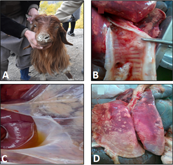
Clinical signs and post mortem examination of naturally infected goats Suspected for CCPP. (A) Mucus plug in nasal cavities, (B) frothy discharges in lower tracheal portion, (C) straw coloured pleural fluid in thoracic cavity, (D) various degree of bilateral hepatisation of lungs.

PPLO broth showing the growth of mycoplasma based on colour change of broth medium (1-4) and 5 negative control.
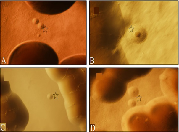
(A, B, C, D) Growth of mycoplasma colonies (colonies with star) on PPLO agar plate on day 4th-7th after the incubation.
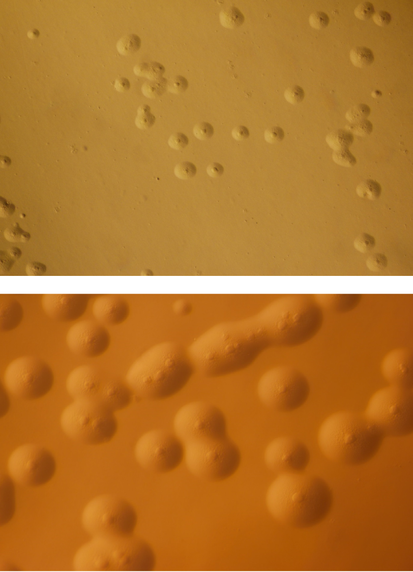
Pure single type of colonies of Mycoplasma mycoides cluster members after 4th passage of the primary culture.
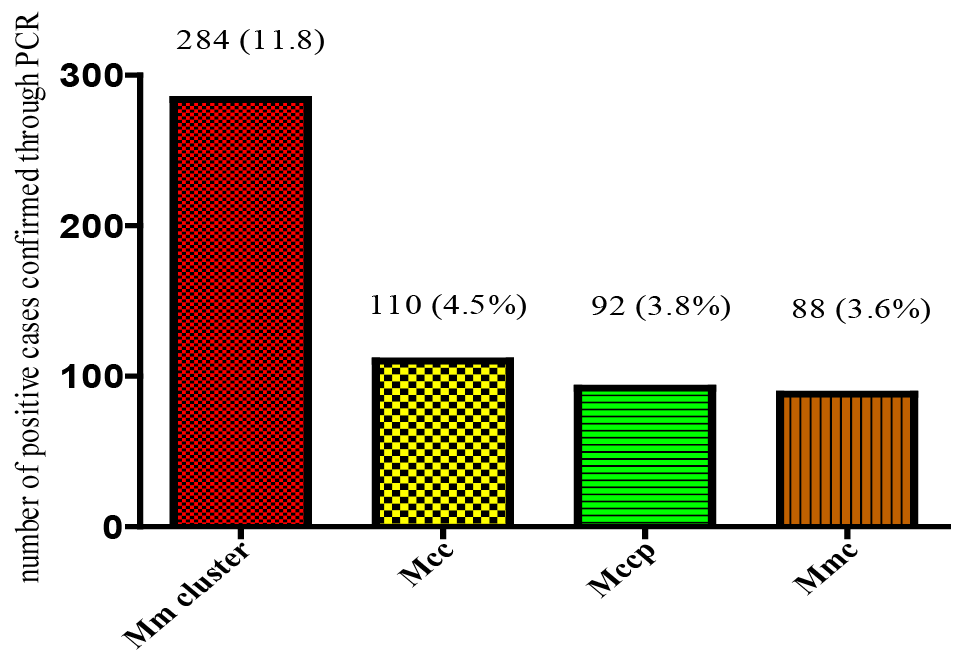
Overall prevalence of the Mycoplasma mycoides cluster and its members in four different zones.
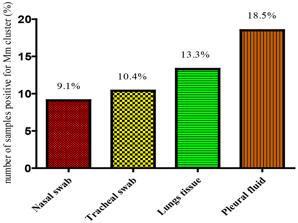
Mycoplasma mycoides cluster recovered from different source of sample. Total number of samples was 2400. Nasal swab n=800, Tracheal swab n=800, Lungs tissue n=400, Pleural fluid n=400.
References
- Abegunde, T., Adler, H., Farver, T. and DaMassa, A., 1981. A serologic survey of Mycoplasma putrefaciens infection in goats. Am. J. Vet. Res., 42: 1798-1801.
- Abrhaley, A., Ejo, M. and Fentie, T., 2019. Seroprevalence and risk factors associated with contagious caprine pleuropneumonia in Western Amhara, Northwest Ethiopia. J. Vet. Med., 2019: Article ID 9878365, 7 pages. https://doi.org/10.1155/2019/9878365[CrossRef]
- Ahmad, F., Khan, H., Khan, F.A., Carson, B.D., Sadique, U., Ahmad, I. and Rehman, H.U., 2021. The first isolation and molecular characterization of Mycoplasma capricolum subsp. capripneumoniae Pakistan strain: A causative agent of contagious caprine pleuropneumonia. J. Microbiol. Immunol. Infect., 54: 710-717. https://doi.org/10.1016/j.jmii.2020.06.002[CrossRef]
- Awan, M., Abbas, F., Yasinzai, M., Nicholas, R., Babar, S., Ayling, R. and Ahmed, Z., 2009. Prevalence of Mycoplasma capricolum subspecie capricolum and Mycoplasma putrefaciens in goats in Pishin district of Balochistan. Pak. Vet. J., 29. http://www.pvj.com.pk/pdf-files/29_4/179-185.pdf
- Awan, M., Abbas, F., Yasinzai, M., Nicholas, R.A., Babar, S., Ayling, R.D. and Khan, F.A., 2010. First report on the molecular prevalence of Mycoplasma capricolum subspecies capripneumoniae (Mccp) in goats the cause of contagious caprine pleuropneumonia (CCPP) in Balochistan province of Pakistan. Mol. Biol. Rep., 37: 3401-3406. https://doi.org/10.1007/s11033-009-9929-0[CrossRef]
- Awan, M., Abbas, F., Yasinzai, M., Tariq, M.M., Bajwa, M., Attique, M. and Shafee, M., 2012. Prevalence of Mycoplasma species by polymerase chain reaction (PCR) directly from the nasal swab samples of goats. Pak. J. Life Soc. Sci., 10: 5-12.
- Ayling, R., Bashiruddin, S. and Nicholas, R., 2004.Mycoplasma species and related organisms isolated from ruminants in Britain between 1990 and 2000. Vet. Rec., 155: 413-416. https://doi.org/10.1136/vr.155.14.413[CrossRef]
- Bascuñana, C.R., Mattsson, J.G., Bölske, G. and Johansson, K.E., 1994. Characterization of the 16S rRNA genes from Mycoplasma sp. strain F38 and development of an identification system based on PCR. J. Bact., 176: 2577-2586. https://doi.org/10.1128/jb.176.9.2577-2586.1994[CrossRef]
- Chazel, M., Tardy, F., Le, Grand, D., Calavas, D. and Poumarat, F., 2010. Mycoplasmoses of ruminants in France: Recent data from the national surveillance network. BMC Vet. Res., 6: 1-8. https://doi.org/10.1186/1746-6148-6-32[CrossRef]
- Cottew, G., Breard, A., DaMassa, A., Ernø, H., Leach, R., Lefevre, P. and Smith, G., 1987. Taxonomy of the Mycoplasma mycoides cluster. Isr. J. med. Sci., 23: 632-635.
- Economic Survey of Pakistan, 2022-23. Finance division, economic advisors wing, Ministry of Finance, Govt. of Pakistan, Islamabad. Chapter-2, pp: 34-35.
- Fauzia, B., Ferhat, A., Awan, M., Aayesha, R., Khan, I., Zafar, A. and Baig, R., 2016. Molecular survey on the prevalence of Caprine mycoplasma in the goats of Quetta City of Pakistan. Int. J. Vet. Sci., 5: 158-163.
- Fischer, A., Shapiro, B., Muriuki, C., Heller, M., Schnee, C., Bongcam-Rudloff, E. and Jores, J., 2012. The origin of the ‘Mycoplasma mycoides cluster’coincides with domestication of ruminants. PLoS One, 7: e36150. https://doi.org/10.1371/journal.pone.0036150[CrossRef]
- Gyles, C.L., Prescott, J.F., Songer, J.G. and Thoen, C.O., 2008. Pathogenesis of bacterial infections in animals. John Wiley and Sons.
- Khan, M.A., Sattar, A., Parveen, S., Rauf, A.M. and Niazi, N., 1989. Mycoplasmosis in Pakistan: A study of contagious caprine pleuropneumonia organism. Pak. J. Vet. Sci., 1: 47-50.
- Kipronoh, A.K., Ombui, J.N., Kiara, H.K., Binepal, Y.S., Gitonga, E. and Wesonga, H.O., 2016. Prevalence of contagious caprine pleuro-pneumonia in pastoral flocks of goats in the Rift Valley region of Kenya. Trop. Anim. Hlth. Prod., 48: 151-155. https://doi.org/10.1007/s11250-015-0934-0[CrossRef]
- Lorenzon, S., Wesonga, H., Ygesu, L., Tekleghiorgis, T., Maikano, Y., Angaya, M. and Thiaucourt, F., 2002. Genetic evolution of Mycoplasma capricolum subsp. capripneumoniae strains and molecular epidemiology of contagious caprine pleuropneumonia by sequencing of locus H2. Vet. Microbiol., 85: 111-123. https://doi.org/10.1016/S0378-1135(01)00509-0[CrossRef]
- Manso-Silván, L., Perrier, X. and Thiaucourt, F., 2007. Phylogeny of the Mycoplasma mycoides cluster based on analysis of five conserved protein-coding sequences and possible implications for the taxonomy of the group. Int. J. Sys. Evol. Microbiol., 57: 2247-2258. https://doi.org/10.1099/ijs.0.64918-0[CrossRef]
- McMartin, D., MacOwan, K. and Swift, L., 1989. A century of classical contagious caprine pleuropneumonia from original description to aetiology. Br. Vet. J., 136: 507-515. https://doi.org/10.1016/S0007-1935(17)32196-6[CrossRef]
- Mekuria, S. and Asmare, K., 2010. Cross-sectional study on contagious caprine pleuro pneumonia in selected districts of sedentary and pastoral production systems in Southern Ethiopia. Trop. Anim. Hlth. Prod., 42: 65-72. https://doi.org/10.1007/s11250-009-9386-8[CrossRef]
- Noah, E., Kusiluka, L., Wambura, P. and Kimera, S., 2011. Field isolation of Mycoplasma capripneumoniae in central zone of Tanzania. Int. J. Anim. Vet. Adv., 3: 434-442.
- Ongor, H., Kalin, R. and Acik, M.N., 2011. Detection of Mycoplasma ovipneumoniae from goats with nasal discharge by culture and polymerase chain reaction. Pak. Vet. J., 31: 244-248.
- Ozdemir, U., Loria, G.R., Godinho, K.S., Samson, R., Churchward, C., Ayling R.D. and Nicholas, R.A.J., 2006. Effect of danofloxacin (Advocin A180) on goats affected with contagious caprine pleuropneumonia. Trop. Anim. Hlth. Prod., 38: 533-540. https://doi.org/10.1007/s11250-006-4427-z[CrossRef]
- Peacock, C., 1996. Improving goat production in the tropics: A manual for development workers: Oxfam. https://doi.org/10.3362/9780855987732.000[CrossRef]
- Rahman, H.U., Saddique, U., Shakoor, A., Shah, M.K., Shah, S.S.A., Khan, F.A. and Rahman, S.U., 2018. The predominant incidence of Mycoplasma mycoides subsp. capri in suspected cases of contagious caprine pleuropneumonia in sheep and goats of northern Pakistan. Pakistan J. Zool, 50: 1995-1998. https://doi.org/10.17582/journal.pjz/2018.50.5.sc9[Full text][CrossRef]
- Regassa, F., Netsere, M. and Tsertse, T., 2010. Sero-prevalence of contagious caprine pleuropneumonia in goat at selected woredas of Afar region. Ethiop. Vet. J., 14: 83-90.
- Sadique, U., Chaudhry, Z., Younas, M., Anjum, A., Hassan, Z., Idrees, M. and Sabtain, S., 2012. Molecular characterization of contagious caprine pleuropneumonia (CCPP) in small ruminants of Khyber Pakhtunkhwa, Pakistan. J. Anim. Pl. Sci., 22: 33-37.
- Samiullah, S., 2013. Contagious caprine pleuropneumonia and its current picture in Pakistan: A review. Veterinarnimedicina, 58: 389-398. https://doi.org/10.17221/6977-VETMED[CrossRef]
- Schumacher, V.L., Hinckley, L., Liao, X., Tulman, E., Geary, S.J. and Smyth, J.A., 2011. Meningitis caused by Mycoplasma mycoides subspecies capri in a goat. J. Vet. Diag. Invest., 23: 565-569. https://doi.org/10.1177/1040638711403413[CrossRef]
- Shah, M.K., Saddique, U., Ahmad, S., Iqbal, A., Ali, A., Shahzad, W. and Shah, S.S.A., 2017. Molecular characterization of local isolates of Mycoplasma capricolum sub species capripneumoniae in goats (Capra hircus) of Khyber Pakhtunkhwa, Pakistan. Pak. Vet. J., 37: 90-94.
- Shahzad, W., Munir, R., Khan, M.S., Shakil, M., Iqbal, M. and Rashid, A., 2012. Characterization, molecular diagnosis and prevalence of caprine mycoplasmosis in different areas of Pakistan. Pakistan J. Zool., 44: 559-568. http://zsp.com.pk/pdf44/559-562%20_36_%20PJZ-792-11%20559%20DD.pdf
- Shahzad, W., Yaqoob, T., Mukhtar, N., Munir, R., Ahmad, R., Khan, M. and Hussain, A., 2016. Sero-prevalence of Mycoplasma capricolum subsp. capripneumoniae in goats through cELISA in different districts of Punjab, Pakistan. J. Anim. Pl. Sci., 26: 931-937.
- Sherif, M., Addis, M. and Tefera, M., 2012. Contagious caprine pleuropneumonia: Serological survey in selected Districts of Jijiga zone, Ethiopia. Asian J. Anim. Sci., 6: 309-315. https://doi.org/10.3923/ajas.2012.309.315[CrossRef]
- Shiferaw, G., Tariku, S., Ayelet, G. and Abebe, Z., 2006. Contagious caprine pleuropneumonia and Mannheimia haemolytica-associated acute respiratory disease of goats and sheep in Afar Region, Ethiopia. Rev. Sci. Tech. Off. Int. Epizoot., 25: 1153. https://doi.org/10.20506/rst.25.3.1723[CrossRef]
- Studdert, V., Gay, C. and Blood, D., 2007. Saunders comprehensive veterinary dictionary 3rd. Saunders Elsevier, St. Louis, Missouri, USA.
- Thiaucourt, F. and Bolske, G., 1996. Contagious caprine pleuropneumonia and other pulmonary mycoplasmoses of sheep and goats. Rev. Sci. Tech. Off. Int. Epizoot., 15: 1397-1414. https://doi.org/10.20506/rst.15.4.990[CrossRef]
- Tigga, M., Choudhary, B., Ghosh, R. and Malik, P., 2014. Mycoplasmosis: An emerging threat to developing livestock industry. Int. J. Adv. Res., 2: 558-564.
- Verbisck-Bucker, G., González-Candela, M., Galián, J., Martín-Atance, P. and León-Vizcaíno, L., 2008. Epidemiology of Mycoplasma agalactiae infection in free-ranging Spanish ibex (Capra pyrenaica) in Andalusia, southern Spain. J. Wildl. Dis., 44: 369-380. https://doi.org/10.7589/0090-3558-44.2.369[CrossRef]
- Wang, H., Ni, L., Yang, H., Xu, L., Ma, N. and Ding, H., 2014. Isolation and identification of Mycoplasma mycoides cluster strains from goats in Chongqing, China. Bull. Vet. Inst. Pulawy, 58: 11-15. https://doi.org/10.2478/bvip-2014-0002[CrossRef]
- Wazir, I., Hussain, I., Khan, M.A., Ali, M.I., Rahman, H.U., Ashraf, F. and Ullah, Q., 2016. Seroepidemiological analysis of contagious caprine pleuropneumonia through cELISA in selected districts of Khyber Pakhtunkhwa-Pakistan. Am. Sci. Res. J. Eng. Tech. Sci., 26: 274-281.
- WOAH, 2018. Contagious caprine pleuropneumonia, OIE Terrestrial Manual. Chapter 2.7.6. Office International Des Epizootics, Paris, France. pp. 704-719.
- Yousuf, E., Melaku, A. and Bogale, B., 2012. Seroprevalence of contagious caprine pleuropneumonia in Dire Dawa provisional administrative council, Eastern Ethiopia. J. Vet. Med. Anim. Hlth., 4: 93-96.
Services
Article usage statistics
Loading…