ResearchersLinks

Research Article · Volume 12, Issue 1, 10 January 2024 · Open Access

Detection of ESBL Encoding Gene and the Virulence Factors of Klebsiella pneumoniae from Dairy Cattle Farms in South Sulawesi

· ·

Affiliations & Notes ⌄
  1. 1Postgraduate Student of Medical Microbiology, School of Veterinary Medicine and Biomedical Sciences, IPB University, Bogor, 16680, Indonesia
  2. 2Division of Medical Microbiology, School of Veterinary Medicine and Biomedical Sciences, IPB University, Bogor, 16680, Indonesia. *Correspondence | Safika Safika, Division of Medical Microbiology, School of Veterinary Medicine and Biomedical Sciences, IPB University, Bogor, 16680, Indonesia
  3. 3Email: safika@apps.ipb.ac.id
Article Info ⌄
Journal:
Advances in Animal and Veterinary Sciences
Published:
10 January 2024

From the article

  • Three isolates that encoded the blaSHV gene and were expressed in udder rinsed water and udder swabs (40% and 25%, respectively).
  • The frequency of this gene is lower than the other two ESBL coding genes in K. pneumoniae.
  • In conclusion, K. pneumoniae isolated in South Sulawesi dairy cattle farms indicated the presence of the ESBL and virulence factors gene.
  • All of the samples of water sources, udder rinses water, and udder swabs have the potential for gene transfer between species of ubiquitous bacteria in the same environment.

Selected from the article's own text.

Abstract

One of the multi-resistance mechanisms commonly found in dairy farms is ESBL (Extended Spectrum Beta Lactamase). This study aimed to investigate the antibiotic resistance and virulence factors of Klebsiella pneumoniae from Dairy Farm isolates in South Sulawesi, Indonesia. Twelve isolates obtained in the field were tested for phenotype resistance profile with four antibiotics of β-lactams using the Kirby-Bauer method. The Polymerase Chain Reaction (PCR) was used to detect the resistance genes and virulence factors of K. pneumoniae. Samples isolated from water sources showed a resistance profile to cefotaxime (100%), ceftazidime, ampicillin, and amoxicillin (50%). Samples isolated from udder rinses water were resistant to amoxicillin, cefotaxime, ampicillin (100%), and ceftazidime (60%). Samples obtained from milker hand swabs results were resistant to amoxicillin, cefotaxime, and ampicillin (100%). The cattle udder swab samples showed resistance to ampicillin and amoxicillin (100%), cefotaxime (75%), and ceftazidime (50%). Gene resistance detection found that water samples encoded blaTEM gene (100%), Udder rinses water encoded blaTEM (80%), blaSHV (20%), blaCTX-M (20%) and udder swab samples detected the presence of blaTEM genes (40%). K. pneumoniae virulence factor genes: mrkD detected in all isolates (100%), entb and wabG were found in all water sources, milker hand, and udder swabs samples (100%) except samples from udder rinses water that encoded gene virulence factors 60% and 80%, respectively. All samples were negative for rmpA and magA genes. The study results showed K. pneumoniae of Dairy farms indicated the presence of ESBL resistance and virulence factors genes with different frequencies.
 
Keywords | β-lactam, Dairy, ESBL, K. pneumoniae, Virulence factors, Resistance
INTRODUCTION

An increase in domestic consumption of milk and its products may improve the national economy and face international trade competition (Jahroh et al., 2020). The growth of the middle and upper class as potential consumers is considered to have played a role in increasing the consumption of milk by the public . Nevertheless, until recently Indonesia’s dairy industry has been dominated by smallholder farmers . One of the obstacles related to the quality of products produced by community-scale dairy farms is hygiene which includes increased incidence of mastitis, the lack of availability of clean water, and contamination of milk . Environmental bacteria such as E. coli, E. feundeii, E. aerugenes, and K. pneumoniae can lead to a decrease in the quality of dairy products and exacerbate the infection (Zalizar et al., 2018).

K. pneumoniae is an opportunistic pathogenic bacteria from the Enterobacteriaceae family, Gram-negative, short rod-shaped, and has a capsule . This bacteria is responsible for infection reports in hospitals which are associated with compromised immune systems, exacerbate local infections, and agents of foodborne illness ; . Virulence factors are naturally encoded in bacteria and have the ability to cause clinical symptoms which help the process of bacterial attachment, invasion, colonization, and the formation of biofilms . The existence of virulence factors is exacerbated by an increase in the ability of bacterial resistance. K. pneumoniae as a health-threatening bacteria has ability to acquire Mobile Genetic Element (MGE) which encodes antibiotic resistance genes.

Enterobacteriaceae bacteria, particularly K. pneumoniae and E. coli are environmental bacteria that encode ESBL gene. ESBL-producing bacteria can hydrolyze β-lactam rings and make antibiotic works ineffectively . In general, ESBL-producing bacteria are encoded by 3 main genes: Cefotaxime-Munich (CTX-M), Sulfhydryl variable (SHV), and Temoneira Enzyme (TEM). ESK(C)APE is an acronym for several names of Gram-negative and Gram-positive bacteria (Enterococcus faecium, Staphylococcus aureus, Klebsiella pneumoniae, Clostridium difficile, Acinetobacter baumannii, Pseudomonas aeruginosa, and Enterobacter species) that are capable of developing Multidrug-Resistant which recorded an increase rapidly in the last decade and can increase the potential for bacterial infection and reduces the choice of treatment therapy ; WHO, 2021).

Livestock acts as a potential reservoir in the environment from humans and animals (Ibebkweet al., 2023). The research conducted by Podschun et al (2001) shows that the strains in the field are as virulent as clinical isolates. The existence of antibiotics resistance genes and virulence factors that are encoded naturally by K. pneumoniae have ability to develop resistance and makes these bacteria more difficult to treat. Research over the decade was extensively done with K. pneumoniae as ESBL-producing bacteria isolated from human and clinical isolates. However, research related to antibiotic resistance in the environment and dairy farms especially in β-lactam antibiotics as an important factor, is still lacking. This study aims to identify K. pneumoniae bacteria that show resistance to several β-lactam antibiotics and virulence factors that support bacteria to attack the host potentially. This study reports the K. pneumoniae ESBL resistance profile and virulence factors gene of K. pneumoniae isolated from dairy farms in South Sulawesi, Indonesia.

MATERIALS AND METHODS

Study area

This research was located at a total of five dairy farms that are located close to each other in Enrekang Regency South Sulawesi Province. These 5 farms are still traditional community-scale farms with a total population of no more than 10 dairy cows in each farm. The research was conducted time from December 2022 to August 2023.

Sample collection

The samples from this research were collected in the morning to minimize bacterial contamination before milking with a description of the total sample as follows: udder swabs from cattle (n=11) from 5 farms, samples from the environment in the form of water sources (n=5) and udder rinse water (n=11) and skin swab from dairy farmers (n=5). All of the water samples were taken using a bottle with a volume of 250 ml while the swab sample was taken using a transport swab (Amies). Swab samples on the milker’s hand are taken from the entire surface of the hand and between the fingers and for each udder swab sample is taken from 4 quarters of the udder. The samples were sealed using wrap and taken using the coolbox at 4°C for further bacteria culture and analysis at the Research Laboratory, Division of Medical Microbiology, School of Veterinary Medicine and Biomedicine Sciences, IPB University.

Biochemical analysis and molecular confirmation of K. pneumoniae bacteria

Samples were isolated with Macconkey Agar (MAC) (Oxoid, UK) as differential selective media and for 24 hours were incubated at 37°C. Macroscopically, K. pneumoniae bacteria colonies are characterized by their ability to ferment glucose and mucoid . The bacteria were then cultivated using Tryptic Soy Agar (TSA) media (Oxoid, UK). Cultured bacteria from TSA media continued for iMVIC test (simmon citrate, methyl red, indole, and voges-proskauer). Confirmation of suspected bacteria (K. pneumoniae) was done molecularly by finding the presence of rpoB gene. Forward primer: AACCAGTTCCGCGTTGGCCTGG and Reverse primer: CCTGAACAACACGCTCGGA .

Antibiotic sensitivity test (AST)

The Kirby-Bauer method was used as phenotype confirmation of antibiotic sensitivity test in this study. The suspension was homogenized and the result of the turbidity was adjusted to McFarland standard solution (0.5 or 1, 5 x 10⁸ CFU/ml). The suspension was spread into Mueller Hinton Agar (MHA) using glass spreader and placing paper discs containing antibiotics with certain concentrations (Oxoid, UK) to incubated at 37°C for 18 hours. Ampicillin (10µg/disk), amoxicillin (10µg/disk), cefotaxime (30µg/disk), and ceftazidime (10µg/disk). After incubation, the clear zone that formed was then measured by vernier calipers and adjusted according to the CLSI Standard (2021).

Table 1: Genes used in research.

Gene Target

Nucleotide sequence (5’-3’)

Amplicon (bp)

Annealing (°C)

Reference

rpoB

F:AACCAGTTCCGCGTTGGCCTGG

R: CCTGAACAACACGCTCGGA

1090

54

Almeida et al. (2023)

blaSHV

F: CCTGTTAGCCACCCTGCC

R: CCGCAGATAAATCACCAC

768

52

Momtaz et al. (2012)

blaTEM

F: ATCAGCAATAAACCAGC

R: CCCCGAAGAACGTTTTC

516

54

Colom et al. (2003)

blaCTX-M

F: ATGATGAAAAAATCGTTATGC

R: CAGCATCTCCCAGCCTAAT

551

57

Lyimo et al. (2016)

magA

F: GGTGCTCTTTACATCATTGC

R: GCAATGGCCATTTGCGTTAG

1.283

59

El-Fertas Aissani et al. (2016)

rmpA

F: ACTGGGCTACCTCTGCTTCA

R: CTTGCATGAGCCATCTTTCA

535

50

mrkD

F: CCACCAACTATTCCCTCGAA

R: ATGGAACCCACATCGACATT

240

54

entB

F: ATTTCCTCAACTTCTGGGGC

R: AGCATCGGTGGCGGTGGTCA

371

57

wabG

F: CGGACTGGCAGATCCATATC

R: ACCATCGGCCATTTGATAGA

683

54

Brisse et al. (2009)

DNA extraction

Extraction of bacterial DNA was done by using the boiling method with several modifications. Colonies bacteria that incubated were cultured to the Tryptic Soy Broth (TSB) media and continued to be centrifuged with 25,000 rpm for 30 minutes. Pellets from the centrifuged process were added to nuclease-free water with a total volume of 100 µl and placed in the water bath for 6 minutes; at 95-100°C and vortexed. The suspension was centrifuged at 12,000 rpm for 7 minutes. Supernatant was taken as master stock DNA.

Detection of virulence factors and antimicrobial resistance genes of K. pneumoniae

Detection of resistance β-lactams genes in this study using PCR focused on detecting the blaTEM, blaCTX-M, and blaSHV as the main genes of ESBL. The reaction volume was made up of 12 µl consisting of 6 µl MyTaqTM HS RedMix (Bioline), 1 µl for each primer consisting of Forward and Reverse (Genetika Science), 2 µl dH2O (DNase, RNAse free) (Invitrogen™, USA), and made up to 12 µl using 2 µl as DNA template. The Reaction of PCR was done in a Thermal Cycler T100TM (Bio-Rad, California, USA) and visualized on 1% agarose. The electrophoresis was performed for 35 minutes at 60 volts using 5 µl samples and 1 µl FloroSafe DNA Stain (1st base). All of the primers used in this study are listed in Table 1.

Amplification resistance gene using protocol as follows: blaTEM: 95°C for 1 minute, followed by 30 cycles that consisting of 95°C for 15 seconds, 54°C for 15 seconds, 72°C for 1 minute and final elongation 72°C for 10 minutes; blaCTX-M: 95°C for 5 minutes, followed by 30 cycles, consisting of 95°C for 30 seconds, 57°C for 1 minute, 72°C for 1 minute, and final elongation 72°C for 5 minutes; blaSHV: 95°C for 5 minutes, followed by 30 cycles that consisting of 95°C for 30 seconds, 52°C for 1 minute seconds, 72°C for 45 seconds, and final elongation 72°C for 5 minutes.

Amplification virulence factors under the condition as follows: rmpA, magA, mrkD, and entB: denaturations start at 94°C for 4 minutes, followed by 30 cycles that consisting of 94°C for 30 seconds, annealings a range of 54-59°C, 72°C for 1 minute, and final elongation at 72°C for 10 minutes; wabG gene detection condition: denaturation at 94°C for 5 minutes followed by 35 cycles that consisting of 94°C for 30 seconds, 54°C for 30 seconds, 72°C for 1 minute and final elongation for 1 minute.

Data analysis

The study result of phenotype confirmation by kirby-bauer method, virulence factors, and resistance genes of K. pneumoniae are shown with tables and analyzed descriptively.

RESULT AND DISCUSSION

Biochemical and molecular confirmation result

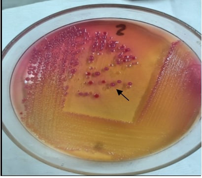
Twelve out of a total of thirty-two isolates obtained at the Dairy farm, Enrekang Regency, South Sulawesi were positive for K. pneumoniae bacteria. The colonies of klebsiella sp are shown in Figure 1. The results of biochemical confirmation are shown in Table 2. This accordance with researches by Effendi et al. (2018),

Table 2: Biochemical test interpretation of suspect Klebsiella sp.

Test result

Methyl red

Indole

Simmon’s citrate

Voges proskauer

TSIA

Suspect klebsiella sp.

-

-

+

+

A/A, (+) Gas, (-) H2S

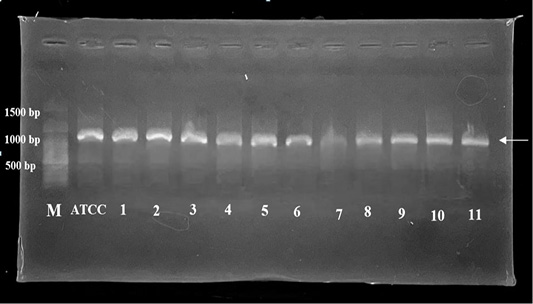
Dita et al. (2019); Salaudin et al. (2019), Permatasari et al. (2020). Bacteria that were suspected as K. pneumoniae biochemically continue to be confirmed by detecting the rpoB gene which is known to encode RNA polymerase β-subunit . Relatively, detection using rpoB is a more appropriate way than using the 16rRNA gene. Some researchers suggest that K. pneumoniae detection is effective by using the rpoB gene ; . The amplicon of rpoB gene was interpreted in Figure 2. From this results, it was found that the dominant isolate of K. pneumoniae was from water sources on farms. This was based on K. pneumoniae bacteria which are ubiquitous in aquatic environments such drinking water ; , Lakes , wastewater , Rivers ; and Seawater .

Table 3: β-lactam antibiotic resistance profile of K. pneumoniae isolated in dairy farm.

No

Samples code

β-lactam antibiotics

AMX

AMP

CTX

CAZ

Water sources

1

4.2

S

I

R

S

2

10.2

R

R

R

R

n = 2

1

1

2

1

50%

50%

100%

50%

Udder rinse water

1

2.3

R

R

R

R

2

6.1

R

R

R

S

3

14.2

R

R

R

I

4

15.1

R

R

R

R

5

16.3

R

R

R

R

n = 5

5

5

5

3

100%

100%

100%

60%

Milker hand swab

1

J.2

R

R

R

S

n = 1

100%

100%

100%

0

Udder swabs

1

B.3

R

R

R

S

2

E.2

R

R

I

S

3

H.1

R

R

R

R

4

O.3

R

R

R

R

n = 4

100%

100%

75%

50%

S: Susceptible; I: Intermediete; R: Resistant

Antibiotic resistance

Twelve isolates were confirmed as K. pneumoniae by PCR and then tested to show the profile resistance with the four β-lactam antibiotics given. Phenotypic confirmation of ESBL-producing bacteria by the Kirby-Bauer Disc method showed a fairly high resistance status. The clear zone of inhibition that formed was adjusted per the CLSI Standard (2021) to determine the antibiotic sensitivity. The phenotype confirmation of β-lactam resistance results are shown in Table 3. Samples from water sources showed that K. pneumoniae were resistant to ceftazidime, ampicillin, amoxicillin, (50%), and cefotaxime (100%). The udder rinses water signified resistance condition to cefotaxime, ampicillin, amoxicillin (100%), and ceftazidime (60%). Milker hand swab samples indicated resistance to cefotaxime, ampicillin, and amoxicillin (100%). The cattle udder swab samples represented resistance to amoxicillin, ampicillin (100%), cefotaxime (75%), and ceftazidime (50%). In general, the resistance of β-lactam antibiotics in isolates shows a percentage above 50%. Reports of β-lactam resistant bacteria have been a concern this decade. The presence of high-resistance bacteria was shown in research by Jelic et al. (2019) found K. pneumoniae isolated from water samples were resistant to β-lactam, specifically to penicillin and all cephalosporin generations. Geographically, the 5 farms that are used as sampling sites are located in adjacent areas that may allow the spread of resistance genes from one source to another place. Water is one of the reservoirs that have the ability to spread resistance genes from the livestock to the environment which live ubiquitously in aquatic environments .

In general, the process of β-lactam binding to the specific site results in blocking the activity of transpeptidase enzyme. Veterinary medicine reports widely used β-lactam antibiotics because of their high specificity, lower toxicity, and generally good bactericidal effect . The use of antibiotic therapy for dairy cows on farms was dominated by β-lactams belonging to the cephalosporin group ; ; FDA, 2022) for clinical mastitis and respiratory infections caused by Klebsiella spp. use the ampicillin, tetracycline, and oxytetracycline (FDA, 2022). In addition, A report by Schrag et al. (2020) showed the use of cephalosporin and penicillin in dairy farm have a higher frequency than other antibiotics for mastitis treatment.

Enterobacteriaceae bacteria has a natural ability to hydrolyze β-lactam ring from several antibiotics by producing Extended Spectrum Beta Lactamase (ESBL) enzyme . This enzyme is coded chromosomally and plasmid. In general, resistance in K. pneumoniae involves inactivation and enzymatic modification by bacteria, changes in antibiotic targets, porin mutations, increased efflux pump expression, and formation of biofilm . Some cases of K. pneumoniae as ESBL bacteria have been found in hospitals , zoos , animal products , pets , and dairy farms ; . The natural ability of K. pneumoniae to raise resistance combined with the ubiquitous habitat such as soil, vegetation, and water makes these bacteria have a higher potential for public health risks.

Antibiotic resistance gene

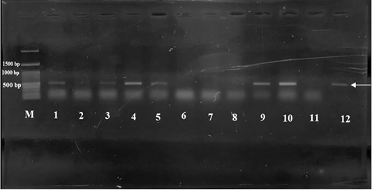
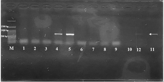
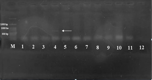
Confirmation of antibiotic resistance gene using blaTEM, blaSHV, and blaCTX-M. This study found that the frequency of gene presence from water sources, udder rinses water, and udder swabs were as follows: blaTEM (100%; 80%, 50%), blaSHV (0; 40%; 25%), blaCTX-M (0; 20%; 0) and the milker hand swab samples did not show any resistance gene from this study (Figures 3, 4, 5, Table 4). The blaTEM gene has the highest frequency compared to all the genes detected in the isolates. The blaTEM gene is encoded by a plasmid, responsible for resistance in the new generation of antibiotics, and was the main cause of pathogenesis of clinical isolates such as Urinary Tract Infection and resistance to antibiotics from K. pneumoniae (Sarshar et al., 2021). Previously, studies reported high frequencies in food-producing and clinical animal samples (90% and 100%, respectively) ; .

Table 4: Profile of rpoB, genes encoding ESBL and virulence factors of K. Pneumoniae.

No

Samples code

rpoB

ESBL Coding gene

Virulence factors of K. pneumoniae

blaTEM

blaSHV

blaCTX-M

rmpA

magA

mrkD

entB

wabG

Water sources

1

4.2

+

+

-

-

-

-

+

+

+

2

10.2

+

+

-

-

-

-

+

+

+

n = 2

2/2

100%

2/2

100%

0

0

0

0

0

0

0

0

2/2

100%

2/2

100%

2/2

100%

Udder rinses water

1

2.3

+

+

-

-

-

-

+

-

-

2

6.1

+

+

+

-

-

-

+

-

+

3

14.2

+

-

-

-

-

-

+

+

+

4

15.1

+

+

-

-

-

-

+

+

+

5

16.3

+

+

+

+

-

-

+

+

+

n = 5

5/5

100%

4/5

80%

2/5

40%

1/5

20%

0

0

0

0

5/5

100%

3/5

60%

4/5

80%

Milker hand swab

1

J.2

+

-

-

-

-

-

+

+

+

n = 1

Udder swabs

1/1

100%

0

0

0

0

0

0

0

0

0

0

1/1

100%

1/1

100%

1/1

100%

1

B.3

+

-

-

-

-

-

+

+

+

2

E.2

+

+

-

-

-

-

+

+

+

3

H.1

+

+

+

-

-

-

+

+

+

4

O.3

+

-

-

-

-

-

+

+

+

n = 4

4/4

100%

2/4

50%

1/4

25%

0

0

0

0

0

0

4/4

100%

4/4

100%

4/4

100%

(+): Positive result for the gene which being tested; (-): Negative result.

Another gene of resistance that was also found in this study is blaSHV gene. Three isolates that encoded the blaSHV gene and were expressed in udder rinsed water and udder swabs (40% and 25%, respectively). In addition to the water, the udder of dairy cattle can allow the transmission of bacteria that carry resistant genes from the environment to humans . The cattle shed floor is one of the common contamination from coliform bacteria in the environment . The blaSHV gene is responsible for resistance to the antibiotics ceftazidime, ampicillin, and penicillin .

Previous studies show that the CTX-M gene was found in clinical isolates of pneumonia patients , livestock, and slaughterhouses in Asia and is responsible for the resistance of the cephalosporin antibiotic group . Over time, K. pneumoniae that produces blaCTX-M increased by 1.7% during 2005-2009 to 26.4% during 2010-2012. This is inconsistent with the results of a study that found that there is just one isolate (20%) encoded the blaCTX-M gene from all of the samples. The frequency of this gene is lower than the other two ESBL coding genes in K. pneumoniae. Other supporting research is from Wang et al. (2013) which discovered only 2 isolates (1.7%) of 121 ESBL bacteria encode the blaCTX-M gene found in clinical samples of K. pneumoniae.

Research conducted by Imasari et al. (2017) found there was a transmission of transfer of ESBL bacteria in the dairy farming environment and cattle breeders by 79.1% through direct or indirect contact. A fairly high percentage indicate the potential for cross-transfer from the environment to livestock. K. pneumoniae is capable of obtaining the Mobile Genetic Elements (MGE) as plasmids that contain antibiotic resistance genes. The undetected genes in this study indicates that several possibilities affect the expression gene such as other resistance genes that were not tested in the study, mutations in the genes, and low in vitro expression .

Virulence factors gene

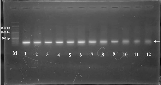
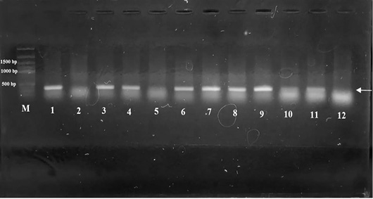
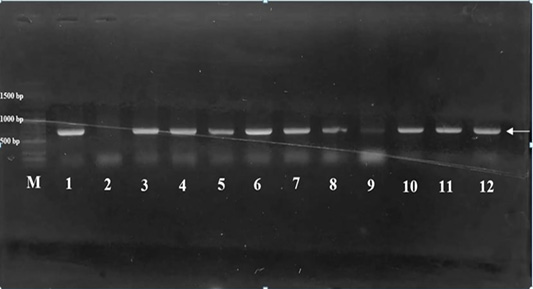
Five genes of virulence factor were tested in this study: rmpA, magA, mrkD, entB, and wabG. The study result shows the prevalence of K. pneumoniae isolates encoding the mrkD gene was 100% in all types of samples. The gene of wabG and entB gene detection for water sources, milker hand swabs, and udder swabs had a percentage of 100% each while the same gene in udder rinses water samples showed percentages of 60% and 80%, respectively. All the isolates show negative results of rmpA and magA detection (Figures 6, 7, 8, Table 4).

The pathogenicity of K. pneumoniae has a major role in increasing infection in the host. The mrkD gene encodes cell surface factor and can mediate the attachment of biotic and abiotic surfaces and adequate biofilm formation which can inhibit the efficiency of antibiotic therapy . Several studies indicate a higher prevalence of mrkD gene in field and clinical isolates and it has been widely studied ; ; . The wabG gene is responsible for LPS formation and plays a role in clinical symptoms such as sepsis and immune modulation during infection . The enterobactin (entB) gene acts as the main system of iron uptake in K. pneumoniae and maintains the life of bacteria in the host . Through this study, it did not detect the presence of the magA and rmpA genes. The magA gene is encoded chromosomally and prevents the bacteria from being recognized by the immune system whereas rmpA is encoded by plasmid, and acts as a regulator of extracapsular polysaccharide . This is presumably because the rmpA and magA genes are commonly found in clinical isolates that lead to liver abscesses, invasive infections and as markers of hvKP ; . 

Antimicrobial resistance (AMR) due to excessive and incorrect use of human, environmental, and animal has become a serious global health threat The presence of K. pneumoniae bacteria that have ESBL coding genes from field samples is a potential concern for farmers and can increase the contribution of bacteria to affect host tissues . Resistance that is related to the plasmids occurs in the environment which can also carry virulence factor determinants through horizontal transmission . Commensal and pathogenic K. pneumoniae show a diversity of geographical/climatic conditions, use of antibiotics, and interactions between bacterial species cause bacteria to acquire virulence factor genes and have the potential to become pathogens. The presence of virulence factors and resistance genes and their relations have been conducted by some researchers ; ; .

CONCLUSIONs and Recommendations

In conclusion, K. pneumoniae isolated in South Sulawesi dairy cattle farms indicated the presence of the ESBL and virulence factors gene. All of the samples of water sources, udder rinses water, and udder swabs have the potential for gene transfer between species of ubiquitous bacteria in the same environment. The existence of virulence factors present in all samples is a concern because of its affect on effectiveness of antibiotics as well as its capabilty to increase the virulence of bacteria. 

ACKNOWLEDGEMENT

The authors give the highest gratitude to the School of Veterinary Medicine and Biomedical Sciences and Dinas Peternakan dan Perikanan Kabupaten Enrekang, South Sulawesi, Indonesia for all of the facilities that were given to the author during the research.

NOVELTY STATEMENT

The study is the first time an ESBL resistance test of K. pneumoniae isolated from dairy farms in Enrekang, South Sulawesi.

The first study conducted to detect ESBL resistance genes and virulence factors genes of K. pneumoniae isolated from dairy farms in Enrekang, South Sulawesi

AUTHOR’S CONTRIBUTION

NM contributed to collecting samples, doing research, data analysis, and preparing the manuscript. SS and AI contributed to revising the manuscript and supervised the research.

Ethical approval

Ethical approval on this study was obtained from Health Research Ethics Commission of Hasanuddin University Hospital Number: 105/UN4.6.4.5.31/PP36/2021.

Conflict of interest

The authors have declared no conflict of interest.

Figures

Figure 1:
Figure 1:.
K. pneumoniae bacteria shown mucoid, pink and convex colonies in MAC media. Arrow: single colonies of K. pneumoniae.
Figure 2:
Figure 2:.
Electrophoresis results on molecular confirmation of K. pneumoniae (1090 bp). M: Marker; ATCC: positive control; 1-11: positive isolates of K. pneumoniae.
Figure 3:
Figure 3:.

Detection of amplicon blaTEM gene (516 bp). M: 100 bp DNA Marker. Isolates 1-5,9,10,12 were positives for blaTEM gene.

Figure 4:
Figure 4:.

Detection of amplicon blaSHV gene (768 bp). M: 100 bp DNA Marker. Isolates 4,5, and 12 were positives for blaSHV genes.

Figure 5:
Figure 5:.

Detection of amplicon blaCTX-M gene (551 bp). M: 100 bp DNA Marker. Isolate 4 were positive for blaCTX-M gene.

Figure 6:
Figure 6:.

Detection of amplicon mrkD gene (240 bp). M: 100 bp DNA Marker.  Isolates 1-12 were positives for mrkD gene.

Figure 7:
Figure 7:.

Detection of amplicon entB gene (371 bp). M: 100 bp DNA Marker. Isolates 1,3,4,6-11 were positives for entB gene.

Figure 8:
Figure 8:.

Detection of amplicon wabG gene (683 bp). M: 100 bp DNA Marker. Isolates 1, 3-12 were positives for wabG gene.

References

  1. Ahmadi M, Benzadi P, Ranjbar R (2021). Virulence factors, antibiotic resistance patterns, and molecular types of clinical isolates of Klebsiella pneumoniae. Exp. Rev. Anti Infect. Ther., 20(3): 463-472. https://doi.org/10.1080/14787210.2022.1990040[CrossRef]
  2. Almeida OAC, Urajo NO, Mulato ATN, Persinoti GF, Sforca ML, Calderan-Rodrigues MJ, Oiveira JVC (2023). Bacterial volatile organic compounds (VOCs) promote growth and induce metabolic changes in rice. Front. Plant Sci., pp. 134. https://doi.org/10.3389/fpls.2022.1056082[CrossRef]
  3. Arafa AA, Hedia RH, Dorgham SM, Ibrahim ES, Bakry MA, Abdalhamed AM dan Abuelnaga ASM (2022). Determination of extended-spectrum β-lactamase-producing Klebsiella pneumoniae isolated from horses with respiratory manifestation. Vet. World, 15(4): 827-833. https://doi.org/10.14202/vetworld.2022.827-833[CrossRef]
  4. Aromolaran O, Amodu OA (2021). Antibiotic susceptibility pattern of Klebsiella pneumoniae and Pseudomonas aeruginosa isolated from some drinking wells in Ondo town southwest Nigeria. J. Appl. Sci. Environ. Manage., 25(1): 59-63. https://doi.org/10.4314/jasem.v25i1.8[CrossRef]
  5. Bakhtiari R, Javadi A, Aminzadeh M, Molaee-Aghaee, Shaffaghat Z (2021). Association between presence of rmpA, mrkA, and mrkD genes and antibiotic resistance in clinical Klebsiella pneumoniae isolates from hospitals in Tehran, Iran. Iran. J. Publ. Hlth., 50(5): 1009-016. https://doi.org/10.18502/ijph.v50i5.6118[CrossRef]
  6. Bartley PS, Domitrovic TN, Moretto VT, Santos CS, Ponce-Terashima R, Reis MG, Barbosa LM, Blanton RE, RA Bonomo, dan F Perez (2019). Antibiotic resistance in Enterobacteriaceae from surface waters in Urban Brazil highlights the risks of poor sanitation. Am. J. Trop. Med. Hyg., 100(6): 1369-1377. https://doi.org/10.4269/ajtmh.18-0726[CrossRef]
  7. Brisse S, Fevre C, Passet V, Issenhuth-Jeanjean S, Tournebize R, Diancourt L, Grimont P (2009). Virulent clones of Klebsiella pneumoniae: identification and evolutionary scenario based on genomic and phenotypic characterization. PLoS One, 4(3): 1-13. https://doi.org/10.1371/journal.pone.0004982[CrossRef]
  8. Chang D, Sharma L, Cruz CSD, Zhang D (2021). Clinical epidemiology, risk factors, and control strategies of Klebsiella pneumoniae infection. Front. Microbiol., Sec. Infect. Agents Dis., pp. 2012-2021. https://doi.org/10.3389/fmicb.2021.750662[CrossRef]
  9. Chilupuri P, Gudisi S, Prakash K (2021). Detection of mrkD gene in clinical isolates of biofilm producing Klebsiella pneumoniae. Medpulse Int. J. Microbiol., 19(2): 25-28. https://doi.org/10.26611/10081922[CrossRef]
  10. CLSI (Clinical and Laboratory Standards Institute). 2021. Performance Standard for Antimicrobial Susceptibility Testing: 31st Edition. Partners available at https://clsi.org/about/press-releases/clsi-publishes-m100-performance-standards-for-antimicrobial-susceptibility-testing-31st-edition/ (accessed 11 January 2023).
  11. Darmawan T (2013). Marketing strategy and competitiveness of local milk production towards ASEAN economic community 2015. Development of partnership structure of small-medium enterprise milk industry towards ASEAN Economic Community 2015. Bogor (Indonesia): ICARD.
  12. Dita RF, Agustina D, Rachmawati DA, Suswati E, Mufida DC, Shodikin MA (2019). Peran protein pili 38,6 kda Klebsiella pneumoniae sebagai protein hemaglutinin dan adhesin yang berfungsi sebagai faktor virulensi. J. Agromed., 5(2): 69-75.
  13. Diwyanto K, Iskandar S (1999). Livestock industries of Indonesia prior to the asian financial crisis [Internet]. Available from: https://www.fao.org/3/ab986e/ab986e.pdf (accessed 01 August 2023).
  14. Dong L, Meng L, Liu H, Wu H, Hu H, Zheng N, Wang J, dan Schroyen M (2020). Effect of therapeutic administration of β-lactam antibiotics on the bacterial community and antibiotic resistance patterns in milk. J. Dairy Sci., 104: 7018–7025. https://doi.org/10.3168/jds.2020-20025[CrossRef]
  15. Effah CY, Sun T, Liu S, Wu Y (2020). Klebsiella pneumoniae: An increasing threat to public health. Ann. Clin. Microbiol. Antimicrobiol., 19(1): 1-9. https://doi.org/10.1186/s12941-019-0343-8[CrossRef]
  16. Effendi MH, Bintari IG, Aksonoc EB, Hermawan IP (2018). Detection of blaTEM Gene of Klebsiella pneumoniae Isolated from Swab of Food Producing Animals in East Java. Trop. Anim. Sci. J., 41(3): 174-178. https://doi.org/10.5398/tasj.2018.41.3.174[CrossRef]
  17. El Fertas-Aissani R, Messai Y, Alouach S, Bakour R (2013). Virulence profiles and antibiotic susceptibility patterns of Klebsiella pneumoniae strains isolated from different clinical specimens. Pathol. Biol., (61): 209–211. https://doi.org/10.1016/j.patbio.2012.10.004[CrossRef]
  18. Enferad E, S Mahdavi (2020). Antibiotic resistance pattern and frequency of some beta lactamase genes in Klebsiella pneumoniae isolated from raw milk samples in Iran. J. Hell Vet. Med. Soc., 71(4): 2455-2462. https://doi.org/10.12681/jhvms.25925[CrossRef]
  19. FDA (Food Drug Administration) (2022). Antimicrobial use and resistance in animal agriculture in the United States: 2016-2019. Partners available at https://www.fda.gov/media/159544/download (accessed 15 January 2023).
  20. Gelalcha BD, Dego OK (2022). Extended-spectrum beta-lactamases producing enterobacteriaceae in the USA dairy cattle farms and implications for public health. Antibiotics, 11: 1313. https://doi.org/10.3390/antibiotics11101313[CrossRef]
  21. Guntoro, Budi, Rakhman, Nur A, Suranindyah, Yuni Y (2016). Innovation of dairy goat farmers in Yogyakarta, Indonesia. Int. J. Environ. Agric. Res., 2(2): 98-109.
  22. Hager R, Khattab MA (2022). Detection of rmpA and magA genes in Hypervirulent Klebsiella pneumoniae isolates from tertiary care hospitals. Egypt. J. Med. Microbiol., 31(3): 43-50. https://doi.org/10.21608/ejmm.2022.247186[CrossRef]
  23. Hamel J, Zhang Y, Wente N, Kromker V (2021). Heat stress and cow factors affect bacteria shedding pattern from naturally infected mammary gland quarters in dairy cattle. J. Dairy Sci., 104(1): 786-794. https://doi.org/10.3168/jds.2020-19091[CrossRef]
  24. Hasan JM, Aburesha RA (2021). Determination of beta lactam resistance of Klebsiella pneumoniae isolated from clinical specimens and water samples. Biochem. Cell. Arch., 21(1): 1463-1469.
  25. Hasibuan M, Suryanto D, Kusumawati RL (2018). Phenotypic and molecular detection of blaCTX-M gene Extended-Spectrum Beta-Lactamases in Escherichia coli and Klebsiella pneumoniae of North Sumatera isolates. IOP Conf. Ser. Earth Environ. Sci. 130(2018). https://doi.org/10.1088/1755-1315/130/1/012032[CrossRef]
  26. Henriot CP, Martak D, Cuenot Q, Loup C, Masclaux H, Gillet F, Bertrand X, Hocquet D dan Bornette G (2019). Occurrence and ecological determinants of the contamination of floodplain wetlands with Klebsiella pneumoniae and pathogenic or antibiotic-resistant Escherichia coli. FEMS Microbiol. Ecol., 95: 1-37. https://doi.org/10.1093/femsec/fiz097[CrossRef]
  27. Hoque MN, Istiaq A, Clement RA, Gibson KM, Saha O, Islam OK, Abir RA, Sultana M, Siddiki AZ, Crandall KA, Hossain MA (2020). Insights into the resistome of bovine clinical mastitis microbiome, a key factor in disease complication. Front. Microbiol., 11(860). https://doi.org/10.3389/fmicb.2020.00860[CrossRef]
  28. Ibebkwe AM, Bhattacharjee AS, Phan D, Ashworth D, Schmidt MP, Murinda SE, Obayiuwana A, Murry MA, Schwartz G, Lundquist T, Ma J, Karathia H, Fanelli B, Hasan NA, Yang CH (2023). Potential reservoirs of antimicrobial resistance in livestock waste and treated wastewater that can be disseminated to agricultural land. Sci. Total Environ., https://doi.org/10.1016/j.scitotenv.2023.162194[CrossRef]
  29. Imasari T, Wiwik T, Wasito EB, Kuntaman K (2017). The prevalence and patterns of gene among bacterial gut flora of dairy cows and people around farm in Surabaya. J. Vet. Udayana, 19(3): 313-320.
  30. Jahroh S, Atmakusuma J, Harmini H, Fadillah A (2020). Comparative analysis of dairy farming management and business model between East Java and West Java, Indonesia. J. Manaj. Agribus., 17(1): 96–107. https://doi.org/10.17358/jma.17.1.96[CrossRef]
  31. Jelic M, Hrenovic J, Dekic S, Goic-Barisi I, Andrasevic AT (2019). First evidence of KPC producing ST258 Klebsiella pneumoniae in river water. J. Hosp. Infect., 103: 147-150. https://doi.org/10.1016/j.jhin.2019.04.001[CrossRef]
  32. Junior JCR, Tamanini R, Soares BF, Oliveira AM, Silva FG, Silva FF, Augusto NA, Beloti V (2016). Efficiency of boiling and four other methods for genomic DNA extraction of deteriorating spore-forming bacteria from milk. Semina Ciências Agrárias. 37(5): 3069-3078. https://doi.org/10.5433/1679-0359.2016v37n5p3069[CrossRef]
  33. Kakuta N, Nakano R, Nakano A, Suzuki Y, Masui T, Horiuchi S, Kakuta R, Tsubaki K, Ogawa M, Yano H (2020). Molecular characteristics of extended-spectrum β-lactamase-producing Klebsiella pneumoniae in Japan: Predominance of CTX-M-15 and emergence of hypervirulent clones. Int. J. Infect. Dis., 98: 281-286. https://doi.org/10.1016/j.ijid.2020.06.083[CrossRef]
  34. Klaper K, Hammerl JA, Rau J, Pfeifer Y dan Werner G (2021). Genome-based analysis of Klebsiella spp. isolates from animals and food products in Germany: 2013–2017. Pathogens, 10(573): 1-11. https://doi.org/10.3390/pathogens10050573[CrossRef]
  35. Kock R, Daniels-Haardt I, Becker K, Mellman A, Friedrich AW, Mevius D, Schwarz S, Jurke A (2018). Carbapenem-resistant Enterobacteriaceae in wildlife, food-producing, and companion animals: A systematic review. Clin. Microbiol. Infect., 24(2018): 1241-1250. https://doi.org/10.1016/j.cmi.2018.04.004[CrossRef]
  36. Lin H, Ma J, Sun J, Qin Z, Jiang B, Li W, Wang Q, Su Y, Lin L, Liu C (2023). Identification and characterization of Klebsiella pneumoniae from farmed American Bullfrogs (Rana catesbeiana). Vet. Microbiol., 11(1): 1-13. https://doi.org/10.1128/spectrum.03579-22[CrossRef]
  37. Liu X, Zhang J, Li Y, Shen Q, Jiang W, Zhao K, He Y, Dai P, Ni Z Xu X, Zhou Y (2019). Diversity and frequency of resistance and virulence genes in blaKPC and blaNDM co-producing Klebsiella pneumoniae strains from China. Infect. Drug Resist., 12: 2819-2826. https://doi.org/10.2147/IDR.S214960[CrossRef]
  38. Lyimo B, Buza J, Subbiah M, Smith W, Call DR (2016). Comparison of antibiotic resistant Escherichia coli obtained from drinking water sources in northern Tanzania: A cross-sectional study. BMC Microbiol., 16(25): 1-10. https://doi.org/10.1186/s12866-016-0870-9[CrossRef]
  39. Martin RM, Bachman MA (2018). Colonization, infection, and the accessory genome of Klebsiella pneumoniae. Front. Cell Infect. Microbiol., 8(4): 1-15. https://doi.org/10.3389/fcimb.2018.00004[CrossRef]
  40. Michaelis C, Grohmann E (2023). Horizontal gene transfer of antibiotic resistance genes in biofilms. Antibiotics (Basel), 12(2): 328. https://doi.org/10.3390/antibiotics12020328[CrossRef]
  41. Michodigni NF, Nyacheo A, Akhwale J K, Magoma G, Kimang’a AN (2021). Molecular identification of co-existence of carbapenemase and Extended-Spectrum β-Lactamase genes in Klebsiella pneumoniae clinical isolates, and their phylogenetic patterns in Kenya. Adv. Microbiol., 11(8): 399-415. https://doi.org/10.4236/aim.2021.118030[CrossRef]
  42. Mohammed N, Samad ABA, Hussien A (2023). Molecular detection of virulence genes In Klebsiella pneumoniae isolates from Wasit Province, Iraq. J. Sustain. Food Syst., 10(3): 3638-3648.
  43. Mohammed ES, Flayyih MT (2018). Detection of rmpA and magA genes and hypermucoviscosity phenotype in Klebsiella pneumoniae isolated from water samples in compare with clinical isolates. CRMB. ISSN: 2320-2246. 6(1): 1424-30.
  44. Momtaz H, Karimian A, Madani M, Dehkordi FS, Ranjbar R, Sarshar M, Souod N (2013). Uropathogenic Escherichia coli in Iran: Serogroup distributions, virulence factors and antimicrobial resistance properties. Ann. Clin. Microbiol. Antimicrob., 12(8): 1-12. https://doi.org/10.1186/1476-0711-12-8[CrossRef]
  45. Mulani MS, Kamble EE, Kumkar SN, Tawre MS, Pardesi KR (2019). Emerging strategies to combat ESKAPE pathogens in the era of antimicrobial resistance: A review. Front. Microbiol., 10(539). https://doi.org/10.3389/fmicb.2019.00539[CrossRef]
  46. Naelasari DN, Koendhori EB, Dewanti L, Sulistiawati, Sarassari R, Kuntaman K (2018). The prevalence of extended spectrum β-Lactamase (ESBL) producing gut bacterial flora among patients In dr. Soetomo Hospital and primary Health Center in Surabaya. Fol. Med. Indones., 54(4): 256-262. https://doi.org/10.20473/fmi.v54i4.10708[CrossRef]
  47. Ochoa AM, García MI, Cienfuegos AV, Vásquez-Jaramill L (2022). Isolation of Escherichia coli and Klebsiella pneumoniae strains producing extended-spectrum β-lactamases from dog urine of the metropolitan area of the Aburrá Valley (Antioquia, Colombia). Rev. Med. Vet. Zoot., 69(3): 245-258. https://doi.org/10.15446/rfmvz.v69n3.103805[CrossRef]
  48. Ostria-Hernandez ML, Juárez-de la Rosa KC, Arzate-Barbosa P, Lara-Hernández A, Sakai F, Ibarra JA, Castro-Escarpulli G, Vidal JE (2018). Nosocomial, multidrug-resistant Klebsiella pneumoniae strains isolated from Mexico city produce robust biofilms on abiotic surfaces but not on human lung cells. Microb. Drug Resist., 24(4): 422–433. https://doi.org/10.1089/mdr.2017.0073[CrossRef]
  49. Permatasari DA, Witaningrum AM, Wibisono FJ, Effendi MH (2020). Detection and prevalence of multidrug-resistant Klebsiella pneumoniae strains isolated from poultry farms in Blitar, Indonesia. Biodiversitas, 21(20): 4643-4647. https://doi.org/10.13057/biodiv/d211024[CrossRef]
  50. Podschun R, Pietsch S, Holler C, Ullmann U (2001). Incidence of Klebsiella species in surface waters and their expression of virulence factors. Appl. Environ. Microbiol., 67(7): 3325-3327. https://doi.org/10.1128/AEM.67.7.3325-3327.2001[CrossRef]
  51. Rawy DK, El-Mokhtar MA, Hemida SK, Askora A, Yousef N (2020). Isolation, characterization and identification of Klebsiella pneumoniae From assiut university hospital and sewage water in assiut governorate, Egypt. Assiut. Univ. J. Bot. Microbiol., 49(2): 60-76. https://doi.org/10.21608/aunj.2020.221181[CrossRef]
  52. Remya PA, Shanti M, Sekar U (2020). Characterization of virulence genes associated with pathogenicity in Klebsiella pneumoniae. India J. Med. Microbiol., 37(2): 210-218. https://doi.org/10.4103/ijmm.IJMM_19_157[CrossRef]
  53. Richardson BC Shek R, Van Voorhis, WC, French JB (2022). Structure of Klebsiella pneumoniae adenosine monophosphate nucleosidase. PLoS One, 17(10): e0275023. https://doi.org/10.1371/journal.pone.0275023[CrossRef]
  54. Russo TA and Marr CM (2019). Hypervirulent Klebsiella pneumoniae. Clin. Microbiol. Rev., 32(3): 1-42. https://doi.org/10.1128/CMR.00001-19[CrossRef]
  55. Safika S, Nilasari Z, Pasaribu FH (2022). Detection of antibiotic resistance coding gene in Klebsiella pneumoniae bacteria isolated from broiler chickens in West Java, Indonesia. J. App. Pharm. Sci., 12(7): 190-198. https://doi.org/10.7324/JAPS.2022.120719[CrossRef]
  56. Salauddin M, Akter MR, Hossain MK, Rahma MM (2019). Isolation of multi-drug resistant Klebsiella sp. from bovine mastitis samples in Rangpur, Bangladesh. J. Adv. Vet. Anim. Res., 6(3): 362-365. https://doi.org/10.5455/javar.2019.f355[CrossRef]
  57. Santajit S, Indrawwatana N (2016). Mechanisms of antimicrobial resistance in ESKAPE pathogens. Biomed. Res. Int., 2016: 1-8. https://doi.org/10.1155/2016/2475067[CrossRef]
  58. Sarshar S, Mirnejad R, Babapour E (2021). Frequency of blaCTX-M and blaTEM virulence genes, and antibiotic resistance profiles among Klebsiella pneumoniae isolates in Urinary tract infection (UTI) samples from Hashtgerd, Iran. Rep. Biochem. Mol. Biol., 10(3): 412-419. https://doi.org/10.52547/rbmb.10.3.412[CrossRef]
  59. Schrag NFD, Godden SM, Apley MD, Singer RS, Lubbers BV (2020). Antimicrobial use quantification in adult dairy cows - Part 3 - Use measured by standardized regimens and grams on 29 dairies in the United States. Zoo.Publ. Health, 67(1): 82-93. https://doi.org/10.1111/zph.12773[CrossRef]
  60. Seguel M, Gottdenker NL, Colegrove K, Johnson S, Struve C, dan Howerth W (2017). Hypervirulent Klebsiella pneumoniae in California Sea Lions (Zalophus californianus): Pathologic findings in natural infections. Vet. Pathol., 54(5): 846-850. https://doi.org/10.1177/0300985817705172[CrossRef]
  61. Seiffert SN, Hilty M, Perreten V, dan Endimiani A (2013). Extended spectrum cephalosporin-resistant gram-negative organisms in livestock: An emerging problem for human health? Drug Resist. Updates, 16(1–2): 22–45. https://doi.org/10.1016/j.drup.2012.12.001[CrossRef]
  62. Soekoyo AR, Sulistiawati, Setyorini W, Kuntaman K (2020). The epidemiological pattern and risk factor of ESBL (Extended spectrum β-Lactamase) producing enterobacteriaceae in gut bacterial flora of dairy cows and people surrounding in rural area, Indonesia. Indones. J. Trop. Infect. Dis., 8(3): 144-151. https://doi.org/10.20473/ijtid.v8i3.17553[CrossRef]
  63. Tutelyan AV, Shlykova DS, Voskanyan SL, Gaponov AM, Pisarev VM (2022). Molecular epidemiology of hypervirulent K. pneumoniae and problems of health-care associated infections. Bull. Exp. Biol. Med., 172(5). https://doi.org/10.1007/s10517-022-05424-3[CrossRef]
  64. Urbaniak C, Sielaff AC, Frey K, Allen J, Singh N, Jaing C, Wheeler K, Venkateswaran K (2018). Detection of antimicrobial resistance genes associated with the international Space Station environmental surfaces. Sci. Rep., 8(1): 1-13. https://doi.org/10.1038/s41598-017-18506-4[CrossRef]
  65. Urmi UL, Nahar S, Rana M, Sultana F, Jahan N, Hossain B, Alam MS, Mosaddek ASM, McKimm J, Rahman NAA, Islam S, Haque M (2020). Genotypic to phenotypic resistance discrepancies identified involving β-lactamase genes, blaKPC, blaIMP, blaNDM-1, and blaVIM in uropathogenic Klebsiella pneumoniae. Infect. Drug Resist., 13: 2863-2875. https://doi.org/10.2147/IDR.S262493[CrossRef]
  66. USDA (United States Department of Agriculture) (2017). United States National Animal Health Surveillance System: 2017 Surveillance Activity Report. Partners available at https://www.aphis.usda.gov/animal_health/monitoring_surveillance/nahss-annual-report.pdf (accessed 22 July 2023).
  67. Velazquez-Meza ME, Galarde-Lopez M, Carrilo-Quiroz B, Alpuche-Aranda (2022). Antimicrobial resistance: One health approach. Vet. World, 15(3): 743-749. https://doi.org/10.14202/vetworld.2022.743-749[CrossRef]
  68. Wang G, Huang T, Surendraiah PKM, Wang K, Komal R, Zhuge J, Chern CR, Kryszuk AA, King C, dan Wormser GP (2013). CTX-M β Lactamase–producing Klebsiella pneumoniae in Suburban New York, New York, USA. Emerg. Infect. Diss., 19(11): 1803-1810. https://doi.org/10.3201/eid1911.121470[CrossRef]
  69. Wang G, Zhao G, Chao X, Xie L, Wang H (2020). The characteristic of virulence, biofilm, and antibiotic resistance of Klebsiella pneumoniae. Int. J. Environ. Res. Publ. Health, 17(6278). https://doi.org/10.3390/ijerph17176278[CrossRef]
  70. WHO (World Health Organization) (2021). Antimicrobial resistance. Partners available at https://www.who.int/news-room/fact-sheets/detail/antimicrobial-resistance (accessed 22 July 2023)
  71. Wu Y, Cui E, Zuo Y, Cheng W, Rensing C, Chen H (2016). Influence of two-phase anaerobic digestion on fate of selected antibiotic resistance genes and class I integrons in municipal wastewater sludge. Bioresour. Technol., 211: 414–421. https://doi.org/10.1016/j.biortech.2016.03.086[CrossRef]
  72. Zalizar L, Sujono DI, Soedarsono YA (2015). Kasus mastitis sub klinis pada sapi perah laktasi di Kecamatan Pujon Kabupaten Malang. J. Ilmu-Ilmu Peternakan, 28(1): 35–41. https://doi.org/10.21776/ub.jiip.2018.028.01.03[CrossRef]

Services

Article usage statistics

Loading…