
Anaplasma are zoonotic intracellular bacteria transmitted mainly by ticks. Ticks and tick-borne Anaplasma in the hilly area in Central China are not well studied. During June to July in 2018, ticks were collected from six counties in the hilly area of Funiu Mountainsinin in Central China. All ticks were identified by morphology and confirmed by 16S rDNA gene sequencing. Anaplasma were detected by nested PCR amplification of 16S rDNA gene. In total, 686 ticks including two species Haemaphysalis longicornis (n=683) and Rhipicephalus microplus (n=3) were obtained. All of H. longicornis tick were clustered into 166 pools (n=1-10), and the minimum infection rate (MIR) of Anaplasma was 4.10% (28/683), Anaplasma phagocytophilum carriage rate was 3.66% (25/683) and Anaplasma centrale was 0.44% (3/683). The Anaplasma 16S rDNA gene phylogenetic analysis showed that all Anaplasma-positive samples were divided into four clades (Clade 1, 98.23-100% identity to A. phagocytophilum from Shandong and Zhejiang Province in China; Clade 2, 98.23% identity to A. phagocytophilum from North Korea; Clade 4, 98.48% identity to A. phagocytophilum from South Korea; Clade 3, 100% identity to Anaplasma centrale from South Korea). In the hilly area of Funiu Mountains in Central China, H. longicornis was the dominant tick species with high MIR 3.66% of A. phagocytophilum and low MIR 0.44% of A. centrale. Ticks and tick-borne Anaplasma had high identity to them from other region of Asia. These results indicated that H. longicornis tick carried high prevalence A. phagocytophilum, which may be a challenge for public health in the study area.
Research Article · Volume 57, Issue 6, 24 September 2025 · Open Access
Detection of Anaplasma in Haemaphysalis longicornis from Hilly Area in Central China
· · · · ·
Affiliations & Notes ⌄
- 1School of Medical Technology and Engineering, Zhengzhou Railway Vocational and Technical College, Zhengzhou City, 450046, Henan Province, China. 2College of Medicine, Pingdingshan University, Pingdingshan City, 467000, Henan Province, China. 3College of Medicine, Shihezi University, 832000, Xinjiang, China. * Corresponding author: wangyuanzhi621@126.com
Article Info ⌄
- Journal:
- Pakistan Journal of Zoology
- Published:
- 24 September 2025

From the article
- In the present study, we investigated the ticks and tick-borne Anaplasma in the hilly area of Funiu Mountains in central China.
- H. longicornis was the dominant tick species, and the marker genes had a high identity to the tick and tick-borne Anaplasma in East Asian-Australasian Flyway of migratory birds.
- In addition, high minimum infection rate of Anaplasma especially A. phagocytophilum was detected.
- Our studies suggested ticks and tick borne Anaplasma may be a challenge to public health in study area.
Selected from the article's own text.
Abstract
Introduction
Anaplasma are zoonotic obligate intracellular bacteria mainly transmitted by Ixodes spp. ticks, which causes considerable economic losses in the livestock industry and serious public health concernss . Anaplasma genus includes seven species: A. phagocytophilum, A. ovis, A. bovis, A. centrale, A. marginale A. platys and A. capra ; . A. phagocytophilum can cause human granulocytic anaplasmosis with several features ranging from mild illness such as fever, headache, myalgia, malaise, thrombocytopenia, and leukopenia to severe disease such as gastrointestinal, respiratory distress, myocarditis, neurological complications, septic shock-like disease, and even death ; . A. marginale can cause bovine anaplasmosis with severe anaemia and death in infected cattle and is responsible for economic losses due to high morbidity and mortality, reduced weight gains and milk production, abortions, and treatment costs among cattle worldwide . A. centrale called Anaplasma marginale variety centrale before generally causes a milder, less virulent form of the disease. Due to infection with A. centrale conferring some cross-protection against A. marginale, it has been employed as a live vaccine ; . A. ovis can causes anaplasmosis of sheep, goats and wild ruminants, rarely cattle with mild clinical disease ; ; . Although A. ovis infects the erythrocyte where it is phenotypically similar to, but does not provide protection against A. marginale infection .
Funiu Mountains located in central China is the climate zoning line of the north subtropical and warm temperate zone in China. The mountains in the southeast section usually called hilly area are gradually low and scattered with an altitude of 400-1000 meters, even 200-400 meters, and the slope of the hillside is reduced from 20-40° to 20-35°, even lower than 20°. In the hilly area, traditional sheep, goat and occasionally cattle grazing, and planting of corn, soybean and wheat are both popular. Although ticks and tick-borne Anaplasma were reported in North western China ; ; , in eastern China , in southeastern China , even in Xi’an , in Hebei province , in Xinyang in Dabieshan Mountains , in the Funiu Mountains, especially in the hilly area, ticks and tick-borne pathogens are not well studied. The aim of this study was to investigate ticks and tick-borne Anaplasma in the hilly area of Funiu Mountains in Central China.
Materials and Methods
Ticks collection and identification
In 2018, between June and July, at the tick peak time, the ticks were collected from eleven sites of six counties including Wugang, Baofeng, Lushan, Ruzhou, Jiaxian, and Xincheng in the hilly area of Funiu Mountains (Fig. 1). In summer, average daily temperature of the study area ranges from 23℃ to 34℃ and -2℃ to 10℃ in winter. The average rainfall is about 790 mm/year. In the study area, the altitude of rolling hills ranges mostly from 200 m to 600 m and the altitude of flatlands is less than 70m.
The parasitic ticks were collected over the livestock’s entire body including ears, neck, thorax, armpits, abdomen, interfeminium, crissum and so on . The free-living ticks were collected by the flagging method and artificial trapping method . All the collected ticks were subjected to starvation for 2-3 days, and a stereomicroscope was used to examine their morphological features including back, abdomen, shield plate, gas door plate, false head base, lateral furrow, and genital orifice. One to ten (mean= 4.06, 686/169) ticks of the same species collected from one site were mixed into one pool with no separation of males and females. Then, the pooled ticks were analyzed individually. Partial 16S rDNA sequences of the 60 representative ticks, with 1–8 pool of each tick species from each sampling site were sequenced to validate the findings in the morphology of the ticks . A brief summary of this experiment is illustrated in Table I. Then, ticks were stored at -80 °C until DNA extraction.
DNA extraction
DNA extraction of ticks was carried out following a previously reported procedure . In brief, sterile deionized water and 75% ethanol were used for washing and disinfecting of ticks before extraction of DNA. The TIANamp Genomic DNA Kit (TIANGEN Biotech Co., Ltd., Beijing) was used for the extraction of DNA according to the instructions specified by the manufacture.
Table I. Molecular identification of ticks and tick-borne Anaplasma spp.
| Ticks and tick-borne agent | Target gene | Primer | Sequences (5’-3’) | Tm (℃) | Length (bp) | Ref |
| Ticks | 16S rDNA | 16s F | CTGCTCAATGATTTTTTAAATTGCTGTGG | 54 | 460 | Black and Piesman (1994) |
| 16s R | CCGGTCTGAACTCAGATCAAGT | |||||
| Anaplasma spp. | 16S rDNA | out1 | TTGAGAGTTTGATCCTGGCTCAGAACG | 55 | 650 | Wen et al. (2002) |
| out2 | CACCTCTACACTAGGAATTCCGCTATC | |||||
| HGA1 | GTCGAACGGATTATTCTTTATAGCTTG | 55 | 390 | |||
| HGA2 | TATAGGTACCGTCATTATCTTCCCTAC |
Table II. Number of tick species recovered from the 11 sampling sites.
| Location | Site No. (Date) | Altitude (m) | Origin | H. longicornis/ (♂/♀) | R. microplus/ (♂/♀) |
| Wugang | 1#(2018/6/5) | 269 | Cattle | 6(3/3) | 2(1/1) |
| 2#(2018/6/5) | 234 | Goat | 65(29/36) | 0(0/0) | |
| 3#(2018/6/5) | 221 | Goat | 92(56/36) | 0(0/0) | |
| 4#(2018/6/5) | 215 | Vegetation | 6(2/4) | 0(0/0) | |
| Baofeng | 5#(2018/7/3) | 578 | Goat | 137(39/98) | 0(0/0) |
| Lushan | 6#(2018/7/13) | 476 | Sheep | 110(19/91) | 0(0/0) |
| Jiaxian | 7#(2018/7/13) | 377 | Sheep | 153(31/122) | 0(0/0) |
| 8#(2018/7/13) | 377 | Cattle | 25(3/22) | 0(0/1) | |
| Xinhua | 9#(2018/7/30) | 114 | Sheep | 4(0/4) | 0(0/0) |
| Ruzhou | 10#(2018/7/30) | 209 | Goat | 51(17/34) | 0(0/0) |
| 11#(2018/7/30) | 202 | Sheep | 12(0/12) | 0(0/0) | |
| Total number | 683 | 3 | |||
| Percentage (%) | 99.56% | 0.44% |
Detection of Anaplasma spp. using PCR
The amplification of the 16S rDNA gene (390 bp) was carried out via employing nested PCR and sequencing for the molecular identification of Anaplasma spp. . The PCR products of the 16S rDNA gene for Anaplasma was purified using the TIAN gel Midi Purification Kit (TIANGEN, Beijing, China) and then cloned into the pGEM-T Easy vector and sequenced by ABI 3730 with the Sanger sequencing technique on both strands in the Beijing Genomics Institute. A brief summary of these experiments is illustrated in Table I.
Sequence analysis
The analysis of gene sequences was carried out with the basic local alignment search tool of NCBI (http://blast.ncbi.nlm.nih.gov/Blast.cgi). The phylogenetic tree was constructed using Neighbor-Joining approach in the Mega 5.0 software ; . Bootstrap analysis was tested with 1000 replicates .
Results
Collection and identification of ticks
A total of 686 ticks were collected from eleven sampling sites in the six counties in the hilly area of Funiu Mountains in Central China (Table II). Of them, 683 (pool=166) ticks were identified as H. longicornis and only 3 ticks (pool=3) were identified as R. microplus by morphological methods and confirmed by DNA sequencing of the tick 16S rDNA gene. Compared to the date from GenBank, the 16S rDNA genes of H. longicornis showed 99.51%-100% identity to H. longicornis tick from Henan (KJ652225.1), Hubei (KJ710084.1), Beijing (KC203355.1), Hebei (JF979374.1), Gansu (FJ712721.1), Sichuan (JF979373.1), Shanghai (KP324925.1) from China and Aomori (AB819205.1) from Japan (Fig. 2A). The 16S rDNA genes of R. microplus showed 98.67%-99.76% identity to Henan (KX450285.1) from China and Itanagar (MK621328.1) from Southeast Asia (Fig. 2B).
Detection of Anaplasma
Anaplasma were detected by nested PCR based on the 16S rDNA gene amplification and sequencing. Two species including A. phagocytophilum and A. centrale were detected. Among the 166 pools of H. longicornis, 28 were positive for Anaplasma with MIR (MIR, Minimum Infection Rate, number of positive pools/total specimens tested) 4.10% (28/683) in H. longicornis. The MIR is high for A. phagocytophilum (25/683, 3.67%) and low for A. centrale (3/683, 0.44%) in H. longicornis. In the three pools of R. microplus, Anaplasma was not detected.
Based on the phylogenetic analysis, A. phagocytophilum we detected were classified into three clades including clade 1, 2 and 4 (Fig. 3). Clade 1 (detected at Xinzhuang Village in Yaoshan town in Lushan County, site 6; and at Zhushadong Village in Xincheng County, site 9) revealed 99.74-100% identity to A. phagocytophilum from H. longicornis in Shandong (KT276565.1) and Zhejiang Province (MN044900.1). Clade 2 (detected at Zhushadong Village in Xincheng County, site 9) revealed 98.23% identity to A. phagocytophilum detected in H. longicornis from goats in North Korea (KC422267.1). Clade 4 (detected at Zhushadong Village in Xincheng County, site 9) revealed 98.48% identity to A. phagocytophilum detected in blood of cervids (Chinese water deer) in South Korea (KR611598.1). The A. centrale (Clade 3) we detected at Shizhuang Village in Xiaotun Town in Ruzhou County (site 10) showed 100% identity to A. centrale in H. longicornis from Jeju Island in South Korea (GU064903.1).
DISCUSSION
H. longicornis is widely distributed in Northeast China, Russian Far East, Japan, Republic of Korea, New Zealand, Australia, and certain Pacific Islands . In this study, we performed that in the hilly area of Funiu Mountains in central China, H. longicornis is the most predominant tick species. These results indicated that the local ecological environment is suitable for the survival and reproduction of H. longicornis. In addition, the marker genes of ticks and tick-borne Anaplasma have high identity with them from Shandong Province and Zhejiang Province in China, North Korea, South Korea and Japan. Considering that all of these regions are located in the East Asian-Australasian Flyway of migratory birds ; , these results prompted us that the blood meal and free air tickets provided by migratory birds may promote the marker gene high homology of ticks and tick-borne pathogens in the region of East Asian-Australasian Flyway. In addition to the above factors, the formation of the global distribution pattern of H. longicornis and the pathogens its transmission should also include the ecological environment factors such as low altitude mountainous areas, hot and rainy summer, and sufficient hosts to provide blood meals. However, the recent prevalence of H. longicornis in18 states across eastern USA is maybe different from the above situation, because the initial report found that they may come from imported domestic animals ; .
In the hilly area in Funiu Mountains in Central China, where hemorrhagic fever with renal syndrome (HFRS) was prevalent in the 1970s, have disappeared. In recent ten years, brucellosis in domestic animals, especially grazing sheep, cattle carrying rate is very high. In a county, there are twenty to thirty new cases of human brucellosis each year, most of which come from cattle and sheep farmers. In the previous study, Brucella and Ochrobactrum were detected in ticks collected from the surface of livestock and vegetation . In this study, we reported two species of Anaplama detected from ticks in the hilly area. The local prevalence of these pathogens may be attributed to economic activities, such as increased beef and mutton consumption and increased cattle and sheep breeding, and the local ecological environment.
In the world, A. phagocytophilum was transmitted mainly by several tick species of genus Haemaphysalis and Ixodes ; ; . In addition, the DNA of A. phagocytophilum was detected in Dermacentor albipictus. Meanwhile, A. centrale was transmitted by ticks species mainly belonging to Rhipicephalus genus ; and the DNA of A. centrale was detected in D. reticulatus and Ixodes persulcatus . In this study, we detected A. phagocytophilum and A. centrale in H. longicornis.
Of the genus of Anaplasma, A. phagocytophilum is the agent of human granulocytic anaplasmosis (HGA) . Domestic animals and wildlife can also be infected by A. phagocytophilum ; . In China, since the first suspected human case described in 2006 , an increasing number of HGA cases have been recorded . In the present study, high MIR of A. phagocytophilum were detected in H. longicornis and it is higher than the infection rate of 0.1% from Jiaonan County in Eastern China and 2.4% from South Korea . The diversity could be attributable to different ecological environment of infected ticks or the sensitivity of various primers. Based on our study, high infection rate of A. phagocytophilum were in hilly area in Funiu mountains were detected, which was a challenge for public health and the prevalence of A. phagocytophilum in herders, livestock and wild life need to be further studied.
A. centrale mostly found in wild deer, swine ; , sheep and vectored by several ticks. We detected A. centrale in H. longicornis from grazing livestock in Funiu Mountains in Central China and marker gene 100% identity to A. centrale in H. longicornis from South Korea. These results remind us that the people living here have risk to infection with A. centrale. Even if A.centrale causes a minor infectious disease , it still threatens public health especially herdsmen and A.centrale cannot be completely ignored.
Conclusion
In the present study, we investigated the ticks and tick-borne Anaplasma in the hilly area of Funiu Mountains in central China. H. longicornis was the dominant tick species, and the marker genes had a high identity to the tick and tick-borne Anaplasma in East Asian-Australasian Flyway of migratory birds. These finding remind us that tick ecology related migration of birds maybe is an important strategy to understand ticks and tick-borne disease and regional cooperatives is needed for prevention and control of ticks and tick-borne disease. In addition, high minimum infection rate of Anaplasma especially A. phagocytophilum was detected. Our studies suggested ticks and tick borne Anaplasma may be a challenge to public health in study area. In future, the prevalence of A. phagocytophilum in human and animals need to be further studied.
Declarations
Acknowledgements
We would like to thank the staff of College of Medicine in Shihezi University, Chinese Center for Disease Control and Prevention for their suggestions on improving this study.
Funding
This work was supported by scientific research projects of Zhengzhou Railway Vocational and Technical College (2104230006, 2402230002).
IRB approval
This research was conducted with the approval of the Institutional Review Board (IRB). The IRB ensured that all procedures and protocols adhered to ethical guidelines and protected the rights and welfare of research participants. Their oversight was crucial in maintaining the integrity and validity of the study.
Ethical statement
The study was proved by theAnimal Ethics Committee of Pingdingshan University (PDSUAEC-2018-001).
Data availability
The nucleotide sequences were submitted to Genbank: H. longicornis16S rDNA gene MT555302-MT555306, R. microplus16S rDNA gene MT555307, Anaplasma16S rDNA gene OQ326846-OQ326850 and Rickettsiaceae bacterium 16S rDNA gene OQ383347-OQ383349.
Statement of conflict of interest
The authors have declared no conflict of interest.
Figures
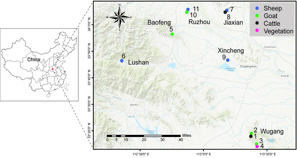
Map of eleven ticks’ collection sites in six counties in the hilly area of Funiu mountains in central China. The purple circle, black circle, green circle, and red circle represent the vegetation, cattle, goat, and sheep, respectively.
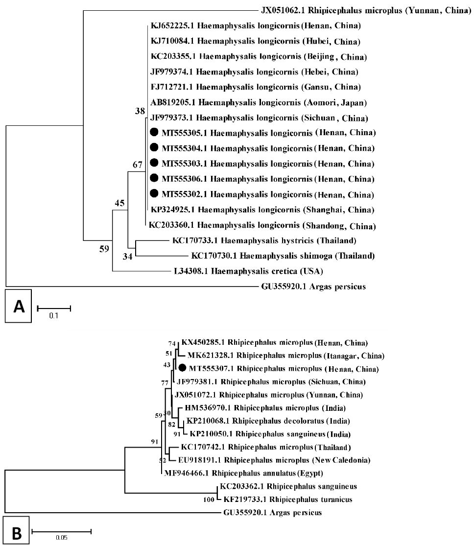
Phylogenetic analysis of ticks from hilly area in the Funiu Mountains in Central China on the basis of on tick 16S rDNA (460 bp). The phylogenetic tree for H. longicornis (A) and R. microplus (B). The bootstrap was 1000 replicates, while at each node, the numbers present bootstrap values. The outgroup is Argas persicus (GU355920).
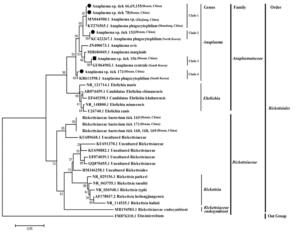
Phylogenetic analysis of Anaplasma spp. based on 16S rDNA (390 bp) sequences. The bootstrap was replicates, while at each node, the numbers present bootstrap values. Elusimicrobium (FM876310.1) is the outgroup.
References
- Bakken, J.S. and Dumler, J.S., 2000. Human granulocytic ehrlichiosis. Clin. Infect. Dis., 31: 554–560. https://doi.org/10.1086/313948[CrossRef]
- Baldridge, G.D., Scoles, G.A., Burkhardt, N.Y., Schloeder, B., Kurtti, T.J. and Munderloh, U.G., 2009. Transovarial transmission of Francisella-like endosymbionts and Anaplasma phagocytophilum variants in Dermacentor albipictus (Acari: Ixodidae). J. Med. Entomol., 46: 625–632. https://doi.org/10.1603/033.046.0330[CrossRef]
- Battilani, M., De Arcangeli, S., Balboni, A. and Dondi, F., 2017. Genetic diversity and molecular epidemiology of Anaplasma. Infect. Genet. Evol., 49, 195-211. https://doi.org/10.1016/j.meegid.2017.01.021[CrossRef]
- Black, W.C., and Piesman, J., 1994. Phylogeny of hard and soft-tick taxa (Acari: Ixodidae) based on mitochondrial 16S rRNA sequences. Proc. natl. Acad. Sci., 91: 10034-10038. https://doi.org/10.1073/pnas.91.21.10034[CrossRef]
- Cao, W.C., Zhao, Q.M., Zhang, P.H., Yang, H., Wu, X.M., Wen, B.H., Zhang, X.T. and Habbema, J.D., 2003. Prevalence of Anaplasma phagocytophila and Borrelia burgdorferi in Ixodes persulcatus ticks from northeastern China. Am. J. trop. Med. Hyg., 68: 547–550. https://doi.org/10.4269/ajtmh.2003.68.547[CrossRef]
- Dumler, J.S., Barbet, A.F., Bekker, C.P., Dasch, G.A., Palmer, G.H., Ray, S.C., Rikihisa, Y. and Rurangirwa, F.R., 2001. Reorganization of genera in the families Rickettsiaceae and Anaplasmataceae in the order Rickettsiales: Unification of some species of Ehrlichia with Anaplasma, Cowdria with Ehrlichia and Ehrlichia with Neorickettsia, descriptions of six new species combinations and designation of Ehrlichia equi and ‘HGE agent’ as subjective synonyms of Ehrlichia phagocytophilum. Int. J. Syst. Evol. Microbiol., 51: 2145–2165. https://doi.org/10.1099/00207713-51-6-2145[CrossRef]
- Dumler, J.S., Madigan, J.E., Pusterla, N. and Bakken, J.S., 2007. Ehrlichioses in humans: Epidemiology, clinical presentation, diagnosis, and treatment. Clin. Infect. Dis., 45 (Suppl 1): S45–S51. https://doi.org/10.1086/518146[CrossRef]
- Dunaj, J., Drewnowska, J., Moniuszko-Malinowska, A., Swiecicka, I. and Pancewicz, S., 2021. First metagenomic report of Borrelia americana and Borrelia carolinensis in Poland. A preliminary study. Annls Agric. Environ. Med., 28: 49-55. https://doi.org/10.26444/aaem/118134[CrossRef]
- Egizi, A., Bulaga-Seraphin, L., Alt, E., Bajwa, W.I., Bernick, J., Bickerton, M., Campbell, S.R., Connally, N., Doi, K., Falco, R.C., Gaines, D.N., Greay, T.L., Harper, V.L., Heath, A.C.G., Jiang, J., Klein, T.A., Maestas, L., Mather, T.N., Occi, J.L., Oskam, C.L., Pendleton, J., Teator, M., Thompson, A.T., Tufts, D.M., Umemiya-Shirafuji, R., VanAcker, M.C., Yabsley, M.J. and Fonseca, D.M., 2020. First glimpse into the origin and spread of the Asian longhorned tick, Haemaphysalis longicornis, in the United States. Zoon. Publ. Hlth., 67: 637-650. https://doi.org/10.1111/zph.12743[CrossRef]
- Fang, L.Q., Liu, K., Li, X.L., Liang, S., Yang, Y., Yao, H.W., Sun, R.X., Sun, Y., Chen, W.J., Zuo, S.Q., Ma, M.J., Li, H., Jiang, J.F., Liu, W., Yang, X.F., Gray, G.C., Krause, P.J. and Cao, W.C., 2015. Emerging tick-borne infections in mainland China: An increasing public health threat. Lancet Infect. Dis., 15: 1467–1479. https://doi.org/10.1016/S1473-3099(15)00177-2[CrossRef]
- Felsenstein, J., 1985. Confidence limits on phylogenies: An approach using the bootstrap. Evolution, 39: 783-791. https://doi.org/10.1111/j.1558-5646.1985.tb00420.x[CrossRef]
- Friedhoff, K.T., 1997. Tick-borne diseases of sheep and goats caused by Babesia, Theileria or Anaplasma spp. Parassitologia, 39: 99–109.
- Guo, L.P., Jiang, S.H., Liu, D., Wang, S.W., Chen, C.F. and Wang, Y.Z., 2016. Emerging spotted fever group rickettsiae in ticks, northwestern China. Ticks Tick Borne Dis., 7: 1146-1150. https://doi.org/10.1016/j.ttbdis.2016.08.006[CrossRef]
- Guo, W.P., Huang, B., Zhao, Q., Xu, G., Liu, B., Wang, Y.H. and Zhou, E.M., 2018. Human-pathogenic Anaplasma spp., and Rickettsia spp. in animals in Xi’an, China. PLoS Negl. Trop. Dis., 12: e0006916. https://doi.org/10.1371/journal.pntd.0006916[CrossRef]
- Hartwig, V., Loewenich, F.D., Schulze, C., Straubinger, R.K., Daugschies, A. and Dyachenko, V., 2014. Detection of Anaplasma phagocytophilum in red foxes (Vulpes vulpes) and raccoon dogs (Nyctereutes procyonoides) from Brandenburg, Germany. Ticks Tick Borne Dis., 5: 277-280. https://doi.org/10.1016/j.ttbdis.2013.11.001[CrossRef]
- Hoogstraal, H., Roberts, F.H., Kohls, G.M. and Tipton, V.J., 1968. Review of Haemophysalis (Kaiseriana) longicornis Neumann (Resurrected) of Australia, New Zealand, New Caledonia, Fiji, Japan, Korea, and Northeastern China and USSR, and its parthenogenetic and bisexual populations (Ixodoidea, Ixodidae). J. Parasitol., 154: 1197-213. https://doi.org/10.2307/3276992[CrossRef]
- Ismail, N., Bloch, K.C. and McBride, J.W., 2010. Human ehrlichiosis and anaplasmosis. Clin. Lab. Med., 30: 261-292. https://doi.org/10.1016/j.cll.2009.10.004[CrossRef]
- Jiang, J.F., Jiang, B.G., Yu, J.H., Zhang, W.Y., Gao, H.W., Zhan, L., Sun, Y., Zhang, X.A., Zhang, P.H., Liu, W., Wu, X.M., Xu, R.M. and Cao, W.C., 2011. Anaplasma phagocytophilum infection in ticks, China–Russia border. Emerg. Infect. Dis., 17: 932–934. https://doi.org/10.3201/eid1705.101630[CrossRef]
- Kasahara, S., Morimoto, G., Kitamura, W., Imanishi, S. and Azuma, N., 2020. Rice fields along the East Asian-Australasian flyway are important habitats for an inland wader’s migration. Sci. Rep., 10: 4118. https://doi.org/10.1038/s41598-020-60141-z[CrossRef]
- Kawahara, M., Rikihisa, Y., Lin, Q., Isogai, E., Tahara, K., Itagaki, A., Hiramitsu, Y. and Tajima, T.J.A., 2006. Novel genetic variants of Anaplasma phagocytophilum, Anaplasma bovis, Anaplasma centrale, and a novel ehrlichia sp. in wild deer and ticks on two major islands in Japan. Appl. Environ. Microbiol., 72: 1102-1109. https://doi.org/10.1128/AEM.72.2.1102-1109.2006[CrossRef]
- Keirans, J.E. and Durden, L.A., 2001. Invasion: Exotic ticks (Acari: Argasidae, Ixodidae) imported into the United States. A review and new records. J. med. Ent., 38: 850-861. https://doi.org/10.1603/0022-2585-38.6.850[CrossRef]
- Kim, C.M., Kim, M.S., Park, M.S., Park, J.H. and Chae, J.S., 2003. Identification of Ehrlichia chaffeensis, Anaplasma phagocytophilum, and A. bovis in Haemaphysalis longicornis and Ixodes persulcatus ticks from Korea. Vector Borne Zoon. Dis., 3: 17-26. https://doi.org/10.1089/153036603765627424[CrossRef]
- Kuttler, K.L., 1981. Infection of splenectomized calves with Anaplasma ovis. Am. J. Vet. Res., 42: 2094–2096
- Li, H., Zheng, Y.C., Ma, L., Jia, N., Jiang, B.G., Jiang, R.R., Huo, Q.B., Wang, Y.W., Liu, H.B., Chu, Y.L., Song, Y.D., Yao, N.N., Sun, T., Zeng, F.Y., Dumler, J.S., Jiang, J.F. and Cao, W.C., 2015. Human infection with a novel tick-borne Anaplasma species in China: A surveillance study. Lancet Infect Dis., 15: 663–670. https://doi.org/10.1016/S1473-3099(15)70051-4[CrossRef]
- Liu, X.Y., Gong, X.Y., Zheng, C., Song, Q.Y., Chen, T., Wang, J., Zheng, J., Deng, H.K. and Zheng, K.Y., 2017. Molecular epidemiological survey of bacterial and parasitic pathogens in hard ticks from eastern China. Acta Trop., 167: 26-30. https://doi.org/10.1016/j.actatropica.2016.12.010[CrossRef]
- Ngnindji-Youdje, Y., Diarra, A.Z., Lontsi-Demano, M., Tchuinkam, T., Parola, P., 2022. Detection of tick-borne pathogens in ticks from cattle in western highlands of cameroon. Microorganisms, 10: 1957. https://doi.org/10.3390/microorganisms10101957[CrossRef]
- Parola, P. and Raoult, D., 2001. Ticks and tick-borne bacterial diseases in humans: An emerging infectious threat. Clin. Infect. Dis., 32: 897–928. https://doi.org/10.1086/319347[CrossRef]
- Portillo, A., Pérez-Martínez, L., Santibáez, S., Santibáez, P., Palomar, A.M. and Oteo, J.A., 2011. Anaplasma spp. in wild mammals and Ixodes ricinus from the North of Spain. Vector Borne Zoon. Dis., 11: 3-8. https://doi.org/10.1089/vbz.2009.0214[CrossRef]
- Potgieter, F.T. and Stoltsz, W.H., 2004. Infectious diseases of livestock. Oxford University Press, Cape Town, South Africa. pp. 594–616.
- Qin, X.R., Han, F.J., Luo, L.M., Zhao, F.M., Han, H.J., Zhang, Z.T., Liu, J.W., Xue, Z.F., Liu, M.M., Ma, D.Q., Huang, Y.T., Sun, Y., Sun, X.F., Li, W.Q., Zhao, L., Hao, Y. and Yu, X.J., 2018. Anaplasma species detected in Haemaphysalis longicornis tick from China. Ticks Tick Borne Dis., 9: 840-843. https://doi.org/10.1016/j.ttbdis.2018.03.014[CrossRef]
- Rehman, A., Conraths, F.J., Sauter-Louis, C., Krücken, J. and Nijhof, A.M., 2019. Epidemiology of tick-borne pathogens in the semi-arid and the arid agro-ecological zones of Punjab province, Pakistan. Transbound. Emerg. Dis., 66: 526-536. https://doi.org/10.1111/tbed.13059[CrossRef]
- Ryff, J.F. and Weibel, J.L., 1964. Thomas GM. Relationship of ovine to bovine Anaplasmosis. Cornell Vet., 54: 407–414.
- Saitou, N. and Nei, M., 1987. The neighbor-joining method: A new method for reconstructing phylogenetic trees. Mol. Biol. Evol., 4: 406-425.
- Shkap, V., Molad, T., Fish, L. and Palmer, G.H., 2002. Detection of the Anaplasma centrale vaccine strain and specific differentiation from Anaplasma marginalein vaccinated and infected cattle. Parasitol. Res., 88: 546-552. https://doi.org/10.1007/s00436-002-0612-9[CrossRef]
- Somveille, M., Manica, A., Butchart, S.H. and Rodrigues, A.S., 2013. Mapping global diversity patterns for migratory birds. PLoS One, 8: e70907. https://doi.org/10.1371/journal.pone.0070907[CrossRef]
- Splitter, E.J., Anthony, H.D., Twiehaus, M.J., 1956. Anaplasma ovis in the United States; experimental studies with sheep and goats. Am. J. Vet. Res., 17: 487–491.
- Tamura, K., Peterson, D., Peterson, N., Stecher, G., Nei, M., Kumar, S., 2011. MEGA5: Molecular evolutionary genetics analysis using maximum likelihood, evolutionary distance, and maximum parsimony methods. Mol. Biol. Evol., 28: 2731-2739. https://doi.org/10.1093/molbev/msr121[CrossRef]
- Theiler, A., 1912. Gallsickness of imported cattle and the protective inoculation against this disease. Agric. J. Union S. Africa., 3: 1–11.
- Wen, B., Jian, R., Zhang, Y. and Chen, R., 2002. Simultaneous detection of Anaplasma marginale and a new Ehrlichia species closely related to Ehrlichia chaffeensis by sequence analyses of 16S Ribosomal DNA in Boophilus microplus ticks from tibet. J. clin. Microbial., 40: 3286–3290. https://doi.org/10.1128/JCM.40.9.3286-3290.2002[CrossRef]
- Wu, R.T., 2013. Parsing and identification of rickettsia of eastern wild deer and ticks insects. J. Inner Mongol. Agric. Univ., 34: 1-6.
- Yan, Y.Q., Jiang, Y.X., Tao, D.Y., Zhao, A.Y., Qi, M. and Ning, C.S., 2020. Molecular detection of Anaplasma spp. in dairy cattle in southern Xinjiang, China. Vet. Parasitol. Reg. Stud. Reports., 20: 100406. https://doi.org/10.1016/j.vprsr.2020.100406[CrossRef]
- Yang, J.F., Liu, Z.J., Guan, G.Q., Liu, Q., Li, Y.Q., Chen, Z., Ma, M.L., Liu, A.H., Ren, Q.Y., Luo J.X., and Yin, H., 2013. Prevalence of Anaplasma phagocytophilum in ruminants, rodents and ticks in Gansu, northwestern China. J. med. Microbiol., 62: 254-258. https://doi.org/10.1099/jmm.0.046771-0[CrossRef]
- Zhang, G., Sun, X., Zhao, Y., Liu, X., Zheng, Z., Sun, Y. and Liu, R., 2013. Prevalence of Anaplasma spp. infection in a desert landscape region of Heshuo, Xinjiang. Chin. J. Epidemiol, 34: 147-151.
- Zhang, K., Li, A.J., Wang, Y., Zhang, J.J., Chen, Y.J., Wang, H.J., Shi, R.X. and Qiu, Y.H., 2021. Investigation of the presence of Ochrobactrum spp. and Brucella spp. in Haemaphysalis longicornis. Ticks Tick Borne Dis., 12: 101588. https://doi.org/10.1016/j.ttbdis.2020.101588[CrossRef]
- Zhang, L., Liu, Y., Ni, D., Li, Q., Yu, Y., Yu, X.J., Wan, K., Li, D., Liang, G., Jiang, X., Jing, H., Run, J., Luan, M., Fu, X., Zhang, J., Yang, W., Wang, Y., Dumler, J.S., Feng, Z., Ren, J. and Xu, J., 2008. Nosocomial transmission of human granulocytic anaplasmosis in China. J. Am. med. Assoc., 300: 2263–2270. https://doi.org/10.1001/jama.2008.626[CrossRef]
- Zhang, Y.Z. and Xu, J.G., 2016. The emergence and cross species transmission of newly discovered tick-borne Bunyavirus in China. Curr. Opin. Virol., 16: 126–131. https://doi.org/10.1016/j.coviro.2016.02.006[CrossRef]
- Zhuang, L., Du, J., Cui, X.M., Li, H., Tang, F., Zhang, P.H., Hu, J.G., Tong, Y.G., Feng, Z.C. and Liu, W., 2018. Identification of tick-borne pathogen diversity by metagenomic analysis in Haemaphysalis longicornis from Xinyang, China. Infect. Dis. Poverty, 7: 45. https://doi.org/10.1186/s40249-018-0417-4[CrossRef]
- Zou, Y.X., Jin, H.T., Wang, Q.Y., Fu, Z.X., Gao, H.W., Liu, P.P., Liu, Q. and Chen, L.F., 2011. Molecular detection of Anaplasma phagocytophilum in Ixodid ticks in Hebei Province, China. Vector Borne Zoon. Dis., 11: 1323-1327. https://doi.org/10.1089/vbz.2010.0253[CrossRef]
Services
Article usage statistics
Loading…