Specail Issue: Artificial Intelligence in Veterinary Medicine: Current Status and Future Prospects · Volume 13, Issue 1, 11 February 2026 · Open Access
Copper Chloride Induces Dose and Time-Dependent Hematotoxicity in Male Rats
·
Affiliations & Notes ⌄
1Department of Biology Sciences, College of Education for Pure Sciences, University of Mosul, Mosul, Iraq. *Correspondence | Ghofran Khadir Mahmood, Department of Biology Sciences, College of Education for Pure Sciences, University of Mosul, Mosul, Iraq
Copper is an essential trace mineral that can be toxic in excess.
Sublethal levels of copper chloride exposure resulted in an increase in total white blood cells, lymphocytes, and monocytes and a decrease in red blood cells, platelets, neutrophils, and eosinophils.
In addition, research should be expanded to include the evaluation of copper chloride effects on blood components in other animal species, with the aim of determining the generalizability of the results and their applicability across different biological systems.
Selected from the article's own text.
Abstract
The effects of sub-lethal dose 160, 235, and 350 mg/kg of copper chloride (CuCl2) for periods 5, 10, 20, and 30 days on the blood parameters of male rat were investigated. Fifty healthy male rats were used in this study. Rats were randomly divided into four groups based on the period and dose concentration. According to the data, there was no significant decrease (P<0.05) in each of the blood parameters of the treated animals with CuCl2 as compared to the control group in the first five days except neutrophils. Rats administered 350 mg/kg B.W. showed a significant decrease (P<0.05) in the number of RBCs, PCV, HB, and platelets after 10 days. During third period (20 days), rats treated with 235 and 350 mg/kg Body Weight (B.W.) of CuCl2 showed a significant decrease (P<0.05) in RBCs, as well as a significant decrease (P<0.05) in PCV% and HB in comparison to the control group. In the fourth period (30 days) there was significant decrease in RBCs, PCV%, HB and Platelets in all experimental groups compared with control. In contrast, there was a significant increase (P<0.05) in white blood cell count, lymphocytes, and monocytes, with the highest values observed at 350 mg/kg. The toxic effect on hematopoietic levels and on Red Blood Cell morphology were shown respectively as it is assessed by Leishman-stained blood smears resulted in echinocytes, acanthocytes, tear drop, stomatocyte and target RBCs. Thus, it was concluded that oral administration of CuCl₂ caused a decrease in the levels of Hb concentration, PCV, RBC, and platelets (PLTS) In addition, alterations in MCV values were noted in relation to red blood cell counts and PCV, while increase in WBCs, Lymphocytes and Monocytes. In addition, anomalies in RBCs morphology occurred, confirming the toxic effect of CuCl2 on blood parameters of male rat.
Keywords | Hematology, RBCs anomalies, Copper chloride, Male rat
INTRODUCTION
An essential mineral and naturally occurring chemical element, copper (Cu) can accumulate in the body to toxic levels, leading to adverse effects , It was a reddish metallic substance which can be found in water, soil, rock sediment, and, to a lesser extent, the atmosphere. The main natural source of copper is the Earth’s crust . Kidney failure, liver damage, and blood issues can result from heavy metal poisoning . Concerns over the possible negative effects of copper’s expanding industrial uses on the environment, human health, and animal health are growing along with these uses .
Copper (Cu) is a necessary trace element for many metabolic functions and micronutrients, However, its widespread use over the past few decades has raised concerns due to its negative impact on the environment and public health . A surplus of Cu in the body can be harmful because it is a necessary component of specific cuproproteins that are in charge of the body’s appropriate growth and development. Heavy metals build up in soil and plants due to anthropogenic factors, a rise in the number of vehicles, and extensive urban and rural infrastructure. These metals are then passed on to people through the food chain . CuCl2, a Cu (II) molecule, causes oxidative alteration of cell components by increasing the production of ROS and reactive nitrogen species (RNS) in RBC .
Oxidative stress, which is caused by prolonged and elevated ROS levels, has been linked to many forms of controlled cell death, including cuproptosis, a copper-dependent regulated cell death mechanism that differs from other recognized regulated cell death forms . Iron-sulfur cluster proteins are lost and mitochondrial lipoylated proteins aggregate as a result of cuproptosis . Cuproptosis results from disruptions to copper homeostasis, which is dependent on the copper transporter. Numerous cardiovascular disorders, including myocardial ischemia/reperfusion (I/R) injury, heart failure, atherosclerosis, and arrhythmias, have been linked to cuproptosis, according to recent research .
Therefore, this study aimed to investigate the effects of sublethal doses of copper chloride (CuCl₂) on hematological parameters and erythrocyte morphology in albino rats.
MATERIALS AND METHODS
Experimental animal
Fifty male albino rats, aged (2-3) months, weighing 250-300 grams, were housed in hanging cages at 25±2ºC, with 14 hours of light and 10 hours of darkness. They were fed regular food and water from the University of Mosul’s Veterinary College’s animal house. Rats were randomly divided into four groups and administered CuCl₂ orally. Each cage has three male rats in each.
Experimental design
The first group received distilled water (control) , while the second, third, and fourth groups received CuCl2 dissolved in water at doses of 160, 235, and 350 mg/kg B.W. respectively. Every group was dissected for five, ten, twenty, and thirty days. Anticoagulant (EDTA) was utilized to collect blood samples, which were then subjected to automated hematological analysis. Leishman stain was used to color the blood smear .
Statistical analysis
data were examined using the mean ± standard deviation. The statistical program Graph Pad v5.0 was used to examine the data. ANOVA in one direction was employed. The Tukey test was used for the post hoc analysis. At P<0.05, mean values were deemed statistically significant, different superscript letters within a row indicate significant differences (P < 0.05) .
RESULTS AND DISCUSSION
Blood parameters
All blood parameters of the treated animals did not significantly decrease (P<0.05) when compared to the control group throughout the first period, according to the data (Tables 1, 2).
Table 1: Effect of CuCl2 s on the blood parameters in male rats during 5 days.
Blood parameters
Control
160 mg/kg B.W.
235 mg/kg B.W.
350 mg/kg B.W.
RBC (X ١٠٦/µl(
٨.35a±0.5
٨.09a±0.3
٨.0٧a±0.٧
٨.0١a±0.٩
Hb g/dl
١٦.٨ a±٠.٤
16.5 a ±٠.٢
١٦ a.٤±٠.٥
١٦.١ a±٠.٣
MCV fL
٥٩.٨٨ a±٠. ٣
٦١.1٨ a±٠. ١
60.9٦ a±٤.٠
61.1٧ a±٢.٠
MCH pg
2.٠٧ a±٢.٠١
٢.٠٣ a±٠.٠٣
2.03 a±٠.٠٦
2.00 a±٠.٠٤
MCHC g/dl
٣٣.٦ a±٤.٠٢
٣٣.٣ a±٦.٠
33.3 a±7.1
32.8 a±٥.٤
Platelets Count)X109/L)
٨٢٣ a±١٠
٨٢٠ a±١٤
٨١٧ a±١٧
٨١٤ a±١٣
Values in the same row with different superscript letters are significantly different (P < 0.05).
Table 2: Effect of CuCl2 on white blood cell count and its types in male rats during 5 Day.
Blood parameters
Control
160 mg/kg B.W.
235 mg/kg B.W.
350 mg/kg B.W.
WBC١٠٣/µl
9.17 a±0.3
9.32 a±٠.1
9.6 a±0.5
10.0 a±0.4
Neutrophils ٪
٥٩.٦١ a±٠.02
58.95 a±0.05
٥٨.0 ab±0.07
٥٧.٧٤ b±0.04
Lym ٪
٣٢.٤ a±1.0
٣٢.٩7 a±1.4
33.88 a±1.6
٣٣.٨٦ a±1.8
Mon٪
٢.٦٩ a±0.4
٢.٧٩ a±0.7
٣.١ a±0.5
٣.٤ a±0.2
Eos %
٥.٣ a±0.06
٥.٢٩ a±0.03
٥.١ a±0.06
٥.٠ a±0.04
Bas %
0.0 a±٠.٠
0.0 a±٠.٠
0.07 a±٠.٠
0.0 a±٠.٠
Values in the same row with different superscript letters are significantly different (P < 0.05).
Table 3: Effect of CuCl2 s on blood parameters in male rats during(10 days).
Blood parameters
Control
160 mg/kg B.W.
235 mg/kg B.W.
350 mg/kg B.W.
RBC ١٠٦/µl
٨.39 a±0.٨
٨.٠٧ a±٠.٦
8.00 ab±٠.٩
7.49 b±٠.٥
٪PCV
٥١.٠ a±٣.٠
49.0 a±١.٢
48.٠ ab±٣.٥
٤٧.٠ b±٢.٢٨
Hb g/dl
١٦.٩ a±٠.٧
١٦.١ a±٠.٩
15.6 ab±٠.٨
15.3 b±١.٠
MCV fL
٦0.7٨ a±٥.٠
٦٠.٧1 a±9.0
٦٠.0 a±٠. ٧
٦٢.٧5 a±4.0
MCH pg
٢.٠١ a±٠.٠٥
١.٩٩ a±٠.٠٨
١.٩٥ a±٠.٠٣
٢.٠٤ a±٠.٠١
MCHC g/dl
٣٣.١٣ a±٤.٠٢
٣٢.٨٥ a±٥.٦
٣٢.٥ a±٣.٩
32.5 a±٥.٨
Platelets count )X109/L)
٨٢٥ a±١٣
٨٠٣ ab±١٢
٧٩٣ bc±١٥
٧٦٤ c±١٨
Values in the same row with different superscript letters are significantly different (P < 0.05).
Table 4: Effect of CuCl2 on white blood cell count and its types in male rats during 10 Day.
Blood parameters
Control
160 mg/kg B.W.
235 mg/kg B.W.
350 mg/kg B.W.
WBC ١٠٣/µl
9.3 b±0.4
١٠.81 ab±0.7
10.93 a±0.8
11.4 a±0.3
٪Neutrophils
٥٩. 7 a±٠.02
57.7 ab±٠.06
٥٤.٤١ c±0.03
٥٠.٩ d±0.01
٪Lym
٣٢.5 a±1.0
٣٤.٨٣ ab±3.2
٣٧.6٩ bc±2.5
٤0.٩ d±1.5
٪Mon
٢.7 c±0.4
٢.٨7 bc±0.5
٣.9 ab±0.7
٤.٥ a±0.4
%Eos
5.1 a±0.06
٤.6 a±0.01
٤.٠ ab±0.05
٣.٧ b±0.03
%Bas
٠.٠ a±0.00
0.٠ a±0
0.٠ a±0
0.٠ a±0
Values in the same row with different superscript letters are significantly different (P < 0.05).
Table 5: Effect of CuCl2 s on blood parameters in rats during (20 days).
Blood parameters
Control
160 mg/kg B.W.
235 mg/kg B.W.
350 mg/kg B.W.
RBC (١٠٦/µl)
٨.44 a±0.9
8.04 a±0.2
7.91 bc±0.4
7.33 c±0.6
٪PCV
٥١.4 a±1.7
47.08 b±1.0
45.1 bc±2.5
43.3 c±3.4
Hb (g/dl)
١٧.٣٣ a±٠.٩
15.7 b±1.6
14.7 bc±2.0
14.0 c±1.8
MCV (fL)
٦٠.٩ c±4.0
58.55 b±2.0
57.0 b±7.0
59.07 a±5.0
MCH (pg)
٢.٠٥ a±٠.٠١
1.95 ab±0.03
1.85 b±0.06
1.90 b±0.05
MCHC (g/dl)
٣٣.٧١a±٢.٣٩
33.3 a±1.9
32.59 a±3.3
32.33 a±5.1
Platelets Count(X109/L)
٨30 a±١٣
790 b±15
787 bc±19
758 cd±17
Values in the same row with different superscript letters are significantly different (P < 0.05).
Table 6: Effect of CuCl2 on white blood cell count and its types in male rats during 20 Day.
Blood parameters
Control
160 mg/kg B.W.
235 mg/kg B.W.
350 mg/kg B.W.
WBC ١٠٣/µl
9.5 c±0.6
11. 8 b±0.9
12.57 ab±0.8
12.57 ab±0.8
٪Neutrophils
٥٩. 8 a±٠. 1
٥3.8 b±1.4
50.1 b±1.3
50.1 b±1.3
٪Lym
٣2.5 a±0.7
37.62 bc±2.3
41.4 cd±1.8
41.4 cd±1.8
٪Mon
٢.9 d±0.07
3.99 cd±0.1
4.6 bc±0.09
4.6 bc±0.09
%Eos
4.8 a±0.06
4.59 ab±0.03
3.9 bc±0.01
3.9 bc±0.01
%Bas
0.0 b±٠.٠
0.0 b±0
0.0 b±0
0.0 b±0
Values in the same row with different superscript letters are significantly different (P < 0.05).
During the second period, there was a significant decrease (P<0.05) in the number of RBCs, PCV, HB, and platelets in rats treated with 350 mg/kg B.W. CuCl2 (Table 3), as well as a significant increase (P<0.05) in the WBC, lymphocytes, and monocytes counts in the groups treated with 235 and 350 mg/kg B.W. in comparison to the control group, while there was a significant decrease (P<0.05) in neutrophils and eosinocytes counts in the groups treated with CuCl2 at 235 and 350 mg/kg B.W. (Table 4).
In comparison to the control group, rats treated with 235 and 350 mg/kg B.W. of CuCl2 exhibited a significant decrease (P<0.05) in RBCs, PCV%, Hb, MCV, and platelets counts throughout the third period (Table 5). While neutrophils and eosinophils counts showed a significant (P<0.05) decrease in comparison to control groups, white blood cells, lymphocytes, and monocytes counts showed a significant (P<0.05) rise (Table 6).
Table 7: Effect of CuCl2 s on blood parameters in rats during(30 days).
Blood parameters
Control
160 mg/kg B.W.
235 mg/kg B.W.
350 mg/kg B.W.
RBC (١٠٦/µl)
٨.٥ a±0.٤
7.٠3 b±0.3
٦.11 b±0.7
٥.8 b±0.4
٪PCV
51.6 a±2.5
44.8 b±1.6
43.1 b±2.0
40.7 c±1.4
HB (g/dl)
١٧.٤ a±٠.٩
14.9 b±1.3
14.0 b±1.6
13.4 bc±1.9
MCV (fL)
٦٠.7 b±0.4
63.72 ab±6.0
70.54 a±3.0
70.17 a±7.0
MCH (pg)
٢.٠4 a±0.01
2.11 a±0.03
2.29 a±0.06
2.3 a±0.04
MCHC (g/dl)
٣٣.٧٢ a±٢.٣٩
33.25 a±5.1
32.48 a±3.3
32.92 a±6.0
Platelets Count (X109/L)
٨34 a±16
782 b±18
767 bc±14
721 d±11
Values in the same row with different superscript letters are significantly different (P < 0.05).
Table 8: Effect of CuCl2 on white blood cell count and its types in male rats during 30 Day.
Blood parameters
Control
160 mg/kg B.W.
235 mg/kg B.W.
350 mg/kg B.W.
WBC (١٠٣/µl)
9.7 d±0.8
14.05 bc±1.1
16.77 b±1.3
18.67 ab±1.9
٪Neutrophils
60. 0 a±٠. 07
50.0 b±0.1
41.9 c±0.4
30.5 d±0.09
٪Lym
٣2.6 d±0.9
41.07 c±1.0
49.06 b±0.9
55.2 a±1.1
٪Mon
2.6 d±0.09
4.33 bc±0.2
5.04 b±0.5
8.5 a±0.7
%Eos
4.8 a±0.06
3.6 b±0.09
3.0 c±0.08
2.8 cd±0.1
%Bas
0.0 c±٠.٠
1.0 b±0.07
1.0 b±0.09
3.0 a±0.1
Values in the same row with different superscript letters are significantly different (P < 0.05).
There was a significant decrease (P<0.05) in PCV% and HB in compared with control group In the fourth period, a significant decrease in RBCs, PCV%, HB and Platelets counts (Table 7), while significant increase (P<0.05) in WBC, lymphocyte, monocytes and basophiles counts, (Table 8).
The results of the current study are consistent with the results of the study conducted by Baqer et al. (2023) who dosed rats with copper at a concentration of (37 mg/kg) of body weight for 45 days and observed a decrease in the recording of red blood cells, hemoglobin and platelets, which was attributed to the causes of general hemolysis.
The results of the current study also agreed with the results of the study by Ghareeb (2023), who dosed rats with copper at a concentration of 300 mg/kg of body weight for 28 days and observed the dissolution of red blood cells. A decrease in the number of red blood cells, hemoglobin concentration, hematocrit percentage, mean corpuscular volume, and platelets causes anemia . The results of the current study are consistent with the study conducted by Fawzy et al. (2021) who dosed rats with different doses of copper at concentrations of (0.1, 0.2, and 0.4) mg/kg of body weight, for 30 days.
Their results showed that high doses of copper led to a decrease in the concentration of hemoglobin and the number of red blood cells, which led to anemia. The results of the study conducted by Sakr et al. (2021) also showed that exposing rats to copper at a concentration of (125 mg/kg) of body weight for a period of 28 days led to changes in blood parameters represented by a significant decrease (P<0.05) in the number of red blood cells, the level of hemoglobin (Hb), and the number of platelets (PLT), compared to the control group. The results of the current study are also consistent with the study conducted by Zhou et al. (2019), who noted that rats that were orally dosed with copper chloride for 28 days showed a significant decrease (P<0.05) in both the percentage of hematocrit (PCV) and platelet counts.
The results of the current study are also consistent with the study conducted by Yahya et al. (2019) who injected rats with copper particles intraperitoneally at a dose of (0.5 mg/kg) of body weight daily for 28 days. Their results showed a significant decrease (P<0.05) in the numbers of red blood cells (RBC), Hb, and platelets, an increase in the mean corpuscular volume (MCV), and a decrease in the mean corpuscular hemoglobin (MCH). High levels of copper ions (Cu++) competitively inhibit iron absorption and utilization, leading to decreased serum iron concentration, impaired hemoglobin synthesis and anemia, The study explained the contribution of copper particles to the occurrence of anemia through two main mechanisms: the first is direct, by stimulating the destruction of red blood cells, and the second is indirect, by increasing their sensitivity to damage caused by oxidative stress and lipid peroxidation.
The increased white blood cell count was interpreted as an inflammatory response to copper exposure . Thrombocytopenia, a condition in which the number of platelets in the bloodstream is lower than normal, is believed to be due to either decreased production by the bone marrow or increased destruction . This may be attributed to damage to cell membranes caused by oxidative stress, and this decrease is an indicator of copper’s ability to cause thrombocytopenia and negatively affect the process of clot formation . Dosing rats with different concentrations of copper (400, 200 and 100) mg/kg of body weight, for 28 days, led to an increase in the percentage of monocytes, and the reason may be attributed to the inflammatory response .
The results of the current study are also consistent with the study of Sa’idu et al. (2022), which confirmed in its results that exposing rats to copper at a concentration of (200 mg/kg) of body weight led to an increase in the number of white lymphocytes. The results of the study conducted by Sakr et al. (2021) also showed that oral administration of copper to rats at a concentration of (125 mg/kg) of body weight for a period of 28 days led to an increase in the percentage of monocytes, thus matching the results of our current study.
Red blood cell (RBCs) morphology
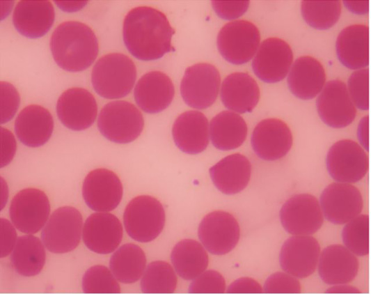
Normal red blood cells have a biconcave disk form and a pallor center (Figure 1). The blood of rats treated with CuCl2 showed several morphological abnormalities in the morphology of RBCs.
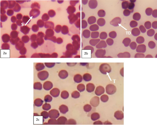
During the first five days, Rats that received 160 mg/kg B.W. had stomatocytes in their erythrocytes (Figure 2a), whereas rats that received 235 mg/kg B.W. had RBCs that resembled teardrop (Figure 2b). RBCs from rats given 350 mg/kg body weight show the target cell (Figure 2c).
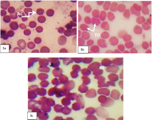
Rats given 160 mg/kg B.W. throughout the second period (10) days had acanthocyte cells in their RBCs (Figure 3a); rats treated with 235 mg/kg B.W. had a teardrop shape (Figure 3b); and rats treated with 350 mg/kg B.W. had Rouleaux formation (Figure 3c).
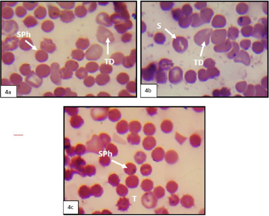
During the third period, which lasted 20 days, rats given 160 mg/kg B.W. RBCs showed spherocytes, as shown in Figure 4a; rats provided 235 mg/kg B.W. RBCs showed stomatocytes and tear drops, as shown in Figure 4b; and rats given 350 mg/kg B.W. Figure 4c shows that RBCs had both target cells and spherocytes.
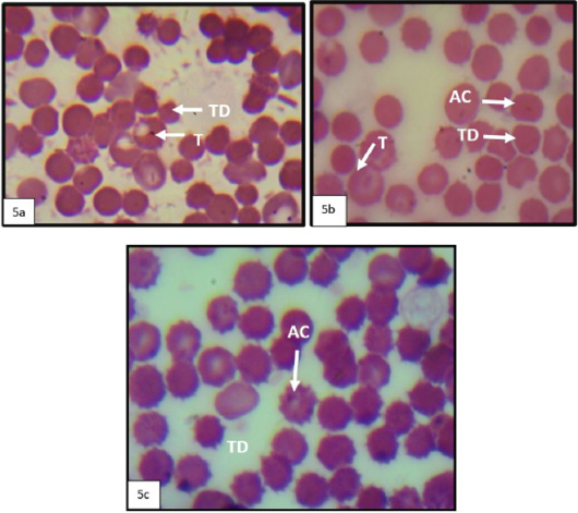
Rats supplied 160 mg/kgm B.W. during the previous 30 days. RBCs showed target cell and tear drop, Figure 5a; rats were administered 235 mg/kg B.W. Rats that received 350 mg/kg B.W. of RBCs were the Acanthocytes and Target cell Figure 5b. Figure 4c shows that RBCs contained acanthocytes and tear drop cells.
Exposure to heavy metals may be responsible for morphological changes in red blood cells, causing cellular abnormalities such as tear drop and kidney shape, which clearly increase with increasing metal concentration and duration of exposure . Morphological changes in red blood cells in fish, including nuclear and cellular abnormalities, significantly increase with increasing heavy metal concentrations, and the severity of these abnormalities also worsens with increasing dose and exposure duration, reflecting a direct relationship between heavy metal exposure and the degree of cellular damage. These changes are likely due to increased lipid peroxidation in the cell membrane, which causes damage to it, increases its permeability to ions, and thus increases the cell’s absorption of toxic substances and causes morphological changes in red blood cells . It was found that oral administration of copper to rats at a concentration of 100 mg/kg body weight for 30 days resulted in changes in the fluidity and deformation of the red blood cell membrane, through disruption of the lipid composition of the membrane, saturation, formation of bonds in phospholipids, and permeability .
CONCLUSION AND RECOMMENDATIONS
Copper is an essential trace mineral that can be toxic in excess. The effects of this increase with the amounts used and exposure duration. Sublethal levels of copper chloride exposure resulted in an increase in total white blood cells, lymphocytes, and monocytes and a decrease in red blood cells, platelets, neutrophils, and eosinophils. For the purpose of obtaining further scientific findings that support and validate the results of the present study, it is recommended that: future studies focus on investigating the effects of sublethal concentrations of copper chloride on various blood parameters in female rats, in order to compare the findings with those obtained in males and to clarify potential physiological differences between the sexes. In addition, research should be expanded to include the evaluation of copper chloride effects on blood components in other animal species, with the aim of determining the generalizability of the results and their applicability across different biological systems.
ACKNOWLEDGEMENT
The present study was supported by the University of Mosul, College of Education for Pure Science, for which the researchers are grateful.
NOVELTY STATEMENT
This study provides a detailed time- and dose-dependent analysis of CuCl₂-induced hematotoxicity. It comprehensively documents a spectrum of erythrocyte morphological abnormalities, such as stomatocytes, acanthocytes, and target cells, offering new insights into the subacute toxic profile of copper chloride in male rats.
Author’s Contribution
The research strategy did by Amal Abdulilah Alkshab, composed the article and conduct the experimental conditions done by Ghofran Khadir Mahmood.
Ethical approval
The University of Mosul, College of Veterinary Medicine’s Institutional Animal Care and Use Committee granted ethical authorization for handling animals with reference number UM.VET.2024.137, dated October 15, 2024.
Generative AI and AI-assisted technology statement
All authors of this work declare that generative AI technologies including large language models (e.g., ChatGPT, Copilot) and text-to-image generators were not utilized in any capacity during the preparation, writing, or editing of this manuscript.
Conflict of interest
The authors have declared no conflict of interest.
Figures
Figure 1:.
Photomicrograph of a blood smear from a control rat showing normal biconcave red blood cells, Leishman stain, 1000× magnification.
Figure 2:.
A: Photomicrograph of a blood smear from a rat treated with 160 mg/kg body weight showing stomatocytes (s), Leishman stain, 1000× magnification. B: Photomicrograph of a blood smear from a rat treated with 235 mg/kg body weight showing Tear Drop(TD), Leishman stain, 1000× magnification. C Photomicrograph of a blood smear from a rat treated with 350 mg/kg body weight showing Target cell (T), Leishman stain, 1000× magnification.
Figure 3:.
A: Photomicrograph of a blood smear from a rat treated with 160 mg/kg body weight showing Acanthocyte (Ac), Leishman stain, 1000× magnification. B: Photomicrograph of a blood smear from a rat treated with 235 mg/kg body weight showing teardrop cell (TD), Leishman stain, 1000× magnification. C: Photomicrograph of a blood smear from a rat treated with 350 mg/kg body weight showing Rouleaux formation (RF), Leishman stain, 1000× magnification.
Figure 4:.
A: Photomicrograph of a blood smear from a rat treated with 160 mg/kg body weight showing teardrop cell (TD) and Spherocytes (SPh), Leishman stain, 1000× magnification. B: Photomicrograph of a blood smear from a rat treated with 235 mg/kg body weight showing teardrop cell (TD) and stomatocyte cell(s), Leishman stain, 1000× magnification. C: Photomicrograph of a blood smear from a rat treated with 350 mg/kg body weight showing target cell (T) and Spheroctyte (SPh), Leishman stain, 1000× magnification.
Figure 5:.
A: Photomicrograph of a blood smear from a rat treated with 160 mg/kg body weight showing teardrop cells(TD) and target cell (T), Leishman stain, 1000× magnification. B: Photomicrograph of a blood smear from a rat treated with 235 mg/kg body weight showing target cell (T), acanthocytes (AC) and Teardrop cell (TD), Leishman stain, 1000× magnification. C: Photomicrograph of a blood smear from a rat treated with 350 mg/kg body weight showing shows acanthocytes (AC) and Teardrop cell (TD), Leishman stain, 1000× magnification.
References
Adele BO, Idama C, Ige AO, Odetola A, Emediong IE, Adewoye EO (2023). Alterations in plasma and erythrocyte membrane fatty acid composition following exposure to toxic copper level affect membrane deformability and fluidity in female Wistar rats. J. Trace Element. Med. Biol., 80: 127316. https://doi.org/10.1016/j.jtemb.2023.127316[CrossRef]
Ban XX, Wan H, Wan XX, Tan YT, Hu XM, Ban HX, Chen XY, Huang K, Zhang Q, Xiong K (2024). Copper metabolism and cuproptosis: Molecular mechanisms and therapeutic perspectives in neurodegenerative diseases. Curr. Med. Sci., 44(1): 28–50. https://doi.org/10.1007/s11596-024-2832-z[CrossRef]
Baqer JM, Abdullal MK, Ibrahim MW (2023). Cumulative toxic effect of heavy metals lead, copper, cadmium and molybdenum in rat blood. J. Hyg. Eng. Design, 44: 19–23.
Culling CF, Allison RT, Barr WT (1985). Cellular pathology technique. 4th ed. New York: Butterworth, pp. 642. https://doi.org/10.1016/B978-0-407-72903-2.50018-6[CrossRef]
Dahiru A, Ibrahim HM, Saidu B, Abdulazeez N, Muawiyyah MM, Sunusi S, Jaafaru IA (2025). Mitigation of copper-induced toxicity in wistar rats ‘serum biochemical profile by cannabis leaf extract. Fudmant. J. Sci., 9(3): 354-357. https://doi.org/10.33003/fjs-2025-0903-2315[CrossRef]
Fawzy MM, Ahmed SM, Khamis T, Hamed A, Abdel-Fattah DM (2022). Effect of copper sulphate pollution and its antidote penicillamine on serum and brain tissues markers of albino rats. Bull. Pharma. Sci. Assiut. Univ., 45(1): 327–337. https://doi.org/10.21608/bfsa.2022.239504[CrossRef]
Garcia-Casal MN, Dary O, Jefferds ME, Pasricha SR (2023). Diagnosing anemia: Challenges selecting methods, addressing underlying causes, and implementing actions at the public health level. Ann. N. Y. Acad. Sci., 1524(1): 37–50. https://doi.org/10.1111/nyas.14996[CrossRef]
Ghareeb OA (2023). Hematotoxicity induced by copper oxide nanoparticles and the attenuating role of giloy in vivo. Cureus, 15(10). https://doi.org/10.7759/cureus.46577[CrossRef]
Henckens MLCM, Worrell E (2020). Reviewing the availability of copper and nickel for future generations. The balance between production growth, sustainability and recycling rates. J. Clean Prod., 264: 121460. https://doi.org/10.1016/j.jclepro.2020.121460[CrossRef]
Husain N, Mahmood R (2020). Mitigation of Cu (II)-induced damage in human blood cells by carnosine: An in vitro study. Toxicology in vitro, 68. https://doi.org/10.1016/j.tiv.2020.104956[CrossRef]
Islam SM, Rohani MF, Zabed SA, Islam MT, Jannat R, Akter Y, Shahjahan M (2020). Acute effects of chromium on hemato-biochemical parameters and morphology of erythrocytes in striped catfish (Pangasianodon hypophthalmus). Toxicol. Rep., 7: 664–670. https://doi.org/10.1016/j.toxrep.2020.04.016[CrossRef]
Maciel-Magalhães M, Medeiros RJ, Bravin JS, Patricio BF, Rocha HVA, Paes-de-Almeida EC, dos Santos LMG, Jacob SC, Savignon TCM, Amendoeira FC (2020). Evaluation of acute toxicity and copper accumulation in organs of Wistar rats, 14 days after oral exposure to copper oxide (II) nano-and microparticles. J. Nanopart. Res., 22(1). https://doi.org/10.1007/s11051-019-4721-0[CrossRef]
Naz S, Hussain R, Guangbin Z, Chatha AMM, Rehman ZU, Jahan S, Khan A (2023). Copper sulfate induces clinico-hematological, oxidative stress, serum biochemical and histopathological changes in freshwater fish rohu (Labeo rohita). Front. Vet. Sci., 10: 1142042. https://doi.org/10.3389/fvets.2023.1142042[CrossRef]
Patriarcheas V, Pikoulas A, Kostis M, Charpidou A, Dimakakos E (2020). Heparin-induced thrombocytopenia: Pathophysiology, diagnosis and management. Cureus, 12(3): e7385. https://doi.org/10.7759/cureus.7385[CrossRef]
Sa’idu B, Hassan IM, Abdulazeez N, Aliyu B, Aliyu BY (2022). Therapeutic effect of methanolic leaves extract of cannabis in copper induced cardio-toxicity in Wistar rats. Int. J. Pharmacogn. Life Sci., 3(1): 15–18. https://doi.org/10.33545/27072827.2022.v3.i1a.40[CrossRef]
Sakr S, Abdelwahab M, Atef M (2021). The oxidative stress mediated toxicity and immunotoxic effect of copper oxide nanoparticles on spleen of adult albino rats. Zagazig J. Forensic Med. Toxicol., 19: 42–59. https://doi.org/10.21608/zjfm.2020.40081.1062[CrossRef]
Steel RG, Torri JH (1980). Principle and procedures of statistics. 2nd ed., McGraw-Hill Company, Inc. London. pp. 38-41.
Thakore KA, Bhadaniya AR, Makwana A, Patel SS, Delvadiya RS, Patel HB, Fefar DT (2024). Nephrotoxic effect of copper oxide nanoparticles in Wistar rats (Rattus norvegicus). Int. J. Adv. Biochem. Res., 8(4S): 431–440. https://doi.org/10.33545/26174693.2024.v8.i4Sf.1003[CrossRef]
Vo TTT, Peng TY, Nguyen TH, Bui TNH, Wang CS, Lee WJ, Chen YL, Wu YC, Lee IT (2024). The crosstalk between copper-induced oxidative stress and cuproptosis: A novel potential anticancer paradigm. Cell Commun. Signal., 22: 353. https://doi.org/10.1186/s12964-024-01726-3[CrossRef]
Wang D, Tian Z, Zhang P, Zhen L, Meng Q, Sun B, Xu X, Jia T, Li S (2023). The molecular mechanisms of cuproptosis and its relevance to cardiovascular disease. Biomed. Pharmacother., 163: 114830. https://doi.org/10.1016/j.biopha.2023.114830[CrossRef]
Yahya RAM, Attia AM, El-Banna SG, El-Trass EES, Azab AE, Jbireal JM, Shkal EMK (2019). Hematotoxicity induced by copper oxide and/or zinc oxide nanoparticles in male albino rats. J. Biotechnol. Bioeng., 3(4): 1–7. https://doi.org/10.22259/2637-5362.0304001[CrossRef]
Yildiz M, Boyacioglu M, Avcioglu M, Elmas S (2024). Changes induced by copper toxicity in the rat liver and the effects of panax ginseng on these changes. Biol. Bull., 50(Suppl 1): S694–S707. https://doi.org/10.1134/S1062359023604731[CrossRef]
Zhou X, Zhao L, Luo J, Tang H, Xu M, Wang Y, Yang X, Chen H, Li Y, Ye G, Shi F, Lv C, Jing B (2019). The toxic effects and mechanisms of nano-Cu on the spleen of rats. Int. J. Mol. Sci., 20(6): 1469. https://doi.org/10.3390/ijms20061469[CrossRef]
Zuheir Z, Al-Kshab AA (2025). Cobalt toxicity induces anomalies in the nucleus of erythrocytes in Coturnix japonica. IOP Conf. Ser. Earth Environ. Sci., 1449(1): 012005. https://doi.org/10.1088/1755-1315/1449/1/012005[CrossRef]