index capacity in cat than rabbit which needed for more velocity.
The back region of cat had 13 thoracic vertebrae but varied in rabbit from 12-13 in numbers that contributed the dorsal boundary of the thoracic cage and articulated ventrolateraly with the ribs (Figs. 1C, D, E, F and 2B). The bodies of first two and last five cat thoracic vertebrae were compressed dorsoventrally, wider and longer than the mid-group (third to eight) (Fig. 2A and 2C). The first one and the last two or three thoracic vertebral bodies in rabbit were compressed dorsoventrally but longer and wider in the last three or four (Fig. 2D and 2F).
The diameter of the cat vertebral canal was gradually decreased caudally along the thoracic series; the vertebral foramen started wide triangular in the first vertebra and ended narrow in the last group (Figs. 2C, E and 3A, C). The intervertebral foramen was appeared wider in the cranial group than the caudal one (Figs. 1E and 3E). Both rabbit vertebral canal and intervertebral foramina were roomy in the cranial and caudal groups than the mid one (Figs. 1F and 3B).
The bodies of cat and rabbit thoracic vertebrae had convex cranial articular surfaces which in cat were centrally depressed especially at the terminal vertebrae, while the caudal articular surfaces were slightly concave (nearly flat) in cat and concave in rabbit (Figs. 3D, F and 4A). The cranial costal facets of thoracic vertebrae present on both sides of the cranial articular surface, except the last four in cat were more extended caudally, for articulation with the heads of the corresponding ribs (Figs. 2C and 4C). These facets were situated ventrally to the first two bodies, but cranially from third to tenth and directed more caudally in the last two or three bodies, in rabbit (Figs. 3D and 4B, D). The last four vertebrae in cat and last three or four in rabbit had no caudal costal facets on both sides of the caudal articular surface (Figs. 4C, D, F and 5A, B).
Both cranial articular processes in cat and rabbit were directed craniodorsally but seem to be extended more cranially preceding the level of cranial articular surface in rabbit preventing any interarcuate space. It represented by two
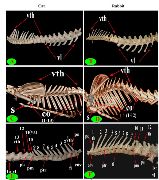
Figure 1: Vertebrae thoracicae (vth), Vertebrae lumbales (vl), Sternum (S), Costae (co), Vertebra anticlinalis (va), Processus spinosus (ps), Processus accessories (pa), Processus mamillaris (pm), Processus transversus (ptr), Foramen intervertebrale (fi) and Corpus vertebrae (cov).
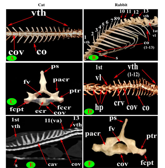
Figure 2: Vertebrae thoracicae (vth), Vertebrae lumbales (vl), Sternum (S), Costae (co), Vertebra anticlinalis (va), Processus spinosus (ps), Processus transversus (ptr), Corpus vertebrae (cov), Foramen vertebrale (fv), Fovea costalis processus transversi (fcpt), Extremitas cranialis (ecr), Fovea costalis cranialis (fccr), Processus articularis cranialis (pacr), Hypapophysis (infraspinous processes) (hp), Crista ventralis (crv) and Canalis vertebralis (cav).
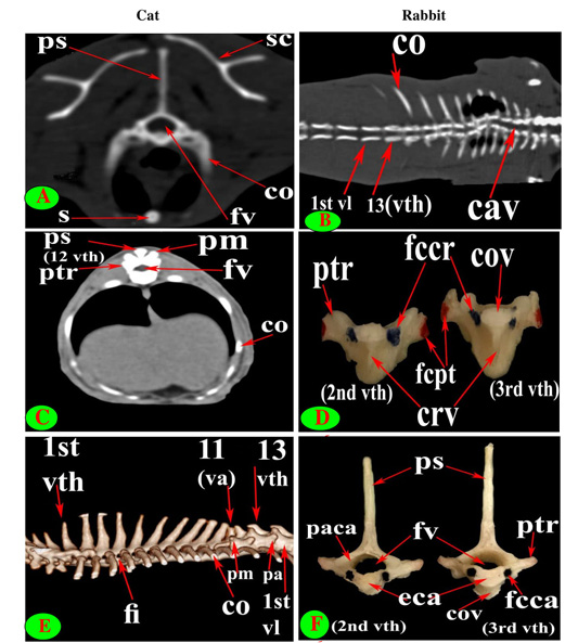
Figure 3: Vertebrae thoracicae (vth), Vertebrae lumbales (vl), Sternum (S), Costae (co), Vertebra anticlinalis (va), Processus spinosus (ps), Processus accessories (pa), Processus mamillaris (pm), Processus transversus (ptr), Foramen intervertebrale (fi), Corpus vertebrae (cov), Foramen vertebrale (fv), Fovea costalis processus transversi (fcpt), Fovea costalis cranialis (fccr), Crista ventralis (crv), Canalis vertebralis (cav), Scapula (sc), Processus articularis caudalis (paca), Extremitas caudalis (eca) and Fovea costalis caudalis (fcca).
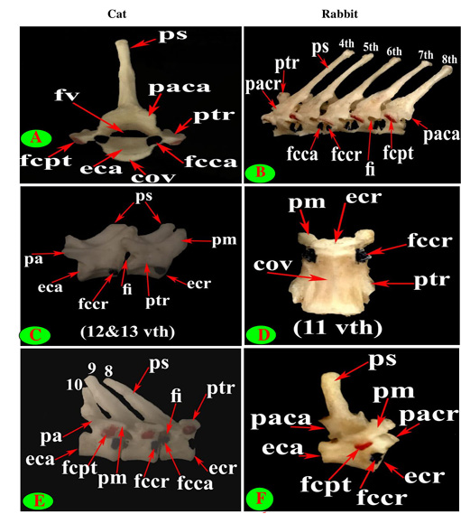
Figure 4: Vertebrae thoracicae (vth), Processus spinosus (ps), Processus accessories (pa), Processus mamillaris (pm), Processus transversus (ptr), Foramen intervertebrale (fi), Corpus vertebrae (cov), Foramen vertebrale (fv), Fovea costalis processus transversi (fcpt), Extremitas cranialis (ecr), Fovea costalis cranialis (fccr), Processus articularis cranialis (pacr), Processus articularis caudalis (paca), Extremitas caudalis (eca) and Fovea costalis caudalis (fcca).
close oval facets on the neural arch except the first two were larger and widely separated in cat (Figs. 2C and 5C), while in rabbit, it is was formed from two widely separated circular facets except the last two or three vertebrae were oval close facets on the neural arch (Fig. 5B and 5D). The caudal articular processes in the form of two close caudoventral facets situated at the root of spinous process and separated from it in the last three vertebrae in cat and last three or four in rabbit (Figs. 3F, 4A and 5A, B).
The spinous process of cat thoracic vertebrae was directed caudally till the anticlinal (11th) vertebra that had vertical spinous process and then inclined cranially in the last two (Figs. 1C, E; 2E and 3E). The rabbit thoracic spines divided into two directions; the first ten directed caudally and then directed cranially in the last two or three (Figs. 1D, F; 2B and 5F). All the cat spines were nearly equal with ill-defined gradual decrease in length, caudally except
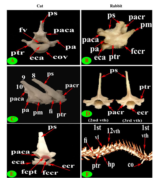
Figure 5: Vertebrae thoracicae (vth), Vertebrae lumbales (vl), Costae (co), Processus spinosus (ps), Processus accessories (pa), Processus mamillaris (pm), Processus transversus (ptr), Foramen intervertebrale (fi), Corpus vertebrae (cov), Foramen vertebrale (fv), Fovea costalis processus transversi (fcpt), Extremitas cranialis (ecr), Fovea costalis cranialis (fccr), Processus articularis cranialis (pacr), Hypapophysis (infraspinous processes) (hp), Processus articularis caudalis (paca) and Extremitas caudalis (eca).
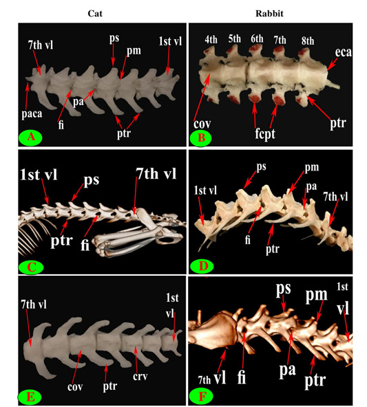
Figure 6: Vertebrae thoracicae (vth), Vertebrae lumbales (vl), Processus spinosus (ps), Processus accessories (pa), Processus mamillaris (pm), Processus transversus (ptr), Foramen intervertebrale (fi), Corpus vertebrae (cov), Fovea costalis processus transversi (fcpt), Crista ventralis (crv), Processus articularis caudalis (paca) and Extremitas caudalis (eca).
notable decline in-between the ninth and tenth (Figs. 1E, 2E, 3E and 4E). In rabbit, the spines gradually increase in length from the first to the third or fourth then gradually decrease caudally till the ninth and then continued with equal length to the last (Figs. 1F, 2B and 5F).
The supraspinous processes were long, narrow and ended by rounded summit in all cat vertebrae except the last three were shorter and wider. The spine had truncated summit in 12th and 13th but that of the eleventh (anticlinal) vertebra was in the form of triangular plate (Figs. 1E, 2E and 3E). The rabbit processes were thinner and ended by pointed summit in all vertebrae except the last four were short, broader and had truncated summit (Figs. 1F, 2B, 4B and 5B, F). All the cat and rabbit spines had cranial and caudal thin borders. The first four cat spines possessed a characteristic crest on both sides of their upper half ended distally by a tubercle (Fig. 2C).
The bodies of thoracic vertebrae devoid of infraspinous processes and represented by faint rough lines in first three and last two vertebrae in cat but this process in rabbit was well defined in the form of ventral crest in the first three and last four or five vertebrae (Fig. 2A and 2D).
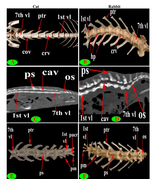
Figure 7: Vertebrae lumbales (vl), Processus spinosus (ps), Processus mamillaris (pm), Processus transversus (ptr), Corpus vertebrae (cov), Processus articularis cranialis (pacr), Hypapophysis (infraspinous processes) (hp), Crista ventralis (crv), Canalis vertebralis (cav) and Os sacrum (os).
The cat and rabbit transverse processes were paired, short and tuberous but stout in cat, it carried costal facets till the tenth one then replaced by low ridge without facets in the rest. The first four transverse costal facets were saddle-shaped then become rounded and convex in cat, while all rabbit tubercular facets were large saddle shape except the ninth and tenth that were small and convex (Figs. 1E, F; 4B, C, E, F; 5B, E and 6B).
The mamillary processes were small and paired that present in all thoracic vertebrae in cat and rabbit except the first two in cat (Figs. 1E, F; 2C; 4F and 5B). These mamillary processes in cat were located above the transverse process from third to eighth then bound with the accessory one in the form of bifid plate in the ninth and tenth vertebrae and separated from it to be beard on the cranial articular process in the last three (Figs. 1E, 4C, E and 5C). In rabbit, the mamillary processes were ill-defined above the transverse processes till be prominent and located in-between the transverse and the cranial articular processes in the tenth then beard on the cranial articular processes in the last three two or three thoracic vertebrae (Figs. 1F, 4F and 5B).
The cat accessory processes were separated caudally from the ninth and tenth mamillary processes to be in-between the caudal articular and transverse processes of last three vertebrae (Figs. 1E, 4C, E and 5C). Last two thoracic vertebrae in rabbit had accessory processes (Figs. 1F and 5B).

