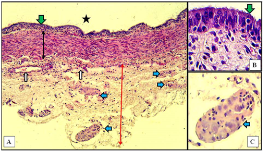
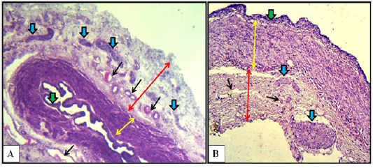
* Significant increased (p<0.05) in the thickness of the vaginal wall of the mature does compared to those of immature does
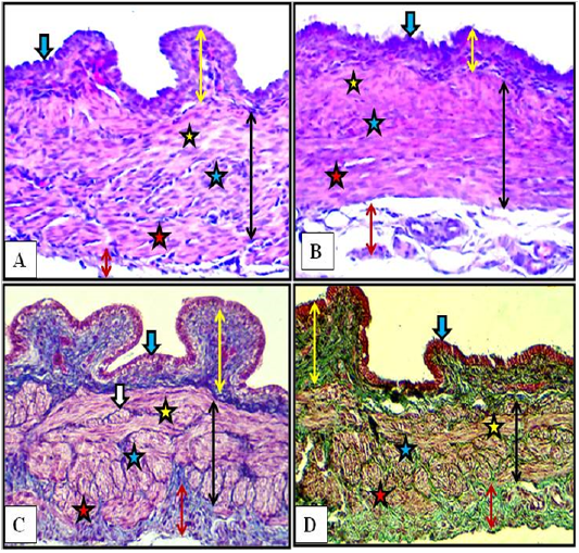
In immature does, the thicknesses of mucosa and muscularis were 41.34 µm and 279.42 µm at the cranial region, 47.72 µm and 240.40 µm at the middle region and at the caudal region were 60.70 µm and 199.30 µm. According to these measurements, the total thickness of mucosa plus muscularis was decreased in the direction cranio – caudally which were 320.76 ± 0.32 SE, 288.12 ± 0.22 SE and 260.00 ± 0.13 SE at the cranial, middle and caudal regions, respectively.
In mature does, the thicknesses of mucosa and muscularis were 44.08 µm and 302.09 µm at the cranial region, 54.15 µm and 281.82 µm at the middle region and at the caudal region were 67.18 µm and 238.26 µm. According to these measurements, the total thickness of mucosa plus muscularis was decreased in the direction cranio – caudally which were 346.17 ± 0.45 SE,335.97 ± 0.32 SE and 305.44 ± 0.27 SE at the cranial, middle and caudal regions, respectively.
Vestibule and Vulva
Microscopic examination of the vestibule showed similar histological structures in kittens, immature and mature does. Actually, there were no changes related to studied ages. As mentioned previously in the gross findings of the present results, the changes in this organ were just macromorphometric in nature. In another aspect, the microscopic picture showed slight changes between the cranial part and the caudal part of the vestibule in all ages of rabbits.
In kittens, the cranial part of the vestibule characterized by mucosa that lined with stratified squamous epithelium. That rest at lamina propria of loose connective tissue and invaded by blood vessels. Some of mucosal invaginations into the lamina propria were detected to develop subsequently into vestibular glands. The tubular structure surrounded by thin muscular layer of smooth muscles bundles arranged circularly. The thick loose connective tunica adventitia was invaded by numerous blood vessels (Figure 7-A). In the caudal part, same mucosa and mucosal invaginations were observed and the lamina propria filled with blood capillaries superficially and large vessels deeply with the presence of spars of circular smooth muscle fibers sunk into the surrounding connective tissue adventitia (Figure 7-B).
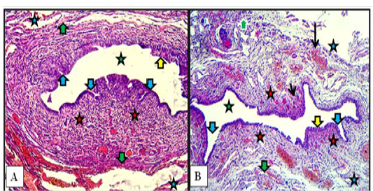
Figure 7: Cranial (A) and caudal (B) parts of vestibule of the female kitten showed mucosa of stratified squamous epithelium (yellow arrows), mucosal invaginations to form vestibular glands (blue arrows), lamina propria of loose connective tissue filled with blood vessels (red stars), superficial blood capillaries (short black arrow) and large vessels deeply (long black arrow), thin circular muscularis (green arrows), adventitia (blue stars) and lumen of vestibule (green stars). A & B: X100, H&E.
In immature does, similar vestibular wall structures were found with slight differences. In the cranial part nearby the urethral opening the circular smooth muscle bundles were more prominent than in that of kittens. In the cranial and caudal parts, some of newly formed vestibular glands were recorded (Figure 8).
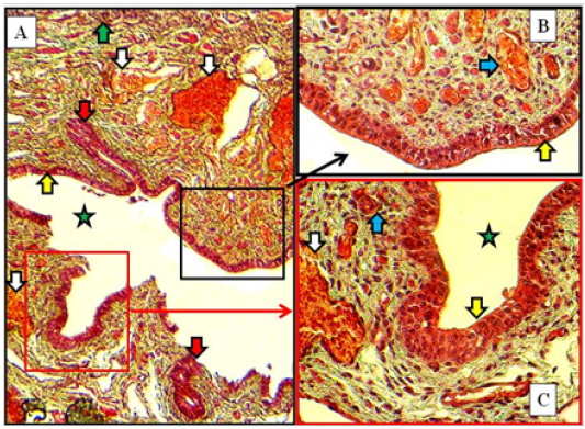
Figure 8: Caudal part of vestibule of immature doe showed stratified squamous epithelium (yellow arrows), lamina propria filled with blood capillaries superficially (blue arrows) and large vessels deeply (white arrows), vestibular glands (red arrow) and lumen (green star). A: X100. B & C: X400, MTC
In mature does, same structures as in immature does but the vestibular glands were increased in number and extended deeply into the lamina propria in both cranial and caudal parts of vestibule. The large blood vessels were prominently observed deeply in lamina propria adjacent to the thin muscularis of smooth muscle bundles (Figure 9).
The, Masson Trichrome and Gomori Trichrome were applied to stain the connective tissue structure present in the wall of the vestibule of the female kittens, immature does and mature does. The stains characterized as well the collagenous fibers which was constituted the lamina propria underneath the lining epithelium of the vestibule. Also they were clearly stained the connective tissue collagenous fibers interspersed between the muscular bundles of the tunica muscularis. The stains also stained the wide loose connective tissue constituents of the tunica adventitia (Figures 8 & 9).
The microscopic examination of the vulva showed no age related changes. Serial cross sections showed that this last part of the reproductive tract was lined with non keratinized stratified squamous epithelium that changed at the labia major into keratinized and pigmented with the presence of hairs bulbs. Thin layer of loose connective tissue under such epithelium was found. The surrounding wall to the vulvar cleft was made of strands of fine smooth muscle fibers rich by cavernous blood vessels. The smooth muscle strands arranged as inner longitudinally and outer circularly fibers (Figure 10).
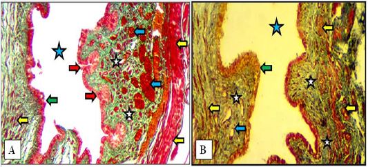
Figure 9: Cranial (A) and caudal (B) parts of the vestibule of the mature doe showed mucosa of stratified squamous epithelium (green arrows), lamina propria of loose connective tissue (white stars) filled with blood vessels (blue arrows), newly formed vestibular glands (red arrows), thin circular muscularis (yellow arrow) and lumen of vestibule (blue stars). A: X100, Gomori Trichrome. B: X200, MTC
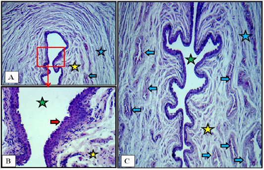
Figure 10: Cross section at the labia minor of vulva of immature doe showed vulvar cleft (green stars), stratified squamous epithelium (red arrows), cavernous vessels (blue arrows), inner longitudinal (yellow stars) and outer circular smooth muscle fibers (blue stars). A: X40. B: X400. C: X40. H&E