Research Article · Volume 10, Issue 7, 18 June 2022 · Open Access
Comparative Effects of Phyllanthus niruri and Plantago major in Carbon Tetrachloride Intoxicated Rats
· · ·
Affiliations & Notes ⌄
1Department of Clinical Pathology, Faculty of Veterinary Medicine, Suez Canal University, 41522, Egypt
2Department of Clinical Pathology, Faculty of Veterinary Medicine, Zagazig University, 44519, Egypt. *Correspondence | Noha Abdelnaeim, Department of Clinical Pathology, Faculty of Veterinary Medicine, Suez Canal University, 41522, Egypt
Additionally, in the PM-CCl4 group, MDA was significantly lower, whereas GSH was numerically higher and SOD was significantly higher.
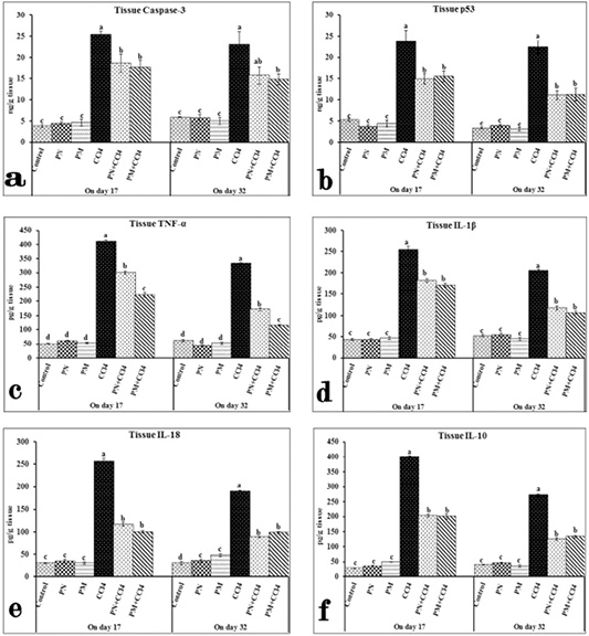
However, the PN-CCl4 and PM-CCl4 groups exhibited a considerably decreased caspase-3 and p53 compared to the CCl4 group.
However, PN pretreatment significantly improved the measured cytokines in the CCl4 intoxicated rats.
However, the PN-CCl4 and PM-CCl4 groups showed considerable amelioration in the hepatic tissue, which could be attributed to inflammatory reaction inhibition.
Selected from the article's own text.
Abstract
Liver damage is recognized as a severe global health issue. Phyllanthus niruri (PN) and Plantago major (PM) are herbal plants that are supposed to have hepatoprotective properties. This study aimed to compare the effects of both medicinal plant extracts on rats intoxicated with carbon tetrachloride (CCl4). A total of 60 male albino rats were equally distributed in six groups. The first group received purified water and was kept as a control. The second and third groups were given oral PN and PM (500 mg/kg/day) for 31 days, respectively. The fourth group was intraperitoneally injected with CCl4 (2 ml/kg/day) on days 15 and 16 of the experiment. The fifth and sixth groups received oral PN and PM (500 mg/kg/day), respectively, for 31 days and were injected with CCl4 on days 15 and 16. On days 17 and 32 of the trial, liver specimens were gathered for estimation of malondialdehyde (MDA), antioxidants, apoptotic markers, cytokines, and histopathological changes. Our results revealed a significantly increased MDA, caspase-3, p53, tumor necrosis factor-α, interleukin (IL)-1β, IL-18 and, IL-10, and a significant decline in glutathione and superoxide dismutase in the CCl4 group. However, intoxicated groups treated with PN and PM showed marked improvement in the measured parameters. Therefore, PN and PM have antioxidant and anti-inflammatory effects, especially PM, which showed better improvement than PN against CCl4 hepatotoxicity in rats.
The liver is vital for metabolizing and detoxifying medicines, environmental toxins, microbial metabolites, and certain pharmaceutical preparations, which are the leading causes of hepatic injury worldwide ; . Carbon tetrachloride (CCl4) is a xenobiotic that is commonly utilized to initiate hepatic tissue damage in laboratory animals . In the liver, cytochrome P450 transformed CCl4 into highly reactive metabolites, resulting in an excess of free radicals and a drop in hepatic antioxidant markers . Consequently, reducing the free radicals could combat hepatotoxicity . Several studies have proved the antioxidant properties of food-derived phenolic compounds, which protect the liver against hepatotoxicity . Hence, herbal alternatives have received much consideration as safe treatments for this issue .
Phyllanthus niruri (PN) is a plant related to the Euphorbiaceae family that possesses various medicinal properties, such as antiulcer, antitumor, anticarcinogenic, hypolipidemic, antiviral, and antioxidant activities . Many disorders can be treated with PN, including dyspepsia, diuretics, jaundice, hyperglycemia, and kidney stones removal . PN is an effective treatment for gallstones and kidney stones .
Plantago major (PM) belongs to the Plantaginaceae family . PM contains a variety of bioactive chemicals, including flavonoids, pectin, terpenoids, iridoid, tannins, and glycosides that have antioxidant and anti-inflammatory properties ; . Previous studies discovered that this herb has other therapeutic properties, such as hepatoprotective , immunomodulating, and anticarcinogenic activities .
This study aimed to assess the effects of PN and PM on the redox state, apoptotic markers, pro-inflammatory, and anti-inflammatory cytokines, as well as histopathological examination in CCl4-intoxicated rats.
MATERIALS AND METHODS
Chemicals
CCl4 was procured from Merck (Darmstadt, Germany). The kits used to assess oxidative stress and antioxidant biomarkers were bought from Biodiagnostic Co. (Giza, Egypt). Caspase-3 and tumor protein P53 were evaluated using the kits from MyBioSource (San Diego, USA). Cytokine kits were acquired from Boster Biological Technology (California, U.S.A).
Preparation of plant extracts
PN was obtained from Madurai (Tamil Nadu, India) and PM from the canal banks of the Nile delta (Egypt). The dried aerial parts of each plant (1500 g) were turned into powder using a mortar and ceramic grinder. Cold extraction was performed on the dried plant matter by soaking it in 70% ethanol for 2 days at ambient temperature with discontinuous shaking. Afterward, it was filtered using Whatman filter paper No. 1 (125 mm). The filtrate was dried in an oven at 40 °C for 2-3 hours daily to obtain a semi-solid mass. The dried extract was then weighed and stored in container at 4 ºC ; .
Animals and experimental design
Sixty healthy albino male rats (150–180 g B.Wt) were purchased from the Laboratory Animal House of the Faculty of Veterinary Medicine, Suez Canal University in Egypt. The animals were kept in separate, clean, and disinfected metal cages (10 rats per cage) with a 12-hour light/darkness cycle and a constant temperature of approximately 25 °C. Water and food were regularly given to the animals. The experimental scheme was accepted by the Research Ethical Committee of the Faculty of Veterinary Medicine, Suez Canal University, Ismailia, Egypt (approval no. 2019015). All possible measures were taken to reduce rat suffering.
All rats were given a 2-week acclimatization period before the start of the study. Six equal groups of rats were formed. The first group received purified water and was kept as a control, while the second group was given PN (500 mg/kg B.Wt) daily by stomach tube for 31 days . The PM was given to the third group at a dosage of 500 mg/kg B.Wt every day by stomach tube for 31 days . The fourth group (CCl4) intraperitoneally received 2 ml/kg B.Wt/day of CCl4 dissolved in olive oil (1:1) on days 15 and 16 of the trial. The fifth and sixth groups were given 500 mg/kg B.Wt of PN and PM extracts, respectively, by stomach tube each day, followed by CCl4 injections on days 15 and 16, and then PN and PM extracts for another 2 weeks.
Tissue sampling
The rats were sedated with isoflurane and then euthanized, and liver tissues were extracted for redox state, apoptotic markers, cytokines, and histological analyses on days 17 and 32 of the trial. One gram of liver sample was collected and homogenized with 5–10 ml of chilled buffer. The homogenate was centrifuged for 30 min at 5000 rpm to eliminate cell debris. The supernatant was stored at -80 ºC to assess the malondialdehyde (MDA) levels, reduced glutathione (GSH), and superoxide dismutase (SOD) levels, in addition to apoptotic markers (caspase-3 and tumor protein P53), and cytokines (tumor necrosis factor [TNF]-α, interleukin [IL-]-1β, IL-18, and IL-10). Liver samples from each group were also taken and preserved in 10% formalin for histological evaluation.
The levels of caspase-3, tumor protein P53 (P53), TNF˗α, IL-1, IL18, and IL10 in tissue homogenates were determined using an enzyme-linked immunosorbent assay as reported by Somade et al. (2020).
Histopathology
Liver tissue specimens were fixed for at least 1 day in 10% formalin. All fixed samples were dehydrated before embedding in paraffin. Then thin paraffin sections were prepared and stained with hematoxylin and eosin as reported by Bancroft et al. (1996).
Statistical analysis
The Statistical Package for the Social Science software version 20.0 was used to collect data and perform a one-way analysis of variance. Then, Tukey’s multiple comparison test was used to compare the means. P-values of ≤0.05 were considered significant. The results were presented as means ± standard errors .
RESULTS
Hepatic MDA and antioxidant biomarkers
Table 1 shows the effects of different treatments on the hepatic MDA and antioxidant biomarkers (GSH and SOD) on days 17 and 32 of the experiment. The groups treated with PN and PM showed no significant changes in the MDA levels compared with the control group. However, in the CCl4 intoxicated group, CCl4 administration resulted in a considerable rise compared to the control. Meanwhile, in the PN-CCl4 and PM-CCl4 groups, the pretreatment with PN or PM caused a marked decrease in liver MDA content compared to the CCl4 group. The hepatic GSH did not display any considerable changes in the PN and PM groups compared with the control group. However, the CCl4 intoxicated group showed a substantial decrease in GSH compared to the control. The PN-CCl4 and PM-CCl4 groups showed numerical elevations compared with the CCl4 group. The PN and PM groups did not show any significant changes in the SOD levels compared to the control. SOD dramatically declined in the CCl4 compared to the control. On day 17, the PN-CCl4 and PM-CCl4 groups revealed a numerical increase in the SOD level compared to the CCl4 group. However, on day 32, SOD numerically increased in the PN-CCl4 group, while it dramatically increased in the PM-CCl4 group compared with the CCl4 group.
Tissue apoptotic markers
The effects of different treatments on apoptotic markers on days 17 and 32 of the experiment are shown in Figure 1. The liver apoptotic marker concentrations (caspase-3 and P53) in the PN and PM groups were not significantly different from those in the control group. Conversely, the CCl4 intoxication significantly raised the apoptotic markers in the CCl4 group compared with the control. On day 17 of the experiment, the PN-CCl4 and PM-CCl4 groups exhibited a marked decrease in caspase-3 and P53 compared to the CCl4 group. However, on day 32 of the experiment, caspase-3 numerically decreased in the PN-CCl4 group but dramatically decreased in the PM-CCl4 group compared to the CCl4 group. P53 significantly decreased in both the PN-CCl4 and PM-CCl4 groups compared to the CCl4 group.
Table 1: Hepatic malondialdehyde and antioxidant biomarkers in different animal groups at days 17 and 32 post-treatment.
Groups
Parameters
On day 17 post treatments
On day 32 post treatments
MDA
(nmol /g tissue)
GSH
(mmol /g tissue)
SOD
(U /g tissue)
MDA
(nmol /g tissue)
GSH
(mmol /g tissue)
SOD
(U /g tissue)
Control
5.47 ± 0.15c
44.67 ± 1.45a
67.33 ± 1.20ab
5.27 ± 0.38c
49.67 ± 0.88a
69.67 ± 1.45a
PN
6.10 ± 0.96c
48.0 ± 1.73a
77.33 ± 2.03a
4.73 ± 0.74c
48.33 ± 0.88a
76.33 ± 2.03a
PM
6.27 ± 0.95c
48.33 ± 3.76a
62.33 ± 2.60b
5.43 ± 0.39c
50.33 ± 1.45a
73.33 ± 3.84a
CCl4
42.33 ± 3.76a
11.70 ± 1.68b
28.33 ± 0.88c
25.33 ± 2.03a
19.33 ± 0.88b
34.0 ± 3.21c
PN+ CCl4
21.27 ± 0.93b
21.27 ± 3.98b
36.67 ± 2.60c
14.17 ± 1.60b
26.0 ± 2.52b
39.33 ± 0.88bc
PM+ CCl4
19.63 ± 2.30b
21.0 ± 2.21b
35.67 ± 3.38c
11.37 ± 0.78b
25.67 ± 2.40b
47.33 ± 0.33b
Data are expressed as means ± SEM (n = 10). Mean values within the same column having different superscript letters are significant at P ≤ 0.05. PN: Phyllanthus niruri; PM: Plantago major; CCl4: carbon tetrachloride.
Cytokines
Figure 1 reveals the influence of CCl4 and different treatments on the pro-inflammatory cytokines (TNF-α, IL-1β, and IL18) and the anti-inflammatory cytokine (IL10) in the hepatic tissues of different animal groups on days 17 and 32 of the experiment. All the measured cytokines showed no significant variations in the PN and PM groups compared to the control group. However, intoxication with CCl4 caused a considerable rise in the cytokines, compared to the control group. All cytokine levels in the PN-CCl4 and PM-CCl4 groups revealed a considerable decrease compared to the CCl4 group.
Histopathology
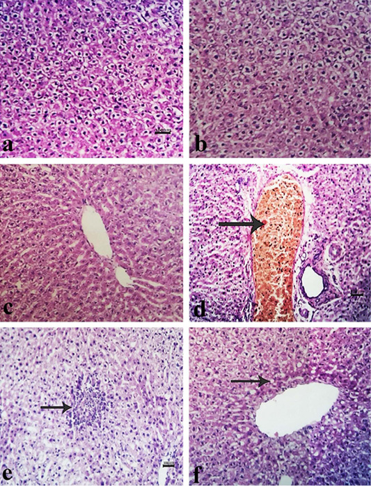
The histopathological changes in separate groups on day 17 of the experiment are presented in Figure 2a-f. The livers of the control group revealed normal tissue architecture and cellular features. The livers of most rats that are given ethanolic PN extract exhibited normal hepatic parenchyma along with mild hepatic blood vessel congestion in a few cases. Hepatic tissue appeared to be normal in the PM group, with slight sinusoidal dilatation in some cases. Liver sections from the CCl4 group, revealed diffuse hepatic cell vacuolation, a periductal proliferation of fibroblast cells, and von kupffer cells with mild perivascular edema and hepatic blood vessel congestion. Mild to moderate hyperplasia of bile ducts and focal necrosis of some hepatic cells were also observed. Liver sections in the PN-CCL4 group exhibited a focal area of fibroblast and von kupffer cell proliferation with only a few mononuclear cell infiltrations and mild to moderate vacuolar degeneration of hepatocytes. Diffuse vacuolar degeneration was seen in the liver sections of the PM-CCL4 group.
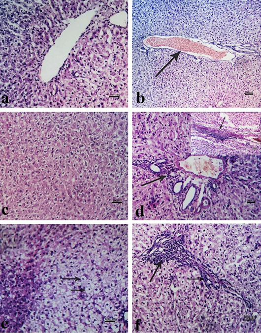
Figure 3a-f illustrates the histopathological alterations in different groups on day 31 of the experiment. The liver of the normal group showed hepatic parenchyma with regular tissue construction and cellular features. On a few occasions, liver sections from the PN group exhibited minor hepatic blood vessel congestion. The hepatic tissue architecture and cellular features in the liver of rats that were given PM ethanolic extract appeared to be normal. Contrastingly, the hepatic tissue from the CCl4 group demonstrated perivascular edema, a focal area of coagulative necrosis, and hepatic blood vessel congestion. A considerably localized aggregation of proliferated fibroblasts and von kupffer cells were seen in some liver sections. However, the liver from the PN-CCL4 group showed a focal region of hepatic vacuolation, which was characterized by hydropic degeneration. A mild focal area of proliferating fibroblasts was seen in a few cases in the PM-CCL4 group, along with mildly dilated sinusoids and moderate vacuolation of hepatocytes.
DISCUSSION
The pathogenesis of liver disorders is thought to be influenced by oxidative stress . Cytochrome P450 biotransforms CCl4 into free radicals in the liver, causing membrane lipid breakdown and lipid peroxidation. Eventually, cell membrane integrity is lost, resulting in liver injury . Our findings revealed that the CCl4 administration induced a considerable rise in the hepatic MDA content and a marked drop in antioxidant biomarker concentrations (GSH and SOD). These findings matched those of Alayunt et al. (2019), who stated that rats intoxicated with CCl4 had higher serum MDA levels and lower GSH concentrations. The PN-CCl4 group exhibited a significant MDA level reduction and a numerical GSH and SOD elevation. Our findings supported previous studies, which found a decline in MDA and a rise in GSH and SOD after using PN ; . This could be attributed to the efficacy of PN in liver disease treatment by inhibiting reactive oxygen species and lipid peroxidation . Additionally, in the PM-CCl4 group, MDA was significantly lower, whereas GSH was numerically higher and SOD was significantly higher. These investigations came in line with Hussan et al. (2015a), who reported an improved MDA and an increased in GSH and SOD after PM administration, which possesses antioxidant action due to the incidence of high phenolic compounds, flavonoids, alkaloids, terpenoids, and vitamin C levels.
Apoptosis is a basic cellular mechanism that helps prevent tumorigenesis by removing damaged cells in different physiological and pathological conditions . Our study revealed that, caspase 3 levels significantly increased in CCl4- intoxicated rats, which may be due to oxidative stress and inflammatory induction . Additionally, rats intoxicated with CCl4 revealed a marked increase in the P53 level, which is accumulated in hepatocytes in liver diseases as a result of hepatocyte apoptosis . Our findings were coming in harmony with Guo et al. (2013), who reported that CCl4 upregulates the p53 expression. However, the PN-CCl4 and PM-CCl4 groups exhibited a considerably decreased caspase-3 and p53 compared to the CCl4 group. This decreased apoptotic markers indicated an improved hepatic injury.
Excessive inflammation is also an essential feature related to CCl4-induced liver damage . The free radicals resulting from CCl4 intoxication are also thought to generate the inflammatory response in the liver by activating macrophages, which then generate TNF and further pro-inflammatory cytokines . These cytokines, particularly TNF- and IL-1β, have a critical function in liver damage, by maintaining hepatic inflammation . Thereby, enhancing the antioxidant system as a key factor in preventing oxidative stress in the liver . Our study revealed considerably increased pro-inflammatory cytokines (TNF-α, IL-1β, and IL18) and anti-inflammatory cytokine (IL10) in the hepatic tissues of CCl4-intoxicated rats. Our results agreed with Zhao et al. (2021), who found an increased TNF-α, IL-1β, and IL-18 blood levels in mice after CCl4 intoxication. Additionally, our results were consistent with Ezzat et al. (2020), who revealed that CCl4 intoxication significantly increased IL10. However, PN pretreatment significantly improved the measured cytokines in the CCl4 intoxicated rats. This could be due to PN’s anti-inflammatory qualities, as indicated by cytokine inhibition as a probable method of hepatoprotection, besides its antioxidant capabilities . Similarly, the PM-CCl4 group exhibited significant cytokine amelioration due to the anti-inflammatory property of the PM . The cytokine results in different groups were confirmed by the histopathological examination. The hepatic tissues of the CCl4 intoxicated rats revealed inflammation signs, such as localized proliferated fibroblast and von kupffer cell aggregation. The persistent inflammatory response has the potential to enhance tissue damage . However, the PN-CCl4 and PM-CCl4 groups showed considerable amelioration in the hepatic tissue, which could be attributed to inflammatory reaction inhibition.
In conclusion, CCl4 produced severe toxicity in rats. PN and PM supplementation curtailed the toxic effects of CCl4 as they exhibited markedly improved in antioxidant status, apoptotic markers, and inflammatory cytokines. Histopathological examination established the ameliorative potentials of both plant extracts as reflected in liver histoarchitecture restoration that was initially distorted by the toxicant. PM was more beneficial than PN. However, both plants have potentials that can be exploited in liver disease management.
Novelty Statement
This study was the first to demonstrate the ameliorative effect of Phyllanthus niruri and Plantago major on the levels of the apoptotic markers (caspase-3 and tumor protein P53) against carbon tetrachloride toxicity. This indicates that these herbal plants have an anti-apoptotic effect.
Author’s Contribution
AK: Sample collection and lab analysis. NA: Write the manuscript. OA & MH: Idea, design and revision.
Conflict of interest
The authors have declared no conflict of interest.
Figures
Figure 1:.
Protective effects of Phyllanthus niruri (PN) and Plantago major (PM) on CCl4 intoxicated rats. (A) Tissue Caspase-3; (B) Tissue P53; (C) TNF-α; (D) IL-1β; (E) IL-18; (F) IL-10. Data show the mean ± SEM (n= 10). Columns with different superscripts refer to significant differences (P ≤ 0.05) between the groups.
Figure 2:.
Liver sections of rats on day 17 of the experiment showing (a, b, c) normal hepatic tissue. Moderate blood vessel congestion (arrow), perivascular edema, bile duct hyperplasia, and degenerated hepatocytes (d). A mild focal area of fibroblastic proliferation (arrow) along with mild to moderate vacuolar degeneration of hepatocytes (e). Mild vacuolation of hepatocytes (arrow) (f). (a) control group; (b) Phyllanthus niruri (PN)group; (c) Plantago major (PM) group; (d) CCl4 group; (e) PN-CCl4 group; (f) PM-CCl4 group.
Figure 3:.
Liver sections of rats on day 32 of the experiment showing (a, b, c) fairly normal hepatic tissue in addition to mild congestion (b). Moderate congestion, bile duct hyperplasia (arrow), degenerated hepatocytes, and severe focal fibrosis (arrow in the window) (d). Mild vacuolation of hepatocytes (arrow) (e). A focal area of fibroblastic proliferation along with moderate hepatocytes vacuolation (arrow) (f). (a) control group; (b) Phyllanthus niruri (PN) group; (c) Plantago major (PM) group; (d) CCl4 group; (e) PN-CCl4 group; (f) PM-CCl4 group.
References
Alayunt N, Aksoy L, Karafakioğlu YS, Sevimli S (2019). Assessment of anti-inflammatory and antioxidant properties of safranal on CCI4-induced oxidative stress and inflammation in rats. Ann. Acad. Bras. Cienc., 91(2): e20181235. https://doi.org/10.1590/0001-3765201920181235[CrossRef]
Bagalkotkar G, Sagineedu S, Saad M, Stanslas J (2006). Phytochemicals from Phyllanthus niruri Linn. and their pharmacological properties. A review. J. Pharm. Pharmacol., 58(12): 1559-1570. https://doi.org/10.1211/jpp.58.12.0001[CrossRef]
Bancroft J, Stevens A, Turner DJM (1996). Theory and practice of histological techniques 4th ed. Churchill Living Stone, New York Edinburgh, 20.
Baskaran M, Periyasamy L, Rajagopalan R (2010). Effect of Phyllanthus niruri on alcohol and polyunsaturated fatty acid induced oxidative stress in liver. Int. J. Pharm. Pharm. Sci., 2(4): 58-62.
Beutler E, Duron O, Kelly BM (1963). Improved method for the determination of blood glutathione. J. Lab. Clin. Med., 61: 882-888.
Chughtai B (2016). Healing in urology: Clinical guidebook to herbal and alternative therapies, World Scientific, Singapore, pp. 280. https://doi.org/10.1142/9758[CrossRef]
El-Sayed YS, Lebda MA, Hassinin M, Neoman SA (2015). Chicory (Cichorium intybus L.) root extract regulates the oxidative status and antioxidant gene transcripts in CCl4-induced hepatotoxicity. PLoS One, 10(3): e0121549. https://doi.org/10.1371/journal.pone.0121549[CrossRef]
Eldesoky AH, Abdel-Rahman RF, Ahmed OK, Soliman GA, Saeedan AS, Elzorba HY, Elansary AA, Hattori M (2018). Antioxidant and hepatoprotective potential of Plantago major growing in Egypt and its major phenylethanoid glycoside, acteoside. J. Food Biochem., 42(5): e12567. https://doi.org/10.1111/jfbc.12567[CrossRef]
Ezzat MI, Okba MM, Ahmed SH, El-Banna HA, Prince A, Mohamed SO, Ezzat SM (2020). In-depth hepatoprotective mechanistic study of Phyllanthus niruri: In vitro and in vivo studies and its chemical characterization. PLoS One, 15(1): e0226185. https://doi.org/10.1371/journal.pone.0226185[CrossRef]
Gressner OA, Weiskirchen R, Gressner AM (2007). Biomarkers of liver fibrosis: clinical translation of molecular pathogenesis or based on liver-dependent malfunction tests. Clin. Chim. Acta, 381(2): 107-113. https://doi.org/10.1016/j.cca.2007.02.038[CrossRef]
Guo XL, Liang B, Wang XW, Fan FG, Jin J, Lan R, Yang, JH, Wang XC, Jin L, Cao Q (2013). Glycyrrhizic acid attenuates CCl₄-induced hepatocyte apoptosis in rats via a p53-mediated pathway. World J. Gastroenterol., 19(24): 3781-3791. https://doi.org/10.3748/wjg.v19.i24.3781[CrossRef]
Hariri AT, Moallem SA, Mahmoudi M, Memar B, Hosseinzadeh H (2010). Sub-acute effects of diazinon on biochemical indices and specific biomarkers in rats: protective effects of crocin and safranal. Food Chem. Toxicol., 48(10): 2803-2808. https://doi.org/10.1016/j.fct.2010.07.010[CrossRef]
Hussan F, Basah RHO, Yusof MRM, Kamaruddin NA, Othman F (2015a). Plantago major treatment enhanced innate antioxidant activity in experimental acetaminophen toxicity. Asian Pac. J. Trop. Biomed., 5(9): 728-732. https://doi.org/10.1016/j.apjtb.2015.06.013[CrossRef]
Hussan F, Mansor AS, Hassan, SN, Kamaruddin TNE, Tasnim TN, Budin SB, Othman F (2015b). Anti-inflammatory property of Plantago major leaf extract reduces the inflammatory reaction in experimental acetaminophen-induced liver injury. Evid. Based Complement. Altern. Med., 2015: 347861. https://doi.org/10.1155/2015/347861[CrossRef]
Kanter M, Coskun O, Budancamanak M (2005). Hepatoprotective effects of Nigella sativa l and Urtica dioica l on lipid peroxidation, antioxidant enzyme systems and liver enzymes in carbon tetrachloride-treated rats. World J. Gastroenterol., 11(42): 6684-6688. https://doi.org/10.3748/wjg.v11.i42.6684[CrossRef]
Landau S, Everitt BS (2003). A handbook of statistical analyses using SPSS, Chapman and Hall/CRC, New York. https://doi.org/10.1201/9780203009765[CrossRef]
Lee KJ, Jeong HG (2002). Protective effect of Platycodi radix on carbon tetrachloride-induced hepatotoxicity. Food Chem. Toxicol., 40(4): 517-525. https://doi.org/10.1016/S0278-6915(01)00104-1[CrossRef]
Li R, Yang W, Yin Y, Ma X, Zhang P, Tao K (2021a). 4-OI attenuates carbon tetrachloride-induced hepatic injury via regulating oxidative stress and the inflammatory response. Front. Pharmacol., 12: 651444. https://doi.org/10.3389/fphar.2021.651444[CrossRef]
Li X, Liu X, Zhang Y, Cheng C, Fan J, Zhou J, Garstka MA, Li Z (2021b). Hepatoprotective effect of apolipoprotein A4 against carbon tetrachloride induced acute liver injury through mediating hepatic antioxidant and inflammation response in mice. Biochem. Biophys. Res. Commun., 534: 659-665. https://doi.org/10.1016/j.bbrc.2020.11.024[CrossRef]
Liu B, Shi Y, Peng W, Zhang Q, Liu J, Chen N, Zhu R (2016). Diosmetin induces apoptosis by upregulating p53 via the TGF-β signal pathway in HepG2 hepatoma cells. Mol. Med. Rep., 14(1): 159-164. https://doi.org/10.3892/mmr.2016.5258[CrossRef]
Liu J, Wang X, Liu R, Liu Y, Zhang T, Fu H, Hai C (2014). Oleanolic acid co-administration alleviates ethanol-induced hepatic injury via Nrf-2 and ethanol-metabolizing modulating in rats. Chem. Biol. Interact., 221: 88-98. https://doi.org/10.1016/j.cbi.2014.07.017[CrossRef]
Manjrekar AP, Jisha V, Bag PP, Adhikary B, Pai MM, Hegde A, Nandini M (2008). Effect of Phyllanthus niruri Linn. treatment on liver, kidney and testes in CCl4 induced hepatotoxic rats. Indian J. Exp. Biol., 46(7): 514-520.
Muhammad AA, Ibrahim RB, Aminu C, Abbas AY, Kabiru A, Bisallah CI (2020). Activity of aqueous extract of Phyllanthus niruri Linn in 2, 4-dinitrophenylhydrazine induced anaemic rats. J. Pharm. Pharmacol., 4(3): 79-95.
Nazarizadeh A, Mikaili P, Moloudizargari M, Aghajanshakeri S, Javaherypour S (2013). Therapeutic uses and pharmacological properties of Plantago major L. and its active constituents. J. Basic Appl. Sci. Res., 3(9): 212-221.
Nishikimi M, Rao NA, Yagi K (1972). The occurrence of superoxide anion in the reaction of reduced phenazine methosulfate and molecular oxygen. Biochem. Biophys. Res. Commun., 46(2): 849-854. https://doi.org/10.1016/S0006-291X(72)80218-3[CrossRef]
Nofal ZAE, El-Maghraby AF, Gad MR, Farghaly AA, Ahmed EM (2016). Evaluation of the protective effect of ethanolic extract of Plantago major plant against CCl4 induced genotoxicity and biochemical disturbance in Rat. Egypt. J. Vet. Sci., 47(1): 83-110. https://doi.org/10.21608/ejvs.2016.1082[CrossRef]
Ohkawa H, Ohishi N, Yagi K (1979). Assay for lipid peroxides in animal tissues by thiobarbituric acid reaction. Anal Biochem., 95(2): 351-358. https://doi.org/10.1016/0003-2697(79)90738-3[CrossRef]
Pandit A, Sachdeva T, Bafna P (2012). Drug-induced hepatotoxicity: A review. J. Appl. Pharm. Sci., 2(5): 233-243. https://doi.org/10.7324/JAPS.2012.2541[CrossRef]
Shin DS, Kim KW, Chung HY, Yoon S, Moon JO (2013). Effect of sinapic acid against carbon tetrachloride-induced acute hepatic injury in rats. Arch. Pharm. Res., 36(5): 626-633. https://doi.org/10.1007/s12272-013-0050-5[CrossRef]
Siegel R, Ma J, Zou Z, Jemal A (2014). Cancer statistics, 2014. CA Cancer J. Clin., 64(1): 9-29. https://doi.org/10.3322/caac.21208[CrossRef]
Somade OT, Ajayi BO, Olunaike OE, Jimoh LA (2020). Hepatic oxidative stress, up-regulation of pro-inflammatory cytokines, apoptotic and oncogenic markers following 2-methoxyethanol administrations in rats. Biochem. Biophys. Rep., 24: 100806. https://doi.org/10.1016/j.bbrep.2020.100806[CrossRef]
Upadhyay G, Singh AK, Kumar A, Prakash O, Singh MP (2008). Resveratrol modulates pyrogallol-induced changes in hepatic toxicity markers, xenobiotic metabolizing enzymes and oxidative stress. Eur. J. Pharmacol., 596(1-3): 146-152. https://doi.org/10.1016/j.ejphar.2008.08.019[CrossRef]
Venkatesh P, Dinakar A, Senthilkumar N (2010). Hepatoprotective activity of ethanolic extract of the stems of Anisochilus carnosus against carbon tetrachloride-induced hepatotoxicity in rats. Int. J. Health Res., 3(3): 179-183. https://doi.org/10.4314/ijhr.v3i3.70303[CrossRef]
Yu H, Zheng L, Yin L, Xu L, Qi Y, Han X, Xu Y, Liu K, Peng J (2014). Protective effects of the total saponins from Dioscorea nipponica Makino against carbon tetrachloride-induced liver injury in mice through suppression of apoptosis and inflammation. Int. Immunopharmacol., 19(2): 233-244. https://doi.org/10.1016/j.intimp.2014.01.019[CrossRef]
Zhang Y, He Y, Yu H, Ma F, Wu J, Zhang X (2015). Liquiritigenin protects rats from carbon tetrachloride induced hepatic injury through PGC-1α pathway. Evid. Based Complement. Altern. Med., 2015: 649568. https://doi.org/10.1155/2015/649568[CrossRef]
Zhao J, He B, Zhang S, Huang W, Li X (2021). Ginsenoside Rg1 alleviates acute liver injury through the induction of autophagy and suppressing NF-κB/NLRP3 inflammasome signaling pathway. Int. J. Med. Sci., 18(6): 1382-1389. http://doi.org/10.7150/ijms.50919[CrossRef]
Zubair M, Nybom H, Lindholm C, Rumpunen K (2011). Major polyphenols in aerial organs of greater plantain (Plantago major L.), and effects of drying temperature on polyphenol contents in the leaves. Sci. Hortic., 128(4): 523-529. https://doi.org/10.1016/j.scienta.2011.03.001[CrossRef]