Research Article · Volume 14, Issue 10, 23 September 2026 · Open Access
Characterization of Canine Parvovirus 2c in Dogs from Vinh Long, Vietnam
· ·
Affiliations & Notes ⌄
1School of Basic Science, Tra Vinh University, Vinh Long 85000, Vietnam
2Animal Science and Veterinary Medicine Department, Agriculture and Aquaculture Faculty, Tra Vinh University, Vinh Long 85000, Vietnam
3Faculty of Veterinary Medicine, College of Agriculture, Can Tho University, Can Tho 94000, Vietnam. *Correspondence | Nguyen Thi Yen Linh, School of Basic Science, Tra Vinh University, Hoa Thuan Ward, Vinh Long 85000, Vietnam
Among 466 enrolled dogs with clinical suspicion of CPV-2-associated enteritis, 103 were PCR-positive, corresponding to a clinic-based suspected-case positivity of 21.6 (17.0–27.0).
Owner-reported vaccination category was strongly associated with CPV-2 positivity in the unadjusted analysis (χ² = 128.56, df = 2, p < 0.0001; Table 4).
Likewise, the lower positivity observed in dogs reported as fully vaccinated should not be interpreted as a direct estimate of vaccine effectiveness.
Among clinic-presenting dogs with suspected parvoviral enteritis in Vinh Long, CPV-2 PCR positivity was 22.1% and was highest in younger dogs and dogs reported as unvaccinated in unadjusted analyses; breed differences were not statistically supported.
Selected from the article's own text.
Abstract
Canine parvovirus type 2 (CPV-2) remains an important cause of acute enteritis in dogs, while current clinic-based molecular data from southern Vietnam remain limited. In this cross-sectional study, CPV-2 DNA was detected in 103 of 466 clinically suspected dogs (22.1%; 95% CI: 18.6–26.1) presented to three veterinary clinics in Tra Vinh Ward and Nguyet Hoa Ward, Vinh Long Province, Vietnam, between March 2025 and February 2026. Rectal swabs were screened by PCR targeting a partial VP2 fragment, and unadjusted positivity was compared across age, breed, and owner-reported vaccination categories. Clinical signs were summarized descriptively among PCR-positive dogs. Nine purposively selected positive samples (9/103, 8.7%) underwent bidirectional Sanger sequencing. Positivity was highest in dogs aged 3–4 months (37.7%) and in dogs reported as unvaccinated (50.3%), whereas the overall breed comparison was not statistically significant. Anorexia, vomiting, lethargy, and diarrhea were common among PCR-positive dogs; these frequencies should be interpreted descriptively because the signs also contributed to the enrollment case definition and were not compared with PCR-negative dogs. All nine sequenced samples were classified as CPV-2c and shared the VP2 426E/447M profile within the analyzed fragment, clustering with contemporary Vietnamese and Chinese CPV-2c references. The cross-sectional association with reported vaccination history does not estimate vaccine effectiveness or establish causality. Rather, the findings are consistent with existing recommendations to complete the primary puppy vaccination series and provide a regional snapshot supporting continued molecular surveillance of circulating CPV-2 variants.
Keywords | Canine parvovirus type 2, Cross-sectional study, Dogs, Phylogeny, Vinh Long Province, Vietnam
INTRODUCTION
Canine parvovirus type 2 (CPV-2) is a small, non-enveloped, single-stranded DNA virus of the genus Protoparvovirus, family Parvoviridae, and remains a major viral enteropathogen of dogs. Infection is most severe in puppies and young dogs because the virus targets rapidly dividing cells, particularly intestinal crypt epithelium, lymphoid tissues, and bone marrow. Typical disease includes anorexia, vomiting, lethargy, dehydration, leukopenia, and either non-hemorrhagic or hemorrhagic diarrhea; severe cases may show intestinal congestion or hemorrhage, fluid-filled bowel segments, and lymphoid depletion, while microscopic lesions commonly include crypt necrosis, villous collapse, and inflammatory or necrotic changes in intestinal and lymphoid tissues ; ; ; . Control therefore relies on timely core vaccination, completion of the puppy primary series, environmental hygiene, and prompt isolation of clinically affected dogs ; .
The VP2 capsid protein is central to host interaction, antigenicity, and molecular classification of CPV-2. CPV-2a, CPV-2b, and CPV-2c are conventionally distinguished in part by VP2 residue 426 (426N, 426D, and 426E, respectively). In practical molecular-epidemiological terms, continued VP2 evolution has produced sequence clusters that are more frequently associated with particular geographic regions and time periods, reflecting viral spread, founder effects, and local diversification; such clustering does not by itself demonstrate differences in virulence, vaccine escape, or clinical phenotype. CPV-2c has been documented globally, including Asia, Europe, the Americas, and Africa, while several contemporary Vietnamese and other Asian CPV-2c sequences carry VP2-447M ; ; ; ; ; ; . The 447M substitution is therefore interpreted here primarily as a molecular lineage marker. Although recent structural modeling has suggested that I447M may alter local VP2 conformation, its direct effects on vaccine efficacy, diagnostic performance, receptor binding, virulence, or disease severity have not been experimentally established .
The rationale for studying Vinh Long was a regional surveillance gap rather than an assumption that the province has a uniquely different CPV-2 ecology. Earlier Vietnamese studies established important national and regional VP2 lineages, but a defined 12-month clinic-based dataset linking a denominator of clinically suspected dogs with age, owner-reported vaccination history, clinical presentation, and local VP2 sequence characteristics had not been reported for these Vinh Long veterinary catchments. The present study therefore adds geographically and clinically contextualized data rather than claiming a first detection of CPV-2c in Vietnam.
Accordingly, this study evaluated CPV-2 PCR positivity among dogs with clinical signs compatible with CPV-2-associated enteritis in Tra Vinh Ward and Nguyet Hoa Ward, Vinh Long Province, from March 2025 to February 2026. The objectives were to compare unadjusted CPV-2 positivity across age, breed, and owner-reported vaccination categories; summarize the clinical profile of PCR-positive dogs; and characterize a purposively selected subset of positive samples by partial VP2 sequencing, pairwise sequence identity, phylogenetic analysis, and deduced amino acid substitutions. The 573-bp assay spans the portion of VP2 containing the conventional genotype-defining residue 426 and residue 447, permitting genotype assignment and regional sequence comparison within this fragment. It does not substitute for full-length VP2 or whole-genome sequencing and cannot assess substitutions outside the amplified region, recombination across the genome, or genome-wide transmission history .
MATERIALS AND METHODS
Study design and area
A clinic-based cross-sectional molecular epidemiological study was conducted from March 2025 to February 2026 to estimate CPV-2 PCR positivity among dogs with clinical suspicion of CPV-2-associated enteritis in Vinh Long Province, Vietnam. The study covered Tra Vinh Ward (9.948056° N, 106.334167° E) and Nguyet Hoa Ward (9.936667° N, 106.298889° E), within the geographic range 9.906791°–9.948056° N and 106.298889°–106.342692° E.
Dogs were recruited from Moon Veterinary Clinic (9.906791° N, 106.342692° E), Nhu Veterinary Clinic (9.929510° N, 106.327702° E), and Mong Huyen Veterinary Clinic (9.932949° N, 106.337894° E). During routine clinical presentation, a dog was eligible when it was examined during the study period, met the predefined clinical case definition, and owner consent for sampling and study use of clinical data was obtained. No population sampling frame, randomization, or probability-based selection was used, the study should therefore be interpreted as clinic-based convenience recruitment of suspected cases. The denominator of all dogs attending the three clinics for any reason was not collected. Samples were transported to the Laboratory of Tra Vinh University for PCR detection and molecular characterization. Accordingly, the study estimates PCR positivity among enrolled clinically suspected dogs and not prevalence in the general dog population or among all clinic attendees.
Study population and case definition
The study population comprised 466 dogs with clinical suspicion of CPV-2 infection. A suspected case was defined as a dog presenting with at least one sign compatible with acute canine parvoviral enteritis, including vomiting, anorexia, lethargy, non-hemorrhagic or hemorrhagic diarrhea, dehydration, or pale mucous membranes. These criteria were based on recognized clinical presentations of canine parvoviral enteritis ; . This deliberately broad screening definition favored sensitive case recruitment rather than clinical specificity; therefore, individual signs were not treated as diagnostic predictors of CPV-2. The data presented in the current manuscript do not include a separate one-sign-versus-multiple-sign analysis or a sign-by-sign comparison between PCR-positive and PCR-negative dogs. Consequently, the clinical frequencies reported in Table 5 are descriptive of PCR-positive dogs only and cannot establish symptom specificity for CPV-2.
Dogs were grouped into four age categories: 1–2 months, 3–4 months, 5–6 months, and >6 months. No restrictions were imposed on breed or sex. Breed was recorded from owner-provided information together with clinical observation. Dogs were then grouped into the breed categories represented in the study population. Age, breed, sex, vaccination history, and clinical presentation were recorded on a standardized case-record form.
Vaccination history was obtained from owner report and supplemented by available clinical or vaccination records when such records could be shown at presentation. Dogs were classified as unvaccinated when no CPV-containing vaccine was reported, partially vaccinated when at least one dose had been administered but the primary series was incomplete at presentation, and fully vaccinated when completion of the primary series recommended by the attending veterinarian or vaccine manufacturer was reported. No serological confirmation, centralized vaccine registry verification, or uniform record validation was performed. These categories therefore represent reported vaccination history rather than confirmed immunological status, and misclassification is possible. Exact vaccine brand, vaccine strain, cold-chain performance, administration quality, timing relative to sampling, and post-vaccination antibody titers were not consistently available. The importance of serial puppy vaccination in the presence of maternally derived antibodies is consistent with current WSAVA guidance .
Because all enrolled animals were privately owned dogs presented to veterinary clinics, the study did not house dogs under standardized experimental conditions. Ambient temperature, relative humidity, feeding and watering systems, ventilation, lighting, rodent/fly control, household cleaning and disinfection, and routine treatment practices were determined by individual owners and were not experimentally controlled or systematically recorded. No study-specific treatment or experimental infection was administered. Consequently, these management and microclimatic variables were not included in the statistical analysis. The study protocol did not include necropsy, gross-lesion recording, histopathology, or mortality follow-up; therefore, no data on these outcomes were collected.
PCR and DNA sequencing assays
Rectal swabs were collected aseptically using sterile swabs, placed in sterile tubes, and transported under cold-chain conditions to the Laboratory of Tra Vinh University; rectal swabs are commonly used for molecular detection of CPV-2 in clinically affected dogs . Samples were processed on arrival or stored at −20 °C until nucleic-acid extraction. Viral nucleic acids were purified from 200 µL of sample suspension using the TopPURE® Serum Viral DNA/RNA Co-Extraction Kit (HI-004; ABT, Vietnam) according to the manufacturer’s instructions. The final eluate was collected in 50 µL of EB buffer and stored at −20 °C until PCR.
A 573-bp partial fragment of VP2 was amplified using CPV-F 5′-TGATGGAGCAGTTCAACCAGA-3′ and CPV-R 5′-TCAGATCTCATAGCTGCTGGA-3′, originally reported for CPV-2 screening . PCR was performed in a final volume of 50 µL containing 100 ng DNA template, 10 pmol of each primer, 25 µL of 2X PCR Master Mix (Bioline, United Kingdom; used at 1× final concentration), and nuclease-free water. Thermal cycling consisted of 94 °C for 4 min; 35 cycles of 94 °C for 30 s, 58 °C for 30 s, and 72 °C for 60 s; followed by a final extension at 72 °C for 5 min. DNA from a commercially available CPV vaccine was used only as a run-level amplification positive control because it contains the target CPV sequence; it was not used to demonstrate field-strain specificity or to distinguish vaccine-derived from field-virus DNA. Nuclease-free water served as the negative control. The assay was applied as a conventional diagnostic PCR targeting the published VP2 region.
PCR products were evaluated by agarose-gel electrophoresis, consistent with previously published CPV-2 PCR workflows . A 1.5% agarose gel was prepared in 1× TAE buffer, PCR products and a DNA ladder (Phusa Genomics, Vietnam) were loaded, and electrophoresis was performed at 100 V for approximately 40 min. After ethidium bromide staining, amplicons were visualized under UV illumination using a GelDoc imaging system (Bio-Rad, USA). A sample was considered PCR-positive when a specific band of approximately 573 bp was present together with a valid positive control and absence of amplification in the negative control.
Sequence and phylogenetic analysis
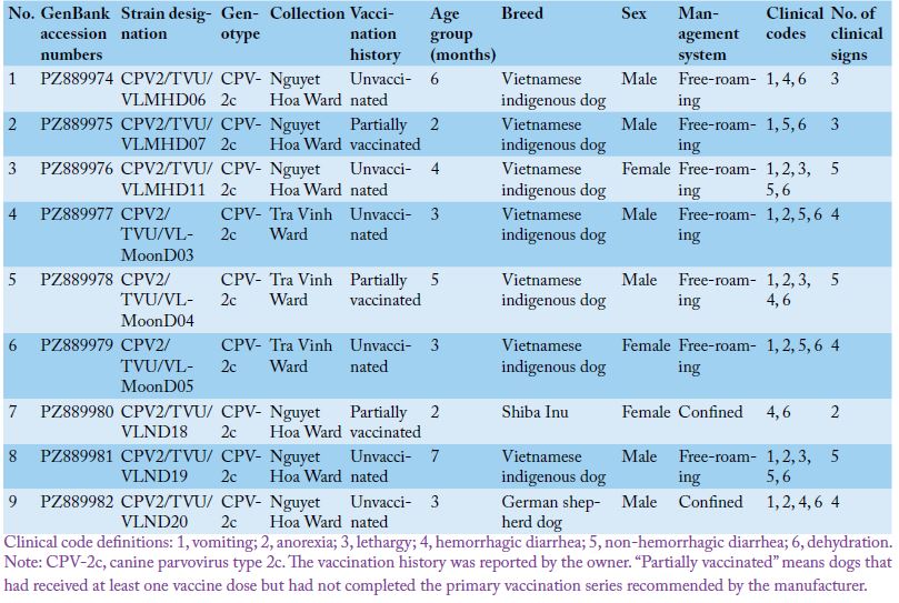
Nine of 103 CPV-2-positive samples (8.7%) were purposively selected for partial VP2 sequencing. Selection required a single, distinct amplicon at approximately 573 bp suitable for bidirectional Sanger sequencing and sought coverage of both collection wards and variation in age, breed, sex, owner-reported vaccination history, management, and clinical presentation (Table 1). The selection description did not use a numerical weighting score or probability-based random sampling; the nine samples should therefore be regarded as a molecularly characterized subset rather than a statistically representative sample of all 103 positives, and additional genotypes or sequence variants among unsequenced positives cannot be excluded. Amplicons were sequenced in both directions by
the Sanger method. Forward and reverse chromatograms were independently inspected, discordant or ambiguous base calls were resolved by chromatogram review, and only concordant consensus sequence positions were retained for genotype assignment and phylogenetic analysis. Consensus sequences were verified as CPV-2 using BLASTn against the NCBI nucleotide database.
Representative CPV-2 sequences of classical CPV-2, CPV-2a, CPV-2b, and CPV-2c were retrieved from GenBank for comparative analysis. Reference selection was restricted to sequences overlapping the homologous partial VP2 region used in this study and was designed to include genotype diversity, temporal and geographic diversity, and multiple contemporary Vietnamese and Chinese CPV-2c sequences relevant to regional comparison. The final 50-reference panel included sequences from Asia, Europe, and the Americas and was intended as a curated comparative dataset rather than an exhaustive global phylogeographic sample; it does not represent all available CPV-2 sequences or every continent. The analyzed study fragment corresponded approximately to VP2 nucleotides 1240–1745 and residues 414–581 relative to CPV-2 reference strain EU659117, thereby including residues 426 and 447. Because this interval represents only part of the approximately 1,755-bp VP2 gene, phylogenetic resolution is inherently lower than that achievable with full-length VP2 or whole-genome data.
Partial VP2 nucleotide sequences were aligned with reference sequences using ClustalW implemented in MEGA version 12 . Deduced amino acid sequences were translated with the standard genetic code and aligned with representative CPV-2 amino acid sequences. BioEdit version 7.7.2 and MEGA 12 were used for sequence inspection, trimming, consensus checking, and pairwise nucleotide and amino acid identity calculations.
For phylogenetic analysis, the nine study sequences and 50 representative CPV-2 reference sequences from GenBank were analyzed in MEGA 12 . The tree was reconstructed with the Neighbor-Joining method under the Tamura three-parameter substitution model , using complete deletion of sites with gaps or missing data and 1,000 bootstrap replicates to assess branch support.
Amino acid substitutions in the analyzed VP2 fragment were compared in BioEdit version 7.7.2 with a focused subset of 31 representative CPV-2 reference strains. Thus, 50 reference sequences were used for phylogenetic reconstruction, whereas 31 were used for the amino acid comparison in Table 6. Genotype assignment was based on the conventional VP2 residue-426 signature together with concordant sequence alignment and phylogenetic placement within the analyzed fragment. This approach is appropriate for partial-VP2 genotype classification but cannot characterize amino acid variation outside residues 414–581 or establish whole-genome lineage ancestry.
Statistical analysis
Data were entered in Microsoft Excel 2019 and analyzed in Minitab version 21. CPV-2 PCR status was treated as a binary outcome (positive = 1, negative = 0). Categorical variables were summarized as counts and percentages, with 95% confidence intervals for proportions calculated by the Wilson score method. Unadjusted associations between CPV-2 positivity and age group, breed, or vaccination category were assessed using Pearson’s chi-square test; Fisher’s exact test was used when expected cell counts were small. When an overall comparison was significant, pairwise comparisons of proportions were used to explore category differences. No multivariable regression model was fitted; therefore, reported associations are interpreted as unadjusted epidemiological associations rather than independent risk factors. Statistical significance was set at p < 0.05.
RESULTS
Among 466 enrolled dogs with clinical suspicion of CPV-2-associated enteritis, 103 were PCR-positive, corresponding to a clinic-based suspected-case positivity of 21.6 (17.0–27.0). This value is not a population prevalence estimate and cannot be interpreted directly as the burden of CPV-2 among all dogs in Vinh Long or among all dogs attending the participating clinics.
CPV-2 positivity differed significantly among age groups (χ² = 49.29, df = 3, p < 0.0001; Table 2). The highest positivity occurred at 3–4 months (57/151; 37.7%), whereas dogs >6 months had the lowest positivity (6/116; 5.2%). In the 1–2-month group, 28 of 98 dogs were PCR-positive (28.6%; 95% CI: 20.6–38.2). Serological measurement of maternally derived antibodies was not reported, and the conventional PCR assay was not designed to distinguish recently shed vaccine virus from field virus. Because PCR positivity can occur transiently after vaccination in puppies, these results should be interpreted together with vaccination timing when such information is available .
CPV-2 was detected in most breed categories, but the overall breed comparison was not statistically significant (χ² = 10.06, df = 6, p = 0.122; Table 3). Because breed-group sizes were unequal and several categories had relatively few observations and wide confidence intervals, this non-significant result should be interpreted as a lack of statistically supported evidence for a breed association in this dataset, not as evidence that breed effects are absent. A post-hoc power analysis is not reported; interpretation is therefore based on the observed group sizes and confidence intervals.
Table 2: CPV-2 positivity by age group.
Age group (months)
No. tested
No. CPV-2 positive
CPV-2 positivity, % (95% CI)
Distribution among positive dogs, %
1–2
98
28
28.6 (20.6–38.2)
27.2
3–4
151
57
37.7 (30.4–45.7)
55.3
5–6
101
12
11.9 (6.9–19.6)
11.7
>6
116
6
5.2 (2.4–10.8)
5.8
Overall
466
103
22.1 (18.6–26.1)
100.0
Note: CPV-2, canine parvovirus type 2; CI, confidence interval. Positivity was calculated as the number of positive dogs within each age group. The 95% CIs were estimated using the Wilson method, and differences among age groups were assessed using the chi-square test. Different superscript letters indicate significant differences among age groups (P < 0.05).
Table 3: CPV-2 positivity by breed.
Breed
No. tested
No. CPV-2 positive
CPV-2 positivity, %
(95% CI)
Distribution among CPV-2-positive dogs, %
Vietnamese indigenous dog
231
56
24.2 (19.2–30.2)
54.4
Poodle
72
22
30.6 (21.1–42.0)
21.4
Chihuahua
20
5
25.0 (11.2–46.9)
4.9
Siberian Husky
34
7
20.6 (10.3–36.8)
6.8
Pug
34
9
26.5 (14.6–43.1)
8.7
German Shepherd Dog
31
1
3.2 (0.6–16.2)
1.0
Shiba Inu
16
3
18.8 (6.6–43.0)
2.9
Overall
466
103
22.1 (18.6–26.1)
100.0
Note: CPV-2, canine parvovirus type 2; CI, confidence interval. Positivity was calculated as the number of positive dogs within each breed. The 95% CIs were estimated using the Wilson method. The overall breed comparison used the chi-square test. Because category sizes were unequal and some confidence intervals were wide, a non-significant overall comparison should not be interpreted as evidence that all breeds have identical risk.
Table 4: CPV-2 positivity by vaccination status.
Vaccination history
No. tested
No. CPV-2 positive
CPV-2 positivity, % (95% CI)
Distribution among CPV-2-positive dogs, %
Unvaccinated
161
81
50.3 (42.7–57.9)
78.6
Partially vaccinated
64
16
25.0 (16.0–36.8)
15.5
Fully vaccinated
241
6
2.5 (1.1–5.3)
5.8
Overall
466
103
22.1 (18.6–26.1)
100.0
Note: CPV-2, canine parvovirus type 2; CI, confidence interval. Positivity was calculated as the number of positive dogs within each vaccination group. The 95% CIs were estimated using the Wilson method. Different superscript letters indicate significant differences among vaccination groups (P < 0.05).
Owner-reported vaccination category was strongly associated with CPV-2 positivity in the unadjusted analysis (χ² = 128.56, df = 2, p < 0.0001; Table 4). Positivity was highest in dogs reported as unvaccinated (81/161; 50.3%) and lowest in dogs reported as fully vaccinated (6/241; 2.5%). Because age, vaccination history, and other exposures may be correlated and no multivariable model was fitted, these percentages do not estimate vaccine effectiveness and should not be interpreted causally.
Clinical manifestations of CPV-2-positive dogs
Among the 103 PCR-positive dogs, anorexia and vomiting were the most frequently recorded signs, followed by lethargy and diarrhea (Table 5). Non-hemorrhagic diarrhea was recorded in 48.5% and hemorrhagic diarrhea in 41.7% of PCR-positive dogs. Because the same clinical features contributed to the broad enrollment case definition and because PCR-negative dogs were not analyzed as a comparison group for individual signs, these frequencies describe the clinical profile of positive cases only; they should not be interpreted as measures of diagnostic specificity or as evidence that any individual sign is uniquely associated with CPV-2.
Table 5: Clinical manifestations in CPV-2-positive dogs.
Clinical manifestation
Clinical code
No. affected
Frequency, % (95% CI)
Anorexia
2
99
96.1 (90.4–98.5)
Vomiting
1
96
93.2 (86.6–96.7)
Lethargy
3
60
58.3 (48.6–67.3)
Non-hemorrhagic diarrhea
5
50
48.5 (39.1–58.1)
Hemorrhagic diarrhea
4
43
41.7 (32.7–51.4)
Dehydration
6
32
31.1 (22.9–40.6)
Pale mucous membranes
7
21
20.4 (13.7–29.2)
Note: CPV-2, canine parvovirus type 2; CI, confidence interval. Frequencies were calculated among 103 CPV-2-positive dogs and the 95% CIs were estimated using the Wilson method. These signs also contributed to the clinical suspicion criteria used for enrollment; no sign-by-sign comparison with PCR-negative dogs was performed. The table is therefore descriptive and does not estimate diagnostic specificity.
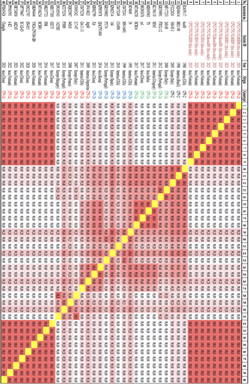
Nucleotide and amino acid identity in pairwise comparisons
The nine study sequences were identical to one another across the analyzed partial VP2 consensus region at both nucleotide and deduced amino acid levels (Figure 1). This finding indicates no detected sequence diversity within this short fragment among the nine selected samples and should not be extrapolated to genome-wide identity. They showed 99.8–100.0% nucleotide identity and 100.0% amino acid identity with contemporary Asian CPV-2c references, particularly strains from Vietnam and China. CPV-2c references from the Americas and Europe showed 98.8–99.3% nucleotide identity and 99.0–99.3% amino acid identity across the same region. Bidirectional chromatograms were concordant at retained consensus positions, and PCR negative controls were required to remain free of amplification for a run to be accepted. These controls reduce, but cannot by themselves eliminate, all possibilities of pre-analytical or laboratory cross-contamination.
Phylogenetic relationships of CPV-2 strains
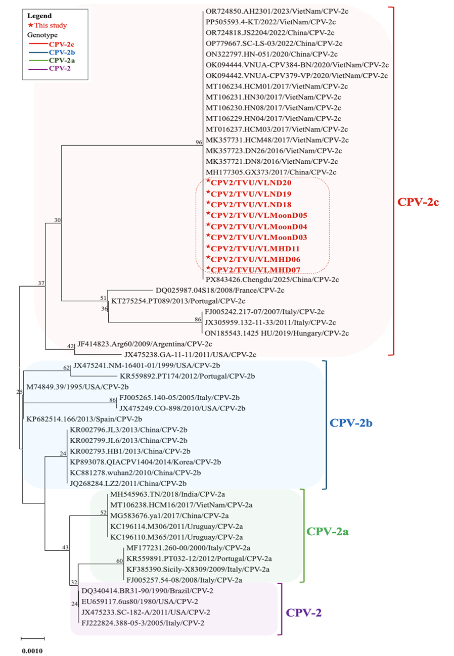
Phylogenetic analysis placed all nine study sequences within the CPV-2c group and adjacent to contemporary Vietnamese and Chinese CPV-2c references (Figure 2). The node grouping the study sequences with the contemporary Asian CPV-2c cluster shown in Figure 2 had 96% bootstrap support. Deeper nodes separating some older CPV-2c references had lower support, which is consistent with the limited phylogenetic information contained in a short partial VP2 fragment. Accordingly, the tree supports regional sequence relatedness within the analyzed interval but should not be interpreted as a robust whole-genome phylogeographic reconstruction.
Amino acid substitution profile VP2
The deduced amino acid profile of the partial VP2 region was consistent across all nine study sequences (Table 6). Each study sequence contained 426E, the conventional residue used to classify CPV-2c, and 447M. For clarity, the amino acids at positions 419, 426, 440, 447, 555, 564, and 568 are shown explicitly for every study strain in Table 6 rather than using dot notation. No variation among the nine
The phylogenetic tree was constructed using partial VP2 gene sequences of the nine study samples and 50 representative CPV-2 references. Study sequences are marked with red stars and highlighted in red; genotype groupings are indicated by colored brackets. Numbers at internal nodes indicate bootstrap support from 1,000 replicates. The node grouping the study sequences with the contemporary Vietnamese/Chinese CPV-2c cluster had 96% bootstrap support, whereas several deeper nodes had lower support. The scale bar represents nucleotide substitutions per site. Because the tree is based on a short partial VP2 region, it is intended for genotype and regional.
Table 6: VP2 amino acid substitutions of CPV-2 strains.
No.
Accession no.
Year
Geographic origin
Genotype
VP2 amino acid residue
419
426
440
447
555
564
568
1
EU659117
1980
America (USA)
CPV-2
N
N
T
I
V
S
G
2
DQ340414
1990
America (Brazil)
CPV-2
.
.
.
.
.
.
.
3
FJ222824
2005
Europe (Italy)
CPV-2
.
.
.
.
.
.
.
4
MF177231
2000
Europe (Italy)
CPV-2a
.
.
.
.
.
.
.
5
KR559891
2012
Europe (Portugal)
CPV-2a
.
.
A
.
.
.
.
6
KC196110
2011
America (Uruguay)
CPV-2a
.
.
A
.
.
.
.
7
MH545963
2018
Asia (India)
CPV-2a
.
.
A
.
.
.
.
8
MG583676
2017
Asia (China)
CPV-2a
.
.
A
.
.
.
.
9
MT106238
2017
Asia (Vietnam)
CPV-2a
.
.
A
.
.
.
.
10
M74849
1995
America (USA)
CPV-2b
.
D
.
.
.
.
.
11
JX475241
1999
America (USA)
CPV-2b
.
D
.
.
.
.
.
12
JX475249
2010
America (USA)
CPV-2b
.
D
.
.
.
.
.
13
KP682514
2013
Europe (Spain)
CPV-2b
.
D
.
.
.
.
.
14
KR559892
2012
Europe (Portugal)
CPV-2b
.
D
.
.
.
.
.
15
KP893078
2014
Asia (Korea)
CPV-2b
.
D
A
.
.
.
.
16
KR002799
2013
Asia (China)
CPV-2b
.
D
A
.
.
.
.
17
JF414823
2009
America (Argentina)
CPV-2c
.
E
A
.
.
.
.
18
JX475238
2011
America (USA)
CPV-2c
.
E
A
.
.
.
.
19
FJ005242
2007
Europe (Italy)
CPV-2c
.
E
.
.
.
.
.
20
DQ025987
2008
Europe (France)
CPV-2c
.
E
.
.
.
.
.
21
KT275254
2013
Europe (Portugal)
CPV-2c
.
E
.
.
.
.
.
22
ON185543
2019
Europe (Hungary)
CPV-2c
.
E
.
.
.
.
.
23
MH177305
2017
Asia (China)
CPV-2c
.
E
.
M
.
.
.
24
MK357721
2016
Asia (Vietnam)
CPV-2c
.
E
.
M
.
.
.
25
MT106234
2017
Asia (Vietnam)
CPV-2c
.
E
.
M
.
.
.
26
OK094444
2020
Asia (Vietnam)
CPV-2c
.
E
.
M
.
.
.
27
ON322797
2020
Asia (China)
CPV-2c
.
E
.
M
.
.
.
28
OP779667
2022
Asia (China)
CPV-2c
.
E
.
M
.
.
.
29
OR724850
2023
Asia (Vietnam)
CPV-2c
.
E
.
M
.
.
.
30
PP505593
2022
Asia (Vietnam)
CPV-2c
.
E
.
M
.
.
.
31
PX843426
2025
Asia (China)
CPV-2c
.
E
.
M
.
.
.
32
VLMHD06 (this study)
2025
Asia (Vietnam)
CPV-2c
N
E
T
M
V
S
G
33
VLMHD07 (this study)
2025
Asia (Vietnam)
CPV-2c
N
E
T
M
V
S
G
34
VLMHD11 (this study)
2025
Asia (Vietnam)
CPV-2c
N
E
T
M
V
S
G
35
VLMoonD03 (this study)
2025
Asia (Vietnam)
CPV-2c
N
E
T
M
V
S
G
36
VLMoonD04 (this study)
2025
Asia (Vietnam)
CPV-2c
N
E
T
M
V
S
G
37
VLMoonD05 (this study)
2025
Asia (Vietnam)
CPV-2c
N
E
T
M
V
S
G
38
VLND18 (this study)
2025
Asia (Vietnam)
CPV-2c
N
E
T
M
V
S
G
39
VLND19 (this study)
2025
Asia (Vietnam)
CPV-2c
N
E
T
M
V
S
G
40
VLND20 (this study)
2025
Asia (Vietnam)
CPV-2c
N
E
T
M
V
S
G
Note: CPV-2, canine parvovirus type 2; VP2, viral protein 2. Amino acid positions were numbered according to CPV-2 reference strain EU659117. Dots indicate residues identical to the reference strain for reference sequences. To remove ambiguity, residues for all nine study sequences are written explicitly; each study strain has the profile 419N/426E/440T/447M/555V/564S/568G within the analyzed region.
study strains was detected at these positions. The 447M residue has been reported in contemporary Vietnamese and other Asian CPV-2c lineages, but the present study did not test its functional effect; it is therefore treated as a molecular lineage marker rather than evidence of altered virulence, vaccine escape, or diagnostic performance.
DISCUSSION
This study provides a defined 12-month clinic-based molecular snapshot of CPV-2 among clinically suspected dogs in two wards of Vinh Long Province and links the diagnostic denominator with age, owner-reported vaccination history, clinical presentation, and partial VP2 sequence data. Its contribution is regional molecular epidemiology rather than demonstration of a new biological mechanism: CPV-2c with a conserved 426E/447M profile was detected in the sequenced subset, and these sequences were most closely related to contemporary Vietnamese and Chinese references within the analyzed VP2 interval. This complements earlier Vietnamese work by adding current clinic-level context from a local catchment that had not been characterized in the same integrated manner ; .
The concentration of PCR positivity in younger dogs is compatible with the recognized susceptibility window during which maternally derived antibodies decline while active immunity may still be incomplete. However, the present data cannot disentangle age from vaccination history or other correlated exposures because only unadjusted analyses were performed. Likewise, the lower positivity observed in dogs reported as fully vaccinated should not be interpreted as a direct estimate of vaccine effectiveness. This study does not provide evidence to change vaccine product type, starting age, dose intervals, or booster schedules. Its vaccination-related implication is limited to consistency with established WSAVA guidance on completing the primary puppy series , not an independently derived vaccination schedule.
The breed comparison did not reach statistical significance, but unequal category sizes and broad confidence intervals limit precision. Therefore, the findings do not support a breed predisposition in this dataset, while also not excluding a smaller breed effect that the study was underpowered to detect. Similarly, the frequencies of anorexia, vomiting, lethargy, dehydration, and diarrhea among PCR-positive dogs should be viewed as a descriptive clinical profile rather than evidence that these signs discriminate CPV-2 from other causes of canine gastroenteritis. A PCR-negative comparison group analyzed by individual clinical sign would be required to estimate diagnostic associations or predictive value.
Molecular findings were concordant across sequence identity, phylogeny, and amino acid analysis within the sequenced region. The 426E residue supports CPV-2c classification, while 447M is notable because Nguyen Manh et al. (2021) reported a Vietnamese CPV-2c sublineage carrying 426E/447M, and related Asian sequences have subsequently been described. CPV-2c itself is not restricted to Asia and has been documented in Europe, the Americas, and Africa ; . Thus, the importance of the present result is not the global uniqueness of CPV-2c, but the close sequence affinity of the Vinh Long partial VP2 sequences to contemporary Asian, particularly Vietnamese and Chinese, references. The biological significance of 447M remains unresolved. In silico structural analyses have suggested that I447M may influence local VP2 conformation, but direct experimental evidence linking 447M to vaccine failure, altered antigenicity, receptor binding, clinical severity, or diagnostic-test performance is lacking . The present data therefore do not support such functional claims.
From a clinical perspective, identifying CPV-2c does not currently imply a different supportive-treatment protocol from that used for other CPV-2 antigenic variants; treatment decisions remain driven by the severity of enteritis, dehydration, systemic complications, and patient response. The value of genotyping is principally epidemiological: documenting variant circulation, comparing regional lineages, detecting sequence change over time, and informing future vaccine-matching or whole-genome studies. Accordingly, the present study supports continued surveillance and adherence to established vaccination guidance, but it does not demonstrate a need to alter vaccine type or schedule.
This study has several limitations that directly constrain interpretation. First, enrollment was restricted to clinic-presenting dogs meeting a broad suspicion definition; therefore, the 22.1% estimate is positivity among enrolled suspected cases, not population prevalence or a direct measure of regional disease burden. Second, vaccination categories relied mainly on owner report and incompletely available records. If misclassification of vaccination status was non-differential with respect to PCR status, it would generally be expected to dilute differences between vaccination categories toward the null; if reporting accuracy differed between PCR-positive and PCR-negative dogs, bias could occur in either direction. A quantitative bias analysis was not possible because the sensitivity and specificity of owner-reported vaccination history were not measured. Third, age, vaccination, breed, and other exposures were analyzed only in unadjusted comparisons, so their independent effects cannot be separated. Fourth, the broad clinical case definition may have included dogs with nonspecific signs, and the available analysis does not provide a one-sign-versus-multiple-sign comparison or sign-by-sign comparisons with PCR-negative dogs. Fifth, serological measurement of maternally derived antibodies was not reported, and the conventional partial-VP2 PCR was not designed to differentiate vaccine-derived DNA from field-virus DNA; molecular positivity can occur transiently after vaccination in puppies and may complicate interpretation when vaccination was recent . Sixth, only nine of 103 positives were purposively sequenced, so other variants could have been missed. Seventh, only a partial VP2 fragment was analyzed and the curated phylogenetic reference panel was not globally exhaustive; whole-genome ancestry, recombination, and fine-scale transmission routes cannot be resolved. Eighth, identical partial VP2 sequences in the nine samples indicate limited detected diversity in that fragment but do not demonstrate genome-wide identity, and routine run controls cannot exclude every possible source of contamination. Finally, privately owned dogs were not maintained under standardized conditions, and necropsy, histopathology, treatment-outcome follow-up, and mortality surveillance were outside the original protocol. Future studies should incorporate broader sampling frames, verified vaccination records and timing, comparison of clinical signs between PCR-positive and PCR-negative dogs, multivariable modeling, vaccine-versus-field strain discrimination when recent vaccination is possible, and full-length VP2 or whole-genome sequencing of a larger randomly selected subset.
CONCLUSION
Among clinic-presenting dogs with suspected parvoviral enteritis in Vinh Long, CPV-2 PCR positivity was 22.1% and was highest in younger dogs and dogs reported as unvaccinated in unadjusted analyses; breed differences were not statistically supported. All nine purposively sequenced samples were CPV-2c and shared the partial-VP2 426E/447M profile, with close relatedness to contemporary Vietnamese and Chinese references. These findings provide regional molecular-surveillance data and are consistent with established recommendations to complete the primary puppy vaccination series, but the cross-sectional design does not estimate vaccine effectiveness, establish causality, or justify changes in vaccine product or schedule.
Acknowledgments
The authors sincerely thank the dog owners for their cooperation and consent during sample collection. We also acknowledge the participating veterinary clinics, veterinarians, and technical staff for their valuable support in clinical examination, sample collection, and laboratory procedures. The authors are grateful to the Laboratory of Tra Vinh University for providing facilities and technical assistance for PCR and molecular analyses.
Novelty Statement
The distinctive contribution of this study is the integration, within a defined clinic-based dataset from Tra Vinh Ward and Nguyet Hoa Ward, of CPV-2 suspected-case PCR positivity, unadjusted age/breed/owner-reported vaccination comparisons, clinical manifestations, and partial VP2-based genotype and phylogenetic characterization. The nine sequenced samples shared a conserved CPV-2c 426E/447M profile and were most closely related to contemporary Asian reference strains within the analyzed region. The study adds region-specific molecular-surveillance evidence for southern Vietnam without claiming first detection, vaccine-effectiveness estimation, or a need to alter established vaccination schedules.
Authors’ Contribution
Nguyen Thi Yen Linh, Nguyen Van Vui, and Tran Ngoc Bich contributed to the conceptualization and design of the study. Nguyen Thi Yen Linh and Nguyen Van Vui performed sample collection, laboratory work, data curation, and data analysis. Tran Ngoc Bich supervised the study, contributed to methodology development, and reviewed the manuscript. Nguyen Thi Yen Linh prepared the original draft. Nguyen Van Vui and Tran Ngoc Bich contributed to manuscript revision, editing, and finalization. All authors read and approved the final version of the manuscript.
Abbreviations
CPV-2, canine parvovirus type 2; CPV-2a/2b/2c, antigenic variants of canine parvovirus type 2; VP2, viral capsid protein 2; PCR, polymerase chain reaction; DNA, deoxyribonucleic acid; CI, confidence interval; NCBI, National Center for Biotechnology Information; TAE, Tris-acetate-EDTA buffer.
Funding
This research was supported by Tra Vinh University under Research Contract No. 15/2025/HĐ.HĐKH&ĐT-ĐHTV.
Ethical statement
The study protocol was reviewed and approved before field sampling by the Science and Education Council of Tra Vinh University under institutional approval No. QT6.2/KHCN2-01.TMĐT and Task Contract No. 15/2025/HĐ.HĐKH&ĐT-ĐHTV (approval date: 10 February 2025). The approved protocol involved sampling privately owned clinical patients and did not include experimental infection, disease induction, or study-specific invasive procedures beyond diagnostic rectal swab collection. Rectal swabs were collected by qualified veterinarians during routine clinical examination with the aim of minimizing handling time and discomfort.
Generative AI and AI-assisted technology statement
Generative AI was used solely for language editing. All scientific content was reviewed and approved by the authors.
Conflict of interest
The authors have declared no conflict of interest.
Figures
Figure 1:.
Pairwise nucleotide and deduced amino acid sequence identity matrix of partial VP2 gene sequences of CPV-2 strains. The upper and lower triangular matrices show amino acid and nucleotide identity, respectively. Values are percentages. Positions were numbered according to CPV-2 reference strain EU659117. The alignment covered VP2 nucleotides 1240–1745, with amino acid comparisons based on VP2 residues 414–581.
Table 1:.
Epidemiological and clinical metadata of CPV-2c strains.
Figure 2:.
Phylogenetic relationship of CPV-2 strains based on partial VP2 gene sequences. The phylogenetic tree was constructed using partial VP2 gene sequences of the nine study samples and 50 representative CPV-2 references. Study sequences are marked with red stars and highlighted in red; genotype groupings are indicated by colored brackets. Numbers at internal nodes indicate bootstrap support from 1,000 replicates. The node grouping the study sequences with the contemporary Vietnamese/Chinese CPV-2c cluster had 96% bootstrap support, whereas several deeper nodes had lower support. The scale bar represents nucleotide substitutions per site. Because the tree is based on a short partial VP2 region, it is intended for genotype and regional.
References
Charoenkul K, Tangwangvivat R, Janetanakit T, Boonyapisitsopa S, Bunpapong N, Chaiyawong S, Amonsin A (2019). Emergence of canine parvovirus type 2c in domestic dogs and cats from Thailand. Transbound. Emerg. Dis., 66(4): 1518–1528. https://doi.org/10.1111/tbed.13177[CrossRef]
Decaro N, Buonavoglia C (2012). Canine parvovirus: A review of epidemiological and diagnostic aspects, with emphasis on type 2c. Vet. Microbiol., 155: 1–12. https://doi.org/10.1016/j.vetmic.2011.09.007[CrossRef]
Hall TA (1999). BioEdit: A user-friendly biological sequence alignment editor and analysis program for Windows 95/98/NT. Nucleic Acids Symp. Ser. 41: 95–98.
Hoang M, Lin WH, Le VP, Nga BTT, Chiou MT, Lin CN (2019). Molecular epidemiology of canine parvovirus type 2 in Vietnam from November 2016 to February 2018. Virol. J., 16(1): 52. https://doi.org/10.1186/s12985-019-1159-z[CrossRef]
Hoelzer K, Parrish CR (2010). The emergence of parvoviruses of carnivores. Vet. Res., 41: 39. https://doi.org/10.1051/vetres/2010011[CrossRef]
Kumar S, Stecher G, Suleski M, Sanderford M, Sharma S, Tamura K (2024). MEGA12: Molecular evolutionary genetic analysis version 12 for adaptive and green computing. Mol. Biol. Evol., 41(12): msae263. https://doi.org/10.1093/molbev/msae263[CrossRef]
Li S, Chen X, Hao Y, Zhang G, Lyu Y, Wang J, Liu W, Qin T (2022). Characterization of the VP2 and NS1 genes from canine parvovirus type 2 (CPV-2) and feline panleukopenia virus (FPV) in Northern China. Front. Vet. Sci., 9: 934849. https://doi.org/10.3389/fvets.2022.934849[CrossRef]
Maganga GD, Labouba I, Milendz Ikapi SZ, Nkili-Meyong AA, Ngonga Dikongo AM, Boundenga L, Ngoubangoye B, Memvie C, Kumulungui BS (2023). Molecular characterization of canine parvovirus variants CPV-2a and CPV-2c, associated with vaccinated dogs at Libreville, Gabon. Viruses, 15(5): 1169. https://doi.org/10.3390/v15051169[CrossRef]
Mazzaferro EM (2020). Update on canine parvoviral enteritis. Vet. Clin. North Am. Small Anim. Pract., 50(6): 1307–1325. https://doi.org/10.1016/j.cvsm.2020.07.008.[CrossRef]
Nguyen Manh T, Piewbang C, Rungsipipat A, Techangamsuwan S (2021). Molecular and phylogenetic analysis of Vietnamese canine parvovirus 2C originated from dogs reveals a new Asia-IV clade. Transbound. Emerg. Dis., 68(3): 1445–1453. https://doi.org/10.1111/tbed.13811.[CrossRef]
Pan S, Man Y, Xu X, Ji J, Zhang S, Huang H, Li Y, Bi Y, Yao L (2024). Genetic diversity and recombination analysis of canine parvoviruses prevalent in Central and Eastern China, from 2020 to 2023. Microorganisms, 12(11): 2173. https://doi.org/10.3390/microorganisms12112173[CrossRef]
Saitou N, Nei M (1987). The neighbor-joining method: A new method for reconstructing phylogenetic trees. Mol. Biol. Evol., 4(4): 406–425. https://www.academia.edu/download/56986116/7sait.pdf
Sarabandi S, Pourtaghi H (2023). Whole genome sequence analysis of CPV-2 isolates from 1998 to 2020. Virol. J., 20(1): 138. https://doi.org/10.1186/s12985-023-02102-2[CrossRef]
Segev G, Yaaran T, Maurice S, Baneth G (2022). Effect of sampling site on the diagnosis of canine parvovirus infection in dogs using polymerase chain reaction. J. Vet. Int. Med., 36(2): 591–598. https://doi.org/10.1111/jvim.16373[CrossRef]
Squires RA, Crawford C, Marcondes M, Whitley N (2024). 2024 guidelines for the vaccination of dogs and cats compiled by the Vaccination Guidelines Group (VGG) of the World Small Animal Veterinary Association (WSAVA). J. Small Anim. Pract., 65(5): 277–316. https://doi.org/10.1111/jsap.13718[CrossRef]
Tamura K (1992). Estimation of the number of nucleotide substitutions when there are strong transition-transversion and G+C-content biases. Mol. Biol. Evol., 9(4): 678–687.
Thompson JD, Higgins DG, Gibson TJ (1994). CLUSTAL W: Improving the sensitivity of progressive multiple sequence alignment through sequence weighting, position-specific gap penalties and weight matrix choice. Nucl, Acids Res., 22(22): 4673–4680. https://doi.org/10.1093/nar/22.22.4673[CrossRef]
Tuteja D, Banu K, Mondal B (2022). Canine parvovirology: A brief updated review on structural biology, occurrence, pathogenesis, clinical diagnosis, treatment and prevention. Comp. Immunol. Microbiol. Infect. Dis., 82: 101765. https://doi.org/10.1016/j.cimid.2022.101765[CrossRef]
Xia Q, Liu J, Gui Y, Xia L, Cao C, Chen B, Yu X, Chen W, Xu F, Wang J, Zhao H (2026). Molecular characteristics and genetic diversity of canine parvovirus in Shanghai, China, from 2016 to 2025. Microorganisms,14(4): 761. https://doi.org/10.3390/microorganisms14040761[CrossRef]
Zhong Z, Liang L, Zhao J, Xu X, Cao X, Liu X, Zhou Z, Ren Z, Shen L, Geng Y, Gu X (2014). First isolation of new canine parvovirus 2a from Tibetan Mastiff and global analysis of the full-length VP2 gene of canine parvoviruses 2 in China. Int. J. Mol. Sci., 15(7): 12166–12187. https://doi.org/10.3390/ijms150712166[CrossRef]