Research Article · Volume 13, Issue 2, 20 January 2025 · Open Access
Biochemical, Hematologic and Oxidative Stress Biomarkers Investigation in Infected Native Breed Calves with Foot and Mouth Disease virus Serotype A
· · · · · ·
Affiliations & Notes ⌄
1Department of Zoology and Entomology, Faculty of Science, Al-Azhar University, Cairo, Egypt
2Department of Botany and Microbiology, Faculty of Science, Al-Azhar University, Cairo, Egypt
3Department of Zoology, Faculty of Science, Al-Azhar University, Cairo, Egypt
4Department of Virology Research, Animal Health Research Institute, Agriculture Research Center, Giza, Egypt
5Department of Veterinary Clinical Pathology, Faculty of Veterinary Medicine, Cairo University, Giza, Egypt
6Department of internal medicine and infectious diseases, Faculty of Veterinary Medicine, Cairo University, Giza, Egypt. *Correspondence | Mohamed I. Oraby, Department of internal medicine and infectious diseases, Faculty of Veterinary Medicine, Cairo University, Giza, Egypt
During the outbreak investigation and sample collection, it was discovered that early-stage FMD-affected animals showed fever, inappetence to anorexia, salivation, and frothiness from the mouth.
Animals with late-stage infections had a reduction in fever with symptoms that were comparable to those in the early stages.
Hemograms of FMD-infected cattle compared to control animals revealed non-significant decreases in Hb%, RBCs count, and PCV% during the early stages of infection on days 1, 4, and 15, as opposed to day 30 during the late stages of infection or following recovery, when Hb%, RBCs count, and PCV% returned to normal range.
Prevention of foot and mouth disease (FMD) has been declared a global priority by the Food and Agricultural Organization of the United Nations (FAO) and the World Organization for Animal Health (OIE).
Selected from the article's own text.
Abstract
This paper presents a comprehensive analysis of biochemistry patterns in cattle during infection with Foot-and-mouth Disease Virus (FMDV) serotype A during FMD outbreaks in 2018 and 2023 in Giza governorate, Egypt. The study investigated the effect of FMDV infection on several blood parameters, including hemogram (HB%, PCV%, RBCs, WBC and platelets count), serum biochemical parameters (glucose, cholesterol, ALT, AST, urea, BUN, creatinine, triglyceride, CK-MB, total proteins, albumin and globulin), oxidative marker (MDA), enzymatic antioxidant (catalase), TAC and trace elements (Zn and Cu). The results showed significant changes in these parameters following FMDV infection, indicating the potential for FMDV to cause systemic effects beyond the respiratory and digestive signs. Overall, this study highlights the importance of monitoring blood parameters as a diagnostic tool for FMDV infection in cattle.
Many cloven-hoofed animals are susceptible to FMD, a highly contagious transboundary viral disease that results in substantial economic losses because of decreased meat and milk production, young animals mortality, medication expenses, and limitations on the movement of animals from endemic areas ; ; ; . Other symptoms include vesicles or blisters on the tongue, severe frothy salivation, fever, dullness, and anorexia. Some infected animals may not show any symptoms, but they still carry the virus and can infect other animals. Although it increases the risk of abortion in pregnant animals, the illness is typically not fatal in adult cattle .
As a member of the Aphthovirus genus, FMDV is a member of the Picornaviridae family. An icosahedral capsid containing around 60 copies of the four structural viral proteins (VP1, VP2, VP3, and VP4) encloses the non-enveloped, single-stranded, positive-sense, non-segmented RNA virus known as FMDV ; . According to Brown et al. (2022); Singanallur et al. (2022); Liu et al. (2021), FMDV is divided into six distinct immunological serotypes: A, O, SAT 1, SAT 2, SAT 3, and Asia. Within these serotypes, over 65 strains, topotypes, and genetic lineages have been identified ; . Africa, Europe-South America (Euro-SA), and Asia are the three topotypes of Serotype A that are known to exist.
Clinical symptoms are often used to make the first diagnosis of the disease. In endemic areas, incomplete signs are often disregarded due to natural or vaccine protection . The amount of specific blood metabolites signals the animal’s illness status and level of stress . However, there is a scarcity of research on the effect of experimental FMD infection on bovine blood and serum profiles, which is critical for developing ameliorative methods to alleviate the animal’s biotic stress caused by the infection. Understanding changes in the pattern of clinical, serum biochemical, and haematological parameters during the course of FMD infection is important in developing therapeutic approaches for circumventing stress and thus reducing the negative effect of the infection in the animal .
Clinical signs are frequently used to obtain an early diagnosis of the illness. In endemic locations, partial symptoms are frequently overlooked because of natural or vaccine protection . The concentration of particular blood metabolites indicates the animal’s disease condition and amount of stress . The impact of experimental FMD infection on cow blood and serum profiles, however, has received little attention. This information is crucial for creating remedial strategies to lessen the biotic stress brought on by the infection. Additionally, it is said that the Malnad Gidda, a native breed of India, is immune to several viral diseases, including FMD; however, no thorough study has been done to substantiate this assertion ; .
Unrestricted animal movement from neighboring countries, the import and transit of live animals and animal products, and the absence of quarantine facilities have all contributed to the discovery of many FMDV serotypes and topotypes in Egypt despite control efforts ; . In order to effectively control the disease, these factors emphasized the need for quick, accurate, sensitive, and efficient diagnostic tests as well as ongoing monitoring of genetic changes in the FMD virus to update FMD vaccine seed strains with currently circulating strains .
Many viral illnesses develop as a result of oxidative stress. Oxidative stress results from an imbalance between oxidants and antioxidants , which causes cellular damage . The stimulation of lipid peroxidation activates protective antioxidant defense systems in cells, resulting in alterations in malondialdehyde (MDA) and antioxidant levels . As a result, antioxidant and oxidative biomarker (MDA) concentrations are assessed to assess oxidative stress in viral infections ; . Stress and pathogenic agents contribute to disease development by increasing free radical generation, reducing antioxidant defences and leading to oxidative stress .
Lipid peroxidation indicates oxidative stress, which impairs cell structure and function. Malondialdehyde (MDA), a breakdown product of lipid peroxidation derived from polyunsaturated fatty acids, may detect cellular damage and oxidative stress ; Khoshvaghtı et al., 2014). Under oxidative stress, the MDA level rises significantly . Oxidative stress contributes to the pathophysiology of several illnesses, including foot and mouth disease. The blood MDA level in the diseased group was thought to have rised as a result of stress and cellular degeneration induced by aphthovirus oxidative damage.
Oxidative stress is a state in which the body cannot scavenge free radicals, leading to tissue damage, alterations, and unfavorable effects on the health of animals . Oxidative processes are linked to a wide range of diseases . Cellular level damage was attributed to this mechanism . Nevertheless, enzymatic (like Catalase) and non-enzymatic (like copper and zinc) antioxidant mechanisms may mitigate these effects . Antioxidants are essential for protecting cells from the damaging effects of reactive oxygen species (ROS) .
Cattle suffer from oxidative stress due to foot and mouth disease, a serious viral infectious disease with a severe clinical course. The number of white blood cells (WBCs) was found to have increased among the haematological parameters tested in infected animals. Among the biochemical markers, MDA, creatinine, and CK enzyme activity were found to have increased, whereas antioxidants, proteins, minerals, and glucose levels were reported to have decreased (Sezar et al., 2024).
To molecularly diagnose and identify the serotype implicated, this study was carried out, with a focus on estimating changes in hematological and serum biochemical markers in cattle affected by FMDV.
MATERIALS AND METHODS
Ethical Statements
This investigation was conducted under the animal ethical guidelines. It was approved by the Veterinary Medicine Cairo University Institutional Animal Care and Use Committee, Faculty of Veterinary Medicine, Cairo University, Egypt (vet cu13102024971).
Blood / Serum Sample Collection
Serum was separated by centrifugation at 3000 g for 10 minutes after 10 mL of blood samples from 25 animals were obtained by jugular venipuncture in a red vacutainer with a clot activator (BD, Franklin, USA). The serum was then maintained at 20°C until serum biochemical parameters were ascertained. A green vacutainer containing heparin as an anticoagulant and RT-PCR for FMDV detection was used to draw a 4 mL blood sample (BD, Franklin, USA). Thorough study of the blood count. A comprehensive blood count analysis was performed using a three-chamber hematology analyzer (BC-3000 Plus, China).
Biochemical Tests
Using a real semi-automated clinical chemistry analyzer (Microlab 300–ELITech Group, Hollanda), commercial kits were used to measure blood glucose, urea, blood urea nitrogen, creatinine, total cholesterol, total proteins, albumin, aspartate transaminase (AST), triglycerides, alanine transaminase (ALT), MDA, Cat, Zn, and Cu. Every Kit was used according to the manufacturer’s instructions. Cardiac damage was measured using the creatine kinase-myocardial band (CK-MB) kit (Coral Clinical Systems, India) and the immune inhibition technique.
Statistical Analysis
The data was coded and entered using SPSS V.22, a statistical program. The Kolmogorov-Smirnov test for normality, the Shapiro-Wilk test for continuous variables, and parametric test assumptions were checked for data. Arcsine Square Root was used to standardize data on probability and percentiles for normalcy. The mean and standard deviation of the data were displayed. The experimental groups’ recorded physiological parameters were subjected to ANOVA analyses. The control group and sick cattle group had at least three replicates of the analysis (on days 1, 4, 15, and 30 after the onset of symptoms). The post-hoc analysis was assessed using Tukey pairwise comparison using MiniTab V 14, and P-values were deemed significant at <0.05. When feasible, data were visualized using R studio V 2022.02.4.
RESULTS AND DISCUSSION
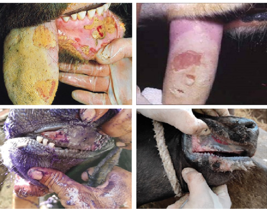
Clinical Examination of FMDV outbreaks examining the animals’ oral cavities revealed sores, ruptured vesicles, ulcerations on the tongue and gum, destruction on the upper and lower pad region, and wear away on the interdigital space. During the outbreak investigation and sample collection, it was discovered that early-stage FMD-affected animals showed fever, inappetence to anorexia, salivation, and frothiness from the mouth. These creatures were noted to be lame, and vesicles were seen in the gaps between their fingers. Animals with late-stage infections had a reduction in fever with symptoms that were comparable to those in the early stages. On day 30, the animals’ clinical symptoms subsided, they resumed their regular diet, and the wounds on their limbs and buccal regions healed (Figure 1). FMD-affected cattle were falling behind healthy ones regarding nutrition and milk output.
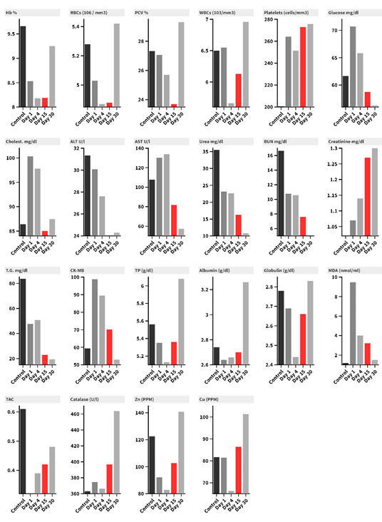
Results of hematology are shown in (Table 1 and Figure 2). Hemograms of FMD-infected cattle compared to control animals revealed non-significant decreases in Hb%, RBCs count, and PCV% during the early stages of infection on days 1, 4, and 15, as opposed to day 30 during the late stages of infection or following recovery, when Hb%, RBCs count, and PCV% returned to normal range. In contrast to control animals, WBCs and platelet counts did not significantly rise during infection.
Table 1: Biochemical, Hematologic and Oxidative stress biomarkers (mean ± SD values) in infected Native breed Calves with FMD virus Serotype A compared with the normal control through the study period.
Control
Day 1
Day 4
Day 15
Day 30
Hb %
9.66 ± 0.32abc
8.53 ± 0.99abc
8.18 ± 1.01bc
8.19 ± 0.73c
9.25 ± 0.91a
RBCs (106 / mm3)
5.28 ± 0.23a
5.03 ± 0.75a
4.87 ± 0.71a
4.88 ± 0.61a
5.42 ± 0.54a
PCV %
27.33 ± 1.24ab
27.06 ± 3.56ab
25.7 ± 3.76ab
23.7 ± 5.74b
29.31 ± 3.63a
WBCs (103/mm3)
6.5 ± 0.48a
6.55 ± 1.66a
5.66 ± 1.51a
6.13 ± 1.36a
6.96 ± 1.35a
Platelets (cells/mm3)
200.66 ± 11.02a
264.15 ± 90.45a
251.46 ± 105.77a
272.84 ± 109.86a
275.76 ± 69.01a
Glucose mg/dl
61.66 ± 4.1a
70.7 ± 18.34a
65.83 ± 19.45a
58.74 ± 15.58a
56.29 ± 15.23a
Cholesterol mg/dl
86.41 ± 7.36a
100.34 ± 23.23a
97.81 ± 33.42a
85.03 ± 13.32a
87.47 ± 8.45a
ALT U/l
31.33 ± 2.86abc
30.07 ± 2.3a
27.61 ± 5.18abc
24 ± 3.41bc
24.3 ± 4.51c
AST U/l
107.66 ± 15.1ab
130.15 ± 30.18a
133.84 ± 32.99a
81.69 ± 17.07bc
57.38 ± 12.67c
Urea mg/dl
35.46 ± 1.16a
23.12 ± 4.38b
22.63 ± 5.31b
16.27 ± 4.42c
10.81 ± 2.25d
BUN mg/dl
16.63 ± 0.57a
10.79 ± 2.04b
10.57 ± 2.49b
7.62 ± 2.04c
5.01 ± 1.03d
Creatinine mg/dl
1.02 ± 0.07ab
1.07 ± 0.19b
1.14 ± 0.14ab
1.27 ± 0.14a
1.3 ± 0.12a
T.G. mg/dl
83.66 ± 5.43a
47.74 ± 27.08a
50.82 ± 25.71a
23 ± 10.7b
19.4 ± 6.81b
CK-MB
59.33 ± 4.64bc
98.76 ± 26.79a
89.53 ± 23.65a
70.15 ± 14.05bc
52.92 ± 16.83c
TP (g/dl)
5.56±0.72a
5.35±0.352b
5.13±0.263b
5.36±0.72a
6.08±0.576a
Albumin (g/dl)
2.74±0.29a
2.64±0.31b
2.66±0.297b
2.70±0.29a
3.26±0.360a
Globulin (g/dl)
2.78±0.45a
2.69±0.351b
2.44±0.362b
2.66±0.45b
2.83±0.293a
MDA (nmol/ml)
1.19±0.601e
9.48±3.79a
4.01±0.874b
3.21±0.634c
1.51±0.77d
TAC
0.61±0.326a
0.32±0.111b
0.39±0.05b
0.42±0.15c
0.48±0.055c
Catalase (U/l)
363.12±101.04a
374.80±81.3a
366.34±135.78a
396.74±105.78a
463.73±151.74a
Zn (PPM)
122.55±50.6b
92.19±20.08d
82.80±22e
102.77±22c
140.71±38.4a
Cu (PPM)
81.68±24.6bc
81.43±26.9bc
66.29±11.3c
86.39±11.3b
101.28±14.1a
*Means in raw that do not share a letter are significantly different.
When comparing the control group to FMD-infected cattle, glucose (mg/d) and cholesterol (mg/dl) did not differ statistically (P>0.05); in fact, the aforementioned parameters did not change substantially (P>0.05) even during the disease (Table 1 and Figure 2).
Day 1 differed substantially (P<0.05) from days 15 and 30, however, the measured levels of ALT (U/l) did not significantly (P>0.05) separate ill and control animals. Conversely, AST (U/l) displays identical results, with the exception that day 30 exhibits a substantial change (P<0.05) in the control group. Day 1 and Day 4 did not substantially differ from each other (P>0.05), but Day 15 and Day 30 did differ statistically (P<0.05) from Day 1 and Day 4 and each other as well. Urea (mg/dl) showed a significant decrease in ill cattle compared to the control (P<0.05). The result for BUN (mg/dl) is the same as that of Urea (mg/dl). Day 1 and Day 4 creatinine (mg/dl) changed (P<0.05) compared to Day 15 and 30, however there was no significant difference (P>0.05) between ill and control cattle. Similar results were noted for T.G. (mg/dl); however, there was a substantial (P<0.05) variation in the control group on days 15 and 30 as well (Table 1 and Figure 2).
The control group’s recorded values of CK-MB revealed that there was no significant relationship (P>0.05) between the records observed on days 15 and 30, but the aforementioned periods differed (P<0.05) with the calculations made on days 1 and 4.
Serum biochemical values analysis for the proteinogram was displayed in (Table 1 and Figure 2). When compared to control, healthy, and recovered cattle on day 30, there was a decrease in total proteins, albumin, and globulin on days 1, 4, and 15.
Changes in the level of oxidants and antioxidants were noted in (Table 1 and Figure 2). Day 1 had a notable increase in MDA, which subsequently steadily dropped between Days 4 and 15, but not at the same rate. Cattle had MDA levels comparable to the control group on day thirty. When compared to the control group, TAC significantly decreased on days 1 and 4, then increased again on days 15 and 30. When compared to the control group, catalase activity did not statistically differ on any of the infection days. Day-1, Day-4, and Day-15 zinc levels significantly decreased in comparison to Day-30 and the control group. Cu level, on the other hand, follows a different trend; it only significantly decreases on day 4 in comparison to the previous infection days and the control group.
All cattle with cloven hooves are known to be susceptible to FMD, a contagious viral illness that can result in enormous financial losses for both the livestock community as a whole and individual animal .
Examination of hematologic parameters reveals a decrease in erythrogram values in diseased animals compared with the control group and these values returned to normal levels in recovered animals after 30 days of infection. Similar findings were recorded in other reports ; however, Nath et al., (2014) reported a change in RBCs only. In the study performed by Kamal and Hassan, (2017), they reported degeneration and necrosis in the kidney cortex and medulla in FMD-affected animals; erythropoietin a hormone produced by the kidney and has a great impact on erythropoiesis might be affected, and hence the reduction in erythrogram.
There are no known serum biochemical benchmarks for cattle that are naturally infected with the FMD virus. According to the current study’s findings, cattle with clinical FMD had considerably greater serum activity of AST, a biomarker of muscle degeneration and necrosis, than calves in good condition.
According to our findings, cattle with acute FMD infection had hyperglycemia. In animals suffering from systemic diseases brought on by stress, hyperglycemia is frequently observed (Paalberg et al., 2002; . The demise of pancreatic ß-cell islets of Langerhans as a result of FMD viral infection may also be the cause of the elevated glucose level .
When the FMD virus infects cattle, it can cause hyperglycemia and diabetes through two distinct mechanisms: either the virus directly destroys the pancreatic ß-cells, or the immune system mounts an attack against the virus . According to Kamal and Abo Heakal (2018), the FMD virus negatively impacts pancreatic cells, as evidenced by a drop in serum insulin levels and amylase activity that leads to hyperglycemia. Additionally, there is a discernible rise in the clinically-infected cattle’s glucose level, which is consistent with the findings of Mousa and Galal (2013); Nasr El-Deen et al. (2017).
In contrast to ; , who noted a marked decrease in blood glucose levels in cows infected with FMD. Hyperglycemia is probably brought on by low serum insulin levels as a result of the FMD virus replicating in the pancreas, which destroys the cells that produce insulin (the pancreatic beta cells). Animal breed variations could be the cause of this discrepancy.
According to the results of this study, which are consistent with those of ; , AST, ALT, BUN, and creatinine levels significantly increased in calves with clinical FMD infection, according to the serum biochemical study. This might be explained by the degenerative alterations brought on by the FMD virus’s detrimental effects on the kidney and liver. Furthermore, the higher BUN levels in FMD-infected cattle compared to the control group of animals are probably due to the negative effects of the FMD virus on the renal cells . Ghanem and Abdel-Hamid (2010), on the other hand, discovered no appreciable differences in AST and ALT levels among clinically infected cattle with FMD.
One of the final products of muscles metabolism is creatinine, a poisonous chemical. Protein catabolism from viral diseases, hunger, and high fever causes serum creatinine levels to rise. It has been noted that when the body loses fluids, blood pressure drops. Thus, renal functions are compromised and prerenal azotemia arises as a result of the glomerular filtration rate decline. Consequently, serum creatinine levels rise ; Sezar et al., 2024). Serum creatinine levels rise in foot and mouth disease based on injury to soft tissues such the kidney, liver, and heart . Because the foot and mouth disease-infected animals may have developed renal impairment, the study’s blood creatinine levels were higher than those of the control group.
The observed variation in serum levels of total protein, albumin, and globulin in clinically infected cattle could potentially be attributed to variations in FMD virus serotypes. This hypoproteinemia could be caused by hypoalbuminemia and hyperglobulinemia, which are brought on by decreased feed intake due to appetite loss, pain in the buccal cavity from lesions in the mouth, and a decrease in liver synthesis due to liver damage, maldigestion and protein absorption from gastrointestinal tract lesions, and plasma loss from ulcer exudation . All tissues, except bone, contain AST; liver and skeletal muscle have the highest concentrations. Conversely, serum AST activity in muscle-injured cows increases gradually and has a longer half-life than CK .
Additionally, AST is found in high concentrations in hepatocytes, and an increase in AST in the serum is frequently regarded as a sensitive indicator of hepatocyte damage, even in cases where the damage is subclinical . The current study’s findings demonstrated that skeletal and cardiac muscles in cattle experience varying degrees of injury throughout FMD. While it is common to find myocardial degeneration and necrosis in suckling calves, lambs, and pigs, along with the corresponding mortality, the mechanism of muscular damage caused by FMDV and Myo tropism remains unclear.
Moreover, there was a significant difference between the serum CK-MB activity and that of normal animals in cattle with FMD. This biomarker indicates myocardial damage. One well-known aspect of FMD is myocarditis, which has a high mortality rate in newborn calves and lambs . According to Ryan et al. (2008), the FMD virus may spread to secondary sites, such as the cardiac muscle of nursing ruminants, following its initial replication in the oropharynx. Viral replication in this tissue is thought to cause cardiac degeneration and necrosis. As markers of myocarditis, CK-MB is released into the bloodstream after cardiac cell damage and its serum levels rise ; . Buffaloes infected with the FMD virus, serotype O, have been shown to have elevated serum CK-MB and AST activities .
When compared to control data, protein profile levels revealed changes in both early and late diseased animals, with a return to nearly normal levels in the recovered group. According to Rousselle et al. (1997), protein catabolism as a consequence of viral infection and fever may be involved; Gokce et al. (2004) suggested that starvation, liver damage, and protein-losing enteropathy could be the causes of hyperproteinemia and hypoalbuminemia. Protein alterations were also connected to modifications in B-cell functions in the pancreas during clinical illness . Furthermore, globulin reduction may be linked to elevated cortisol released during the illness .
The current investigation showed that FMD in cattle raises end products of lipid peroxidation, as evidenced by the higher concentration of MDA in the serum of affected animals than of healthy ones. Polyunsaturated lipids are oxidatively damaged by free radicals, and MDA is one of the biomarkers of lipid peroxidation that is thought to be a good indicator of oxidative stress . Cattle with FMD showed signs of oxidative stress as well as mild myocardial and pancreatic lesions . Cattle with FMD infection exhibited a marked increase in MDA levels. The MDA activity of the animals in the recovered stage was marginally similar to that of the control group. One of the key indicators of the cellular process of oxidative stress is the peroxidation of lipids, which produces a variety of compounds, including MDA (Khoshvaghte et al., 2014). Furthermore, according to Zelnikova et al. (2008), the infection itself may release reactive oxygen species (ROS), which can lead to cellular damage . TAC significantly decreased in diseased animals when compared to the control group, in line with the rise in MDA. An increase in ROS causes the body to produce more antioxidants, which are then depleted in an attempt to counteract the overproduction of lipid peroxidation byproducts, which may be related . TAC provides information about the body’s antioxidant status . Under infection, a decline is anticipated . Catalase activity did not differ statistically significantly from the control group in either the diseased or recovered animals, in contrast to MDA and TAC. One antioxidant enzyme, catalase, was suggested to be affected by FMD . Catalase is important because it helps to destroy H2O2, protecting somatic cells, especially those at inflammatory sites, from the harmful effects of H2O2. .
When compared to the control group, zinc and copper activities in FMD-infected cattle were lower, and in recovered animals, these activities returned to normal. As components of the antioxidant apparatus, zinc, and copper prevent damage caused by free radicals . Zinc can counteract the redox active products, reducing cellular damage; however, the exact mechanism by which zinc exerts its antioxidant effects remains unclear . Additionally, SOD activity decreased in the study conducted by Khoshvaghte et al. (2014), which was linked to FMD infection; copper and zinc are essential for the formation of superoxide dismutase, an antioxidant enzyme that is essential for protecting cells from ROS effects and helping to detoxify “superoxide radicals” ; .
CONCLUSIONS AND RECOMMENDATIONS
While catalase is only slightly altered by FMD, protein and the oxidant-antioxidant system appear to be considerably affected. This impact changes with the stage of the disease. Prevention of foot and mouth disease (FMD) has been declared a global priority by the Food and Agricultural Organization of the United Nations (FAO) and the World Organization for Animal Health (OIE). Effective control tactics, however, need an understanding of FMD distribution and epidemiology, which is why the implementation of simple point-of-care test (POCT) devices for active FMDV detection, monitoring, and characterization is currently an ongoing research endeavor. Affordable livestock imports from Sudan and other sub-Saharan countries are essential to Egypt’s ability to meet the public’s demand for red meat and offset the country’s growing meat shortage caused by the country’s growing population and shrinking meat production sector as a result of the ongoing economic crisis. This need creates a warning against the further spread of animal illnesses, particularly FMDV. Despite the government’s best attempts to avoid it, Egypt is very vulnerable to continued disease introduction because of a lack of quarantining facilities, border-based slaughterhouses, and a breakdown in security forces. Such terrible circumstances might affect the efficacy of disease control efforts and make it harder for the already poor local market to flourish.
ACKNOWLEDGMENTS
We are grateful to the private farms of calves and different owners for allowing us to work on the calves belonging to them and collect samples.
NOVELTY STATEMENTS
This paper presents a comprehensive analysis of hematology, biochemistry and oxidants-antioxidants patterns in cattle during infection with Foot-and-mouth Disease Virus (FMDV) serotype A during FMD outbreaks in 2018 and 2023 in Giza Governorate, Egypt. The study investigated the effect of FMDV infection on several blood parameters, including hemogram (HB%, PCV%, RBCs, WBC and platelets count), serum biochemical parameters (glucose, cholesterol, ALT, AST, urea, BUN, creatinine, triglyceride, CK-MB, total proteins, albumin and globulin), oxidative marker (MDA), enzymatic antioxidant (catalase), TAC and trace elements (Zn and Cu).
AUTHOR’S CONTRIBUTIONS
All authors contributed to the study’s conception, and design. Data collection, clinical examination and experimental study were performed by REE, MAME, AFA and MIO. All biochemical analysis and data analysis were performed by ME and MAE. ARS and ME drafted and corrected the manuscript; REE, AFA, MAE and MIO revised the manuscript. All authors commented on previous versions of the manuscript. All authors read and approved the final manuscript.
Conflict of Interest
The authors declare no conflict of interest.
Figures
Figure 1:.
Clinical signs of FMDV infection in cattle examining the animals’ oral cavities revealed sores, ruptured vesicles, ulcerations on the tongue and gum and destruction on the upper and lower pad region.
Figure 2:.
Column chart represent analyzed the Biochemical, Hematologic, and Oxidative Stress Biomarkers Investigation in Infected Native Breed Calves with Foot and Mouth Disease virus Serotype A compared with normal control through the study period.
References
Agar NS, Sadrzadeh SMH, Hallaway PE, Eaton JW (1986). Erythrocyte catalase: A somatic oxidant defense. J clin Invest 77:319-321. https://doi.org/10.1172/JCI112294[CrossRef]
Akyuz E, Makav M, Kuru M, Bayram P (2021). The effect of Tarantula cubensis extract on gentamicin-induced acute kidney injury in ovariectomized rats. Kafkas Universitesi Veteriner Fakultesi Dergisi, 27: 559-566.
Aslani MR, Mohri M, Movassaghi AR (2013). Serum troponin I as an indicator of myocarditis in lambs affected with foot and mouth disease. Veterinary research forum: An Int. Q. J.;59.
Bozukluhan K, Merhan O, Kiziltepe S, Ergin Egritag H, Akyuz E, Gokce HI (2021). Determination of haptoglobin, some biochemical and oxidative stress parameters in calves with pneumonia. Fresenius Environ. Bull., 30: 9485-9489.
Brown E, Nelson N, Gubbins S, Colenutt C (2022). Airborne transmission of foot-and-mouth disease virus: a review of past and present perspectives. Viruses, 14(5): 1009. https://doi.org/10.3390/v14051009[CrossRef]
Clark Z (2003). Diabetes mellitus in a 6- 6-month-old Charolais heifer calf. The Can. Vet. J., 44(11): 921.
Constable PD, Hinchcliff KW, Done SH, Grunberg W (2017). Veterinary medicine- A textbook of the diseases of cattle, horses, sheep, pigs and goats. Eleven ed. Elsevier Health Sci., 2058–2066.
Das DN, Sri Hari VG, Hatkar DN, Rengarajan K, Saravanan R, Suryanarayana VVS, Murthy LK (2012). Genetic diversity and population genetic analysis of bovine MHC class II DRB3.2 locus in three Bos indicus cattle breeds of Southern India. Int. J. Immunogenet., 39(6): 508-519. https://doi.org/10.1111/j.1744-313X.2012.01126.x[CrossRef]
Diab E, Bazid AHI, Fawzy M, El-Ashmawy WR, Fayed AA, El-Sayed MM (2019). Foot-and-mouth disease outbreaks in Egypt during 2013-2014: Molecular characterization of serotypes A, O, and SAT2. Vet. World, 12(2): 190. https://doi.org/10.14202/vetworld.2019.190-197[CrossRef]
Domingo E, Baranowski E, Escarmıs C, Sobrino F (2002). Foot-and-mouth disease virus. Comp. Immunol. Microbiol. Infect. Dis., 25(5-6): 297-308. https://doi.org/10.1016/S0147-9571(02)00027-9[CrossRef]
EL-Deen NAMN, Neamat-Allah AN, Rizk LG, (2017). Serological, hematological, biochemical and oxidative markers during foot and mouth disease serotype ‘O’infection, Egypt. Bull. Univ. Agric. Sci. Vet. Med. Cluj-Napoca Vet. Med., 74:218-226. https://doi.org/10.15835/buasvmcn-vm:0055[CrossRef]
Erkılıç EE, Öğün M, Kırmızıgül AH, Adalı Y, Ermutlu CȘ, Eroğlu HA, Kükürt A, Çİtİl M, Uzlu E (2017). Determination of some oxidative stress and inflammation markers in serum, blood and CSF in cattle with head-eye form of Malignant Catarrhal Fever. Kafkas Üniv. Vet. Fakültesi Derg., 23(4):515-519.
Gattani A, Gupta K, Koshal JG, Gupta RS (2011). Metabolic profile of foot and mouth disease stressed sheep in semi Arid region . J. St. Physiol Biochem., 7:148-153.
Ghanem MM, Abdel-Hamid O (2010). Clinical, hematological and biochemical alterations in heat intolerance (panting) syndrome in Egyptian cattle following natural foot and mouth disease. Trop. Anim. Health Prod., 42 (6): 1167-1173. https://doi.org/10.1007/s11250-010-9543-0[CrossRef]
Ghiselli A, Serafini M, Natella F (2000). Total antioxidant capacity as a tool to assess redox status: critical view and experimental data. Free Radic. Biol. Med., 29: 1106-1114. https://doi.org/10.1016/S0891-5849(00)00394-4[CrossRef]
Gokce G, GokceHI, Gunes V, Erdogan HM, Citil M (2004). Alterations in some haematological and biochemical parameters in cattle suffering from foot-and-mouth disease. Turk. J. Vet. Anim. Sci., 28:723-727.
Gonzales R, Auclair C, Voisin E, Gautero H, Dhermy D, Boivin P (1984). Superoxide dismutase, catalase and glutathione peroxydase in red blood cells from patients with malignant diseases. Cancer Res., 44: 4137- 4139.
Halliwell B, Gutteridge JM, Cross CE (1992). Free radicals, antioxidants, and human disease: where are we now? J. Lab. Clin. Med., 119(6):598-620.
Halliwell B. and Gutteridge JMC (1999). Free Radicals in Biology and Medicine. 3rd ed., Oxford University Press, New York.
Hassan AM, Zaher MR, Hassanien RT, Abd-El-Moniem MI, Habashi AR, Ibraheem EM, Shahein MA, El Zowalaty ME, Hagag NM (2022a). Molecular detection, phylogenetic analysis and genetic diversity of recently isolated foot-and-mouth disease virus serotype A African topotype, Genotype IV. Virol. J., 19(1): 1-9. https://doi.org/10.1186/s12985-021-01693-y[CrossRef]
Hassanein RT, Abdelmegeed HK, Abdelwahed DA, Zaki AG, Saad AS, Shahein MA, Afify AF, Rohaim MA (2024). Epidemiological and Genetic insights of the circulating Foot-and-Mouth Disease Virus Serotypes in Egypt. Curr. Microbiol., 81(12): 435-446. https://doi.org/10.1007/s00284-024-03944-x[CrossRef]
Kamal SA, Hassan RAER (2017). Advanced virological and clinicopathological studies on cattle suffering from foot and mouth disease virus. Journal of Immunization, 1(1), 33-47.
Kamal E, Abo Heakal N (2018). Alterations in some biochemical parameters in cattle affected with foot and mouth disease in Dakahlia Governorate, Egypt. Mansoura Vet. Med. J., 19 (1): 49-59. https://doi.org/10.21608//mvmj.2018.19.1313[CrossRef]
Kandeil A, El-Shesheny R, Kayali G, Moatasim Y, Bagato O, Darwish M, Ali MA (2013). Characterization of the recent outbreak of foot-and-mouth disease virus serotype SAT2 in Egypt. Arch. Virol., 158(3): 619–627. https://doi.org/10.1007/s00705-012-1529-y[CrossRef]
Khoshvaghti A, Askari A, Nazifi S, Ghane M (2014). Evaluation of some antioxidant enzymes in cattle ınfected with foot and mouth virüs. J. Fac. Vet. Med. İstanbul Univ., 40: 70-75. https://doi.org/10.16988/iuvfd.98796[CrossRef]
Kirkman HN and Gaetani GF (2007). Mammalian catalase: a venerable enzyme with new mysteries. Trends Biochem. Sci., 32: 44–50. https://doi.org/10.1016/j.tibs.2006.11.003[CrossRef]
Kitching RP (2002). Identification of foot and mouth disease virus carrier and subclinically infected animals and differentiation from vaccinated animals. Rev. Sci. Tech., 21(3): 531-535. https://doi.org/10.20506/rst.21.3.1365[CrossRef]
Knight-Jones TJ, Rushton J (2013). The economic impacts of foot and mouth disease–What are they, how big are they and where do they occur?. Prev. Vet. Med., 112(3-4): 161-173. https://doi.org/10.1016/j.prevetmed.2013.07.013[CrossRef]
Krupakaran RP, Porcheziyan T, Sivseelan S (2009). Biochemical and hematological profile of foot and mouth disease affected crossbred cows in Karur district of Tamil Nadu. Vet. Practit., 10(1):37-38.
Liu H, Xue Q, Zhu Z, Yang F, Cao W, Liu X, Zheng H (2021). Foot-and-mouth disease virus inhibits RIP2 protein expression to promote viral replication. Virologica. Sinica, 36 (4): 608–622. https://doi.org/10.1007/s12250-020-00322-2[CrossRef]
Lykkesfeldt J and Svendsen O (2007). Oxidants and antioxidants in disease: Oxidative stress in farm animals. Vet. J, 173, 502-511. https://doi.org/10.1016/j.tvjl.2006.06.005[CrossRef]
Mahmoud EA and Neamat-Allah ANF (2016). Hemato-Biochemical Studies on Egyptian Buffaloes and Calves Naturally Infected with Foot and Mouth Disease Virus Serotype SAT2. Bull. UASVM Vet. Med., 73(2):230-237. https://doi.org/10.15835/buasvmcn-vm:11760[CrossRef]
Marreiro DN, Cruz KJC, Morais JBS, Beserra JB, Severo JS, Oliveira ARS (2017). Zinc and oxidative stress: Current mechanisms. Antioxidants, 6, 24. https://doi.org/10.3390/antiox6020024[CrossRef]
Mousa SA and Galal MK (2013). Alteration in clinical and hemobiochemical and oxidative stress parameters in Egyptian cattle infected with foot and mouth disease. J. Anim. Sci. Adv., 3(9):485-491.
Nasr El-Deen NA (2013). Clinicopathological studies on the effect of foot and mouth disease in Egyptian buffaloes. Zag. Vet. J., 41 (5): 133-141.
Nasr El-Deen NAM, Neamat-Allah ANF, Rizk LG, Fareed RSG (2017). Serological, hematological, Biochemical and Oxidative Markers during Foot and Mouth Disease Serotype ‘O’ Infection, Egypt. Bull. UASVM Vet. Med., 74 (2):218-226. https://doi.org/10.15835/buasvmcn-vm:0055[CrossRef]
Nath R, Prasad RL, Deka SS, Adil A, Senapatti M, Islam M (2014). Impact of foot and mouth disease on ovarian activity in cows. Sch. J. Agric. Vet. Sci., 2(3A):166-168.
Nath R, Prasad RL, Sarma S (2014). ‘Oxidative stress biomarkers in cross bred cows affected with foot and mouth disease’, Indian J. Anim. Res., 48(6): 628–632. https://doi.org/10.5958/0976-0555.2014.00045.4[CrossRef]
Nazifi S, Razavi SM, Kianiamin P, Rakhshandehroo E (2011). Evaluation of erythrocyte antioxidant mechanisms: antioxidant enzymes, lipid peroxidation, and serum trace elements associated with progressive anemia in ovine malignant theileriosis. Parasitol Res.; 109: 275-81. https://doi.org/10.1007/s00436-010-2248-5[CrossRef]
Ozcan O, Erdal H, Cakirca G, Yonden Z (2015). Oxidative stress and its impacts on intracellular lipids, proteins and DNA. J. Clin. Exp. Investig., 6: 331-336. https://doi.org/10.5799/ahinjs.01.2015.03.0545[CrossRef]
Paarlberg PL, Lee JG, Seitzinger AH (2002). Potential revenue impact of an outbreak of foot-and-mouth disease in the United States. J. Am. Vet. Med. Assoc., 220(7): 988-992. https://doi.org/10.2460/javma.2002.220.988[CrossRef]
Powell SR (2000). The antioxidant properties of zinc. J. Nutr., 130 Suppl: S1447-54. https://doi.org/10.1093/jn/130.5.1447S[CrossRef]
Ramesha KP, Saravanan T, Rao MK, Appannavar MM, Obi Reddy A (2002). Genetic distance among south Indian breeds of zebu cattle using random amplified DNA markers. Asian-Australas. J. Anim. Sci., 15(3): 309-314. https://doi.org/10.5713/ajas.2002.309[CrossRef]
Rich KM, Winter-Nelson AE (2007). An integrated epidemiological-economic analysis of foot and mouth disease: Applications to the Southern Cone of South America. Am. J. Agric. Econ., 89(3): 682-697. https://doi.org/10.1111/j.1467-8276.2007.01006.x[CrossRef]
Rowlands GJ, Payne JM, Dew SM, Manston R (1973). A potential use of metabolic profiles in the selection of superior cattle. Vet. Rec., 93(2): 48-49. https://doi.org/10.1136/vr.93.2.48[CrossRef]
Ryan E, Horsington J, Durand S (2008). Foot-and-mouth disease virus infection in young lambs: pathogenesis and tissue tropism. Vet. Microbiol., 127:258-274. https://doi.org/10.1016/j.vetmic.2007.08.029[CrossRef]
Saitou N, Nei M (1987). The neighbor-joining method: a new method for reconstructing phylogenetic trees. Mol. Biol. Evol., 4(4): 406-425.
Salim SAS, Talb AOQ, Saad AMS, Yousif AS (2019). Clinico-pathological and biochemical aspects of foot and mouth disease in calves. Adv. Anim. Vet. Sci., 7: 835-843.
Saravanan S, Umapathi V, Priyanka M, Hosamani M, Sreenivasa BP, Patel BHM, Narayanan K, Sanyal A, Basagoudanavar SH (2020). Hematological and serum biochemical profile in cattle experimentally infected with foot-and-mouth disease virus, Vet. World, 13(3): 426-432. https://doi.org/10.14202/vetworld.2020.426-432[CrossRef]
Sezer K, Keskin M (2014). Role of the free oxygen radicals on the pathogenesis of the diseases. Fırat Üniv. Sağlık Bilimleri Vet. Derg., 28: 149-156.
Sezer M, Gezer T, Merhan O, Akyüz E, Bozukluhan K, Gökçe G (2024). Evaluation of iron, iron binding capacity, transferrin, some oxidative stress markers and hematological parameters in foot and mouth disease in cattle. Vet. J. Mehmet Akif Ersoy Univ., 9 (1): 224-229. https://doi.org/10.24880/maeuvfd.1307672[CrossRef]
Sezer M, Gokce G (2021). Studies on sTREM-1, Mr-Pro-Adrenomedullin (Mr-Pro-Adm): clinical biochemistry and hematology in calves suspected of sepsis. Kafkas Üniversitesi Sağlık Bilimleri Enstitüsü, PhD Thesis, Kars.
Singanallur NB, Ebl´e PL, Ludi AB, Statham B, Bin-Tarif A, King DP, Dekker A, Vosloo W (2022). A vaccine based on the A/ASIA/G-VII lineage of foot-and-mouth disease virus offers low levels of protection against circulating viruses from the A/ ASIA/Iran-05 lineage. Viruses, 14 (1): 97. https://doi.org/10.3390/v14010097[CrossRef]
Singh Z, Karthigesu IP, Singh P (2014). Use of malondialdehyde as a biomarker for assessing oxidative stress in different disease pathologies: a review. Iranian J. Public Health, 43:7-16.
Sobhy NM, Bayoumi YH, Mor SK, El-Zahar HI, Goyal SM (2018). Outbreaks of foot and mouth disease in Egypt: Molecular epidemiology, evolution, and cardiac biomarkers prognostic significance. Int. J. Vet. Sci. Med., 6(1): 22-30. https://doi.org/10.1016/j.ijvsm.2018.02.001[CrossRef]
Soltan MA, Negmaldin AH, El-Diasty MM, Mansour SM, Elbadry MA, Wilkes RP (2017). Molecular characterization of circulating Foot and mouth disease virus (FMDV) serotype O topotype EA-3 and serotype A (African topotype) genotype IV in Egypt, 2016. Vet. Microbiol., 208: 89–93. https://doi.org/10.1016/j.vetmic.2017.07.018[CrossRef]
Soltani H, Aslani MR, Mohebbi A, Mokhtari A (2020). Serum biochemical and oxidative status in Holstein cattle affected with foot and mouth disease. Iran. J. Vet. Sci. Technol., 12(2): 19-24.
Sutmoller P, Barteling SS, Olascoaga RC, Sumption KJ (2003). Control and eradication of foot-and-mouth disease. Virus Res., 91(1): 101-144. https://doi.org/10.1016/S0168-1702(02)00262-9[CrossRef]
Talukder S, Gabai G, Celi P (2015). The use of digital infrared thermography and measurement of oxidative stress biomarkers as tools to diagnose foot lesions in sheep. Small Ruminant Res., 127; 80-85. https://doi.org/10.1016/j.smallrumres.2015.04.006[CrossRef]
Uzlu E, Karapehlivan M, Erdoğan HM, Kızıltepe Ş, Erkılıç EE, Deveci HA, Gökçe E, Kaya İ, Çitil M (2016). Serum and saliva sialic acid and oxidative stress parameters changes in bulls with foot and mouth disease. Kafkas Üniv. Vet. Fakültesi Derg., 22, 321-325.
Valentine JS, Doucette PA, Zittin Potter S (2005). Copper-zinc superoxide dismutase and amyotrophic lateral sclerosis. Annu. Rev. Biochem., (Internet), 74 (1): 563–93. https://doi.org/10.1146/annurev.biochem.72.121801.161647[CrossRef]
Weber J, Zenker M, Koller G (2019). Clinical chemistry investigations in recumbent and healthy German Holstein cows after the fifth day in milk. J. Vet. Res., https://doi.org/10.2478/jvetres-2019-0038[CrossRef]
Zelnikova P, Matiasovic J, Pavlova B, Kudlackova H, Kovaru F, Faldyna M (2008). Quantitative nitric oxide production by rat, bovine, and porcine macrophages. Nitric Oxide, 19 (1): 36-41. https://doi.org/10.1016/j.niox.2008.04.001[CrossRef]
Biochemical, Hematologic and Oxidative Stress Biomarkers Investigation in Infected Native Breed Calves with Foot and Mouth Disease virus Serotype A · ResearchersLinks