
Pregnancy diagnosis is critical to the management of captive breeding of wild animals, as accurate pregnancy diagnosis can improve the reproductive efficiency of captive animals. Pangolins do not show obvious morphological changes during pregnancy, which increases the difficulty of pregnancy diagnosis. In this study, pregnancies were diagnosed using B-ultrasonography in three Sunda pangolins (MJ-X2, MJ-X3, and MJ-X4) that had mated with males. B-ultrasonography revealed embryos in pangolins MJ-X2 and MJ-X3, but not in MJ-X4. Pangolins MJ-X2 and MJ-X3 gave birth to cubs 102 and 123 days, respectively, after B-ultrasound detection; pangolin MJ-X4 did not give birth to a cub. This study showed that B-ultrasound can accurately diagnose pregnancy in Sunda pangolins and detect the developmental status of their embryos and fetuses. As a method of pregnancy diagnosis, B-ultrasound is not only easy to operate, safe, and noninvasive, but also very intuitive and efficient compared with hormone detection methods. Efficient detection methods such as B-ultrasonography are critical to avoid housing already-pregnant pangolins together with males, which may mate with the females again or stress them, resulting in abortion. This approach also helps to conserve male reproductive resources, thereby improving the reproductive efficiency of captive Sunda pangolins. Effective and efficient captive breeding programs are critical for the conservation of Sunda pangolins and other pangolin species.
Short Communication · Volume 53, Issue 4, 8 June 2021 · Open Access
B-ultrasonography: An Accurate and Noninvasive Method for Pregnancy Diagnosis in Pangolins
· ·
Affiliations & Notes ⌄
- 1School of Life Science, South China Normal University, Guangzhou 510631, P.R. China , * Corresponding author: wushibao@163.com
Article Info ⌄
- Journal:
- Pakistan Journal of Zoology
- Published:
- 8 June 2021

From the article
- The B-ultrasound detection results for the three female Sunda pangolins are shown in Figure 1.
- The reproductive status of the three female pangolins further demonstrates the accuracy of the pregnancy diagnosis results obtained using B-ultrasound.
- B-ultrasonography can be used as a noninvasive method for pregnancy diagnosis in Sunda pangolins.
- It can detect and study the embryonic development process in Sunda pangolins and to evaluate and diagnose fetal development and health status.
Selected from the article's own text.
Abstract
Materials and methods
The study animals were three wild female Sunda pangolins (MJ-X2, MJ-X3, and MJ-X4) rescued before May 1, 2012 (exact date unknown). They were housed at the Pangolins Research Base for Artificial Rescue and Captive Breeding of South China Normal University (PRB-SCNU) in Foshan city, namely the Foshan Huaman Pangolin Conservation Breeding Base. Housing conditions for the three Sunda pangolins were the same as those described by Zhang et al. (2015, 2017). The three female pangolins were housed together in the same enclosure with an adult male for mating between September 10, 2012, and November 15, 2012. Pangolins MJ-X2 and MJ-X3 gave birth to their young on March 22 and April 12, 2013, respectively , whereas pangolin MJ-X4 had not given birth by the end of July 2013.
On December 10, 2012, an MSU2 all-digital mechanical fan scanning ultrasonic diagnostic apparatus (for veterinary use; Xuzhou Kaixin Electronic Instrument Co., Ltd., Xuzhou, China) was used to diagnose pregnancy in the three female pangolins. For the examination, the selected pangolin was placed on the operating table. One person controlled its two forelimbs, and another person held its two hindlimbs with both hands and forced them to the rear so that the animal’s body was stretched out while it lay on the operating table. Then, the coupling agent was applied to the lower abdomen of the pangolin and the B-ultrasound probe was used for detection. Because pangolin abdomen hair is scarce, the animals did not need to be shaved. The whole process must be performed gently to avoid unnecessary roughness or stress, which could harm the potential fetus or even lead to abortion. As a result, the pangolin’s body may not be fully stretched out during the procedure.
All experiments and animal housing were conducted in accordance with procedures approved by the Ethics Committee for Animal Research at South China Normal University, and the Guidelines for Animal Care established by the National Institute of Health. All efforts were taken to minimize pain and discomfort to the animal while conducting these experiments.
Results
The B-ultrasound detection results for the three female Sunda pangolins are shown in Figure 1. Hypoechoic pregnancy sacs with clear contours and hyperechoic tissue inside were observed in females MJ-X2 and MJ-X3 (a and b in Figs. 1A and B, respectively); that is, the embryos were clearly visible in the pregnancy sacs. For female MJ-X4, although an obvious hypoechoic entity (c in Fig. 1C) was observed, it contained no hyperechoic tissue; that is, no embryo was detected, which may be because the embryo was too small, or the female was not pregnant and the hypoechoic entity was her bladder. The reproductive information of the two pregnant pangolins is shown in Table I. The gestation length of the Sunda pangolins was 6 months , 2017), and female MJ-X4 had not given birth by the end of July 2013. The reproductive status of the three female pangolins further demonstrates the accuracy of the pregnancy diagnosis results obtained using B-ultrasound.
In addition, based on the gestation length of Sunda pangolins and the delivery dates of the two pregnant female pangolins, these females were about 80 and 60 days pregnant, respectively, when they underwent B-ultrasound
Table I. The records of reproductive information of the two pregnant Sunda pangolins.
| No. | Female ID | B-ultrasonic date | Mating date | Parturition date | Cub ID | Body weight of the cub |
| 1 | MJ-X2 | Dec 10, 2012 | Sep 10-Nov 15, 2012 | Mar 22, 2013 | MJ-X2b | 90g (birth weight) |
| 2 | MJ-X3 | Dec 10, 2012 | Sep 10-Nov 15, 2012 | Apr 12, 2013 | MJ-X3b | 285g (May 8, 2013) |
examination. These results indicate that B-ultrasound can be used to diagnose pregnancy accurately in Sunda pangolins at the gestational timepoint of about 2 months. Furthermore, according to the development statuses of embryos MJ-X2b and MJ-X3b, as well as the pregnancy durations of their mothers, the embryo reaches the fetus stage within the first 2–3 months of pregnancy.
Discussion
Due to the increasing rarity and secretive, solitary, and primarily nocturnal nature of pangolins, combined with difficulties in maintaining them in captivity, little is known about the ecology and biology of these animals. Accurate pregnancy diagnosis and timely grasp pregnancy progress of female pangolin are not only conducive to scientific breeding management in pangolin conservation breeding, but also can provide basic information on pangolins. Pangolin body morphology shows no obvious changes during pregnancy ; , resulting in considerable difficulty with pregnancy diagnosis in captive pangolins. The lack of accurate pregnancy diagnosis methods may result in greater losses in captive breeding efforts. For example, at PRB-SCNU, due to the lack of pregnancy diagnosis methods, pregnant females were not separated from the male in time. Constant harassment from the male caused stress and eventually led to abortion (PRB-SCNU unpublished data). In another case, a female whose pregnancy was not detected was housed in the same enclosure as a male and mated with it (PRB-SCNU unpublished data). Yu et al. (2016) reported a significantly shorter gestation period for a Sunda pangolin than reported elsewhere for this species, which may also have been the result of mating during pregnancy. Although both of these females gave birth to one healthy cub each, the potential risks cannot be ignored. Therefore, as research informing pangolin rescue work progresses, the identification of an accurate, effective, and noninvasive method for pregnancy detection in captive animals is critical.
B-ultrasonography is a safe and harmless method for pregnancy diagnosis. It has been used widely for this purpose in many animals, including cattle , pigs ; , sheep , and alpacas . In this study, B-ultrasound accurately detected Sunda pangolin embryos at the gestational age of about 2 months, indicating that it can be used for pregnancy diagnosis in pangolins. Although Chin et al. (2011) reported the use of serum progesterone levels to monitor gestation length in Chinese pangolins, this method has some limitations. First, the biochemical characteristics of pregnant Chinese pangolins differ among individuals, and several samples were insufficient to establish an accurate index discriminating pregnant from non-pregnant status. Furthermore, progesterone levels in blood fluctuate to a certain extent, which leads to overlap of serum progesterone levels in pregnancy and non-pregnancy; namely, in the range of 10.9–18.5 ng/mL for Chinese pangolins . Levels measured in this range of overlap cannot be used to determine pregnancy status in pangolins. In addition, the gestation length of Chinese pangolins reported by Chin et al. (2011) is also controversial and may be an overestimation . If so, the range of overlapping serum progesterone levels between pregnant and non-pregnant Chinese pangolins would be even larger. Finally, even if progesterone can be used to diagnose pangolin pregnancy status, large samples from different individuals of the same species and taken frequently during the same reproductive cycles from the same individuals are required to establish an accurate and reliable index. The large number of research samples needed and the potential for resulting injuries means that this method is not feasible for these precious animals. Additionally, because of interspecific differences, any index developed would not be applicable to other pangolin species. In comparison, B-ultrasonography is not only harmless to the test subject and not limited by the sample size; it can also be applied universally to the other seven pangolin species.
B-ultrasound examination has become routine for the monitoring of human fetal development and has been used widely to examine fetal development in pigs, alpacas, cows, yaks, and other animals ; ; . B-ultrasound examinations of two pregnant pangolins showed that the embryos, which had developed for about 60–80 days, had just reached the fetal stage. Research suggests that the development of fertilized pangolin eggs is relatively slow during the first half of fetal development, mainly during the organ formation period, whereas fetal development is rapid during the second half of gestation, when organs develop and mature. The results of this study suggest that frequent B-ultrasound monitoring of pregnant pangolins can be used to study the entire fetal development process and all embryonic stages. By monitoring the development process in multiple fetuses, fetal health indicators for this species can be established without damage to the mothers or their fetuses, thereby providing basic data to evaluate the development and health status of pangolin fetuses in the future.
Conclusion
B-ultrasonography can be used as a noninvasive method for pregnancy diagnosis in Sunda pangolins. It can detect and study the embryonic development process in Sunda pangolins and to evaluate and diagnose fetal development and health status. Furthar, B-ultrasonography can be extended to pregnancy diagnosis and fetal development evaluation in other pangolin species.
Acknowledgements
We would like to thank all of those who work at the Pangolin Research Base for Artificial Rescue and Conservation Breeding of South China Normal University in Foshan city, namely the Foshan Huaman Pangolin Conservation Breeding Base. We further acknowledge and thank the Wildlife Rescue Center of Guangdong Province for providing the animal used in this study. This research was supported by the National Natural Science Foundation of China (31572286, 31702029), Science and Technology Program of Guangzhou, China (201804010475), and the Natural Science Foundation of Guangdong Province (S2013010013356).
Statement of conflict of interest
The authors declare there is no conflict of interest.
Figures
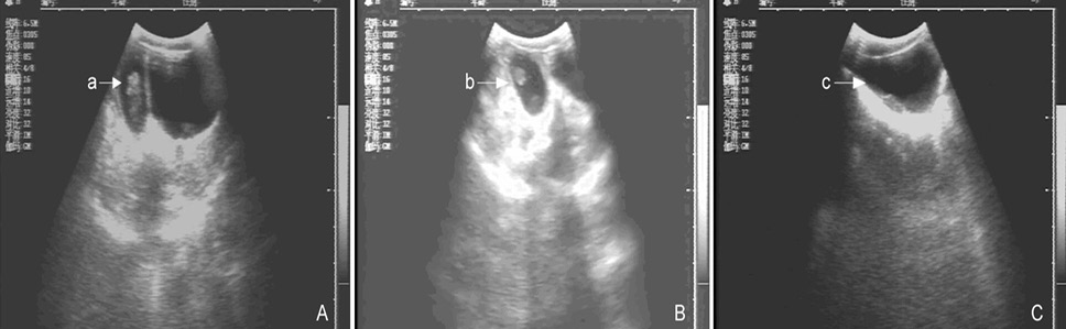
B-ultrasonography images show that female pangolins MJ-X2 (picture A) and MJ-X3 (picture B) are pregnant, but MJ-X4 (picture C) not. Letters a and b show the pregnancy sacs with embryos on Dec 12, 2012. Letter c shows an obvious hypoechoic entity with no embryo.
References
- Challender, D., 2008. Asian Pangolins: How behavioural research can contribute to their conservation. In: Proceeding of the Workshop on Trade and Conservation of Pangolins Native to South and Southeast Asia (eds. S. Pantel and S.Y. Chin) Singapore: Singapore Zoo, pp. 95-102.
- Challender, D., Baillie, J., Ades, G., Kaspal, P., Chan, B., Khatiwada, A., Xu, L., Chin, S., KC, R., Nash, H. and Hsieh, H., 2014a. Manis pentadactyla. The IUCN red list of threatened species. 2014: e.T12764A45222544.
- Challender, D., Nguyen Van, T., Shepherd, C., Krishnasamy, K., Wang, A., Lee, B., Panjang, E., Fletcher, L., Heng, S., Seah Han Ming, J., Olsson, A., Nguyen The Truong, A., Nguyen Van, Q. and Chung, Y., 2014b. Manis javanica. The IUCN red list of threatened species 2014: e.T12763A45222303.
- Chin, S.C., Lien, C.Y., Chan, Y.T., Chen, C.L., Yang, Y.C. and Yeh L.S., 2011. Zoo Biol., 30:1-11.
- CITES. 2019. https://www.cites.org/eng/app/appendices.php (accessed 7 Jun 2019).
- Dong, B., Wu, H., Li, L., Sun, H. and Peng, X., 2017. Anim. Breed. Feed, 12: 4-5.
- Hao, Z. and Yin, L., 2017. Agric. Henan, 12: 47-48.
- Heath, M.E. and Vanderlip, S.L., 1988. Zoo Biol., 7: 293-312. https://doi.org/10.1002/zoo.1430070402[CrossRef]
- IUCN, 2014. Eating pangolins to extinction. https://www.iucn.org/content/eating-pangolins-extinction, (accessed 5 Jul 2019).
- Jia, G., Wang, X., Wang, L., Lv, L. and Yu, L., 2009. Shanghai J. Anim. Husband. Vet. Med., 1: 8-9.
- Mohapatra, R.K. and Panda, S., 2014. Folia Zool., 63:73-80. https://doi.org/10.25225/fozo.v63.i2.a4.2014[CrossRef]
- Ren, Q., Ren, H. and Huang, H., 2015. Chinese Digest Anim. Husband. Vet., 31: 223.
- van Ee, C.A., 1966. Int. Zoo Yearb., 6: 163-164. https://doi.org/10.1111/j.1748-1090.1966.tb01734.x[CrossRef]
- Wang, Q., Xia, K. and Zhang, J., 2018. Hubei J. Anim. Vet. Sci., 39: 9-12.
- Wang, S., 2008. Ultrasonographic assessment of embryonic and fetal development, and adult female genital system changes in the Yak. M. Sc. thesis, Gansu Agricultural University, Lanzhou, China.
- Wu, S.B., Ma, G.Z., Liao, Q.X. and Lu, K.H., 2005. Studies of conservation biology on Chinese Pangolin (Manis pentadactyla). China Forestry Publishing House, China.
- Xie, J., He, J., He, X., Cheng, Z., You, R. and Dong, C., 2010. J. Anhui agric. Sci., 38: 20722-20723.
- Yang, C.W., Chen, S., Chang, C.-Y., Lin, M.F. Block, E., Lorentsen, R., Chin, J.S.C. and Dierenfeld, E.S., 2007. Zoo Biol., 26: 223-230. https://doi.org/10.1002/zoo.20134[CrossRef]
- Yu, J., Peng, J., Liao, G., Jiang, F. and Ma, X., 2016. J. Chongqing Normal Univ. (Nat. Sci.), 33: 33-36.
- Zhang, F., Wu, S., Zou, C., Wang, Q., Li, S. and Sun, R., 2016. Zoo Keys, 618: 129-144. https://doi.org/10.3897/zookeys.618.8886[CrossRef]
- Zhang, F., Yu, J., Wu, S., Li, S., Zou, C., Wang, Q. and Sun, R., 2017. Zoo Biol., 36: 387-396. https://doi.org/10.1002/zoo.21388[CrossRef]
- Zhang, F., Wu, S., Yang, L., Zhang, L., Sun, R. and Li, S., 2015. Folia Zool., 64: 129–135. https://doi.org/10.25225/fozo.v64.i2.a6.2015[CrossRef]
- Zhang, F., Yu, Y., Wang, W., Xu, N. and Wu, S., 2019. Oryx, 53: 411-412. https://doi.org/10.1017/S0030605319000358[CrossRef]
Services
Article usage statistics
Loading…