1Clínica Veterinaria Fauna, Cruz de Malta 553, Hacienda los Pinos, Apodaca, Nuevo León, México
2Dermavet Hospital Veterinario, José de la Luz Blanco, Mz. 187, Lt. 33, Col. Santa Martha Acatitla, Ciudad de México. *Correspondence | Camilo Romero Núñez, Dermavet Hospital Veterinario, José de la Luz Blanco, Mz. 187, Lt. 33, Col. Santa Martha Acatitla, Ciudad de México
The clinical findings observed in this case, such as hyperextensibility, skin fragility and age of onset, were suggestive of cutaneous asthenia.
Cutaneous asthenia is a relatively rare and poorly documented veterinary disease.
To the knowledge of the authors, this is the first report of cutaneous asthenia in a young albino Burmese python in Latin America; and as in other species, histopathological evaluation and clinical findings are useful in the diagnosis of the disease.
Selected from the article's own text.
Abstract
Cutaneous asthenia, dermatosparaxis, Ehlers-Danlos syndrome, or also called skin fragility syndrome, is a hereditary disorder of the connective tissue caused by the defective synthesis or assembly of collagen. This case describes a young albino Burmese python that presented with dry-looking skin, multiple folds and wounds. Skin samples were taken for histopathology and compared with a healthy specimen of the same species and colour characteristics. The diagnosis was cutaneous collagen dysplasia suggestive of cutaneous asthenia. This is the first report of cutaneous asthenia in a baby albino Burmese python in Latin America.
Ehlers-Danlos syndrome (EDS) encompasses a clinically and hereditarily heterogeneous group of connective tissue disorders, clinically involving the tegumental, musculoskeletal, gastrointestinal and cardiovascular systems. EDS is caused by defective collagen synthesis or incorrect assembly of the collagen triple helical structure. It is characterised by a variable combination of skin fragility, skin hyperextensibility, joint hypermobility and fragility of internal organs and vessels ; Chiarelli et al., 2016; ;. which can all contribute to severe disability, compromising patients’ quality of life, and/or early mortality .
Currently, the classification of human EDS distinguishes 14 subtypes, of which 13 have been molecularly elucidated and are caused by defects in 20 different genes, mainly involved in the synthesis and maintenance of collagen and extracellular matrix ; , in particular fibril-forming collagen types I, III, V, or molecules involved in the maturation, folding, secretion or supramolecular organization of these collagens . Depending on the causative variant, EDS may follow an autosomal dominant or autosomal recessive mode of inheritance .
In veterinary medicine, EDS is considered to be rare, although the true prevalence is unknown. EDS have been reported in several species including cattle, horses, cats, rabbits, minks and sheep. Clinical phenotypes vary from mild to life- threatening and usually appear at a young age. Clinical signs associated with EDS in animals are most consistent with human type I, II or VIIa disease ;. The present study describes the clinical and histopathological findings in a young Burmese python affected by cutaneous asthenia.
Case description
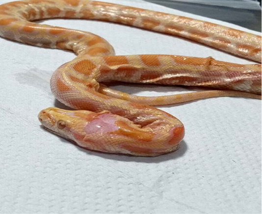
A male albino Burmese python (Python molurus bivittatus), five months old, weighing 72 g and 52 cm long, presented to the Fauna Veterinary Clinic with a history of anorexia. The owner stated that he acquired it at the age of two months from a pet store. It lived in a terrarium without companions and fed on thawed mouse pups. At the time of revision, the patient was noted to have flaccid, dehydrated-appearing skin and multiple generalised folds. A dermatological examination was performed and it was kept in the clinic for evaluation. The lesions found included generalised skin hyperextensibility, generalised skin fragility, cardboard or dehydrated texture, bilateral erythematous macules and an open linear wound in the cranial region (open skin) (Figure 1). There was no apparent itching or pain.
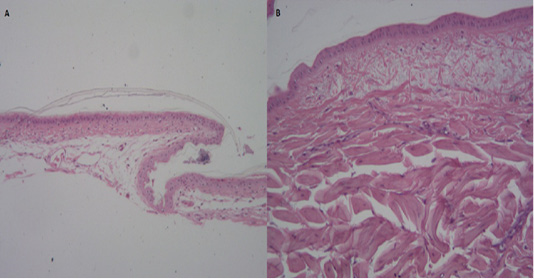
A skin biopsy was taken from the dorsal area of the head. Another sample was obtained from a healthy animal of the same gender and colour characteristics, as a control. Histopathological examination revealed a lower proportion of interlacing of the collagen bands, being more separated from each other, with a blunt and flattened appearance in the sick animal compared to the healthy animal (Figure 2). Among the collagen bands, amorphous acidophilic fibrillar material was observed with an oedematous appearance in some areas. The underlying epidermis also showed moderate thinning and atrophy, with clusters of multifocal melanic pigment grains below it. This provided a diagnosis of collagen dysplasia, suggestive of cutaneous asthenia.
Discussion
The clinical findings observed in this case, such as hyperextensibility, skin fragility and age of onset, were suggestive of cutaneous asthenia. Cutaneous asthenia is a rare genetic abnormality of the dermal connective tissue, mainly affecting young animals , and although it was first described in dogs in 1947, comprehensive studies on the disease have not been conducted in various species . Affected animals usually show open wounds, with little or no bleeding, resulting from minor trauma and multiple atrophic scars mainly involving the back and head ; . The reported Burmese python also evidenced a non-bleeding linear wound in the cranial area, probably associated with self-trauma. Although the disease can be diagnosed based on the clinical signs, the histopathological findings are fundamental, where they usually report disorganisation of the collagen fibres, with shortening, random orientation and a lower proportion .
In the reported case, the microscopic examination clearly showed a lower proportion of interlacing of the collagen bands, separated from each other, with a blunt and flattened appearance, with amorphous acidophilic fibrillar matter with an oedematous appearance between the bands; in comparison with the control animal, where no ultrastructural changes were detected in the collagen fibres and only a surface with a trabecular and laminated appearance with whitish bands was observed. However, it should be noted that not all animals with cutaneous asthenia show changes intense enough to be detected through histopathological examination , or only show the presence of lymphocytic inflammatory infiltrate, with abundant mast cells and neutrophils, some perivascular and reactive fibroblasts , so clinical and histopathological findings are usually essential for diagnosis . The diagnosis of human EDS is based on clinical signs and testing for genetic mutations encoding defective collagen(s) and/or collagen-modifying enzymes . Although confirmatory molecular tests are mandatory to reach a definitive diagnosis , genetic tests for EDS in reptiles are not available, in the present case a diagnosis was made based on clinical history, clinical signs and histopathology.
In humans, mutations in COL5A1 and COL5A2 lead to characteristic skin collagen abnormalities with variability in the diameter of collagen fibrils and the presence of collagen aggregates, termed “collagen cauliflowers” . In veterinary medicine, so far, distinct pathogenic variants in COL5A1 associated with classic EDS have been reported in dogs and cats, while in cattle it has been linked to COL5A2 . Diagnostic techniques to determine the subtype of EDS are not readily available for animals; therefore, it was not possible to determine the subtype of EDS in the present case. There is no specific treatment protocol and despite the poor prognosis, proper management and care are necessary to avoid secondary injuries .
Conclusion
Cutaneous asthenia is a relatively rare and poorly documented veterinary disease. To the knowledge of the authors, this is the first report of cutaneous asthenia in a young albino Burmese python in Latin America; and as in other species, histopathological evaluation and clinical findings are useful in the diagnosis of the disease.
Conflict of Interest
There are no conflicts of interest.
novelty statement
This is the first case report of Asthenia Cutanea in a snake.
Burmese python showing skin hyperextensibility and cranial wound
Figure 2.
Histopathological sections obtained from the disease (A) and healthy (B) specimen
References
Bauer A, Bateman JF, Lamandé, SR, Hassen E, Kirejczyk SGM, Yee M, Ramiche A, Jagannathan V, Welle M, Leeb T, Bateman FL. (2019) Identification of Two Independent COL5A1 Variants in Dogs with Ehlers–Danlos Syndrome. Genes (Basel). 10(10):731. https://doi.org/10.1101/660407[CrossRef]
Chiarelli N, Ritelli M, Zoppi N, Colombi M. (2019) Cellular and Molecular Mechanisms in the Pathogenesis of Classical, Vascular, and Hypermobile Ehlers-Danlos Syndromes. Genes, 10: 609. https://doi.org/10.3390/genes10080609[CrossRef]
de Vasconcelos EA, Gurgel AR, Pachaly JR. (2010) Astenia cutânea canina – Relato de caso. Rev Cient Med Vet Peq Anim e Anim Estim. 8(25): 345-349.
Dokuzeylul B, Altun ED, Ozdogan TH, Bozkurt HH, Arun SS, Or ME. (2013) Cutaneous asthenia (Ehlers–Danlos syndrome) in a cat. Turk. J. Vet. Anim. Sci.37: 245-249.
Halper J. (2013) Connective Tissue Disorders in Domestic Animals. In: Progress in Heritable Soft Connective Tissue Diseases. 231–240. https://doi.org/10.1007/978-94-007-7893-1_14[CrossRef]
Hansen N, Foster SF, Burrows AK, Mackie J, Malik R. (2015) Cutaneous asthenia (Ehlers-Danlos-like syndrome) of Burmese cats. J. Feline Med. Surg. 17: 954-963. https://doi.org/10.1177/1098612X15610683[CrossRef]
Jacinto JGP, Häfliger IM, Veiga IMB, Letko A, Benazzi C, Bolcato M, Drögemüller C. (2020) A Heterozygous Missense Variant in the COL5A2 in Holstein Cattle Resembling the Classical Ehlers–Danlos Syndrome. Animals (Basel). 10(11): 2002. https://doi.org/10.3390/ani10112002[CrossRef]
Paciello O, Lamagna F, Lamagna B, Papparella, S. (2003) Ehlers-Danlos—Like Syndrome in 2 Dogs: Clinical, Histologic, and Ultrastructural Findings. Vet. Clin. Pathol. 32: 13–18. https://doi.org/10.1111/j.1939-165X.2003.tb00306.x[CrossRef]
Seo S, Choi M, Hyun C. (2016) Cutaneous asthenia (Ehlers-Danlos syndrome) in a Korean short-haired cat. Korean J. Vet. Res. 56: 53-55. https://doi.org/10.14405/kjvr.2016.56.1.53[CrossRef]
Syx D, Miller RE, Obeidat AM, Tran PB, Vroman R, Malfait Z, Miller RJ, Malfait F, Malfait Anne-M. (2020) Pain-related behaviors and abnormal cutaneous innervation in a murine model of classical Ehlers-Danlos syndrome. Pain. 161(10): 2274-2283. https://doi.org/10.1097/j.pain.0000000000001935[CrossRef]
Spycher M, Bauer A, Jagannathan V, Frizzi M, De Lucia, M, Leeb T. (2018) A frameshift variant in the COL5A1 gene in a cat with Ehlers-Danlos syndrome. Anim. Gen. 49(6): 641-644. https://doi.org/10.1111/age.12727[CrossRef]
Szczepanik M, Golynski M, Wilkolek P, Popiel J, Smiech A, Pomorska D, Nowakowski H. (2006) Ehlers-Danlos syndrome (cutaneous asthenia)–A report of three cases in cats. Bull Vet Inst Pulawy. 50: 609-612.
Uri M, Verin R, Ressel L, Buckley L, McEwan N. (2015) Ehlers–Danlos Syndrome Associated with Fatal Spontaneous Vascular Rupture in a Dog. J. Comp. Pathol. 152(2-3): 211–216. https://doi.org/10.1016/j.jcpa.2014.12.013[CrossRef]
Vroman R, Malfait Anne-M, Miller RE, Malfait F, Syx D. (2021) Animal Models of Ehlers–Danlos Syndromes: Phenotype, Pathogenesis, and Translational Potential. Front.Genet. 12:726474. https://doi.org/10.3389/fgene.2021.726474[CrossRef]