Research Article · Volume 14, Issue 10, 3 September 2026 · Open Access
Ant’s Nest Plant Infusion and Probiotics as Effective Alternatives to Antibiotic Growth Promoters in Broilers: Modulation of Gut Enterobacteriaceae and Lactic Acid Bacteria
· · · · · · · · · ·
Affiliations & Notes ⌄
1Laboratory of Microbiology and Immunology Veterinary, Faculty of Veterinary Medicine, Universitas Brawijaya. Jl. Puncak Dieng, Kalisongo, Malang Regency 65151, East Java, Indonesia
2Department of Animal Husbandry, Faculty of Veterinary Medicine, Universitas Airlangga, Jl. Dr. Ir. H. Soekarno, Kampus C Mulyorejo, Surabaya 60115, East Java, Indonesia
3Faculty of Agriculture, Islamic University Kadiri, Jl. Sersan Suharmaji No. 38, Manisrenggo, Kediri, 64128, East Java, Indonesia
4Department of Applied Microbiology, Faculty of Science, Ebonyi State University 480211, Abakaliki, Nigeria
5Faculty of Veterinary Medicine, Universitas Brawijaya, Jl. Puncak Dieng, Kalisongo, Malang Regency 65151, East Java, Indonesia. *Correspondence | Fidi Nur Aini Eka Puji Dameanti, Laboratory of Microbiology and Immunology Veterinary, Faculty of Veterinary Medicine, Universitas Brawijaya. Jl. Puncak Dieng, Kalisongo, Malang Regency 65151, East Java, Indonesia
This study characterizes the dominant intestinal bacteria in broiler chickens challenged with avian pathogenic Escherichia coli (APEC) and evaluates the effects of dietary interventions on gut microbial populations.
Dietary supplementation with ant’s nest plant (Myrmecodia sp.) infusion consistently resulted in lower Enterobacteriaceae counts and highest lactic acid bacteria populations compared with other treatment groups under APEC challenge conditions.
Overall, the findings demonstrate that a 20% ant’s nest plant infusion shows strong potential as a non-antibiotic dietary strategy for reducing Enterobacteriaceae and enriching beneficial lactic acid bacteria in broiler chickens.
Nevertheless, the observed microbial modulation supports the potential role of ant’s nest plant infusion as an alternative to antibiotic growth promoters in broiler production systems.
Selected from the article's own text.
Abstract
Broiler chickens are a key source of affordable animal protein in Indonesia; however, intensive production systems increase susceptibility to infectious diseases, often leading to the use of antibiotic growth promoters (AGPs). Prolonged AGP use raises concerns regarding antimicrobial resistance and antibiotic residues, prompting regulatory restrictions in Indonesia. This study evaluated the effects of probiotics, acidifiers, and ant’s nest plant (Myrmecodia sp.) preparations as alternatives to AGPs on gut microbiota modulation in broilers experimentally infected with avian pathogenic Escherichia coli (APEC), with lactic acid bacteria (LAB) abundance as the primary outcome and Enterobacteriaceae counts as a secondary indicator. Broilers were allocated to ten dietary treatments (n= 5 per group). LAB and Enterobacteriaceae populations in the cecum and ileum were quantified using total plate count methods and analyzed using non-parametric statistics. Kruskal–Wallis analysis revealed significant differences among treatments for both Enterobacteriaceae (χ²= 40.809, df= 9, P< 0.001) and LAB counts (χ²= 34.997, df= 9, P< 0.001). Broilers receiving 20% ant nest plant infusion exhibited the highest LAB counts (median 44.6 × 10â· CFU/g), whereas probiotic and acidifier treatments resulted in the lowest Enterobacteriaceae counts (median 8.9–9.0 × 10â· CFU/g). Post hoc pairwise comparisons using the Mann–Whitney U test with Holm–Bonferroni correction did not identify statistically significant differences between individual treatment pairs, treatment effectiveness was interpreted based on median values and mean rank distributions. Overall, ant nest plant infusion at 20% demonstrated the strongest LAB-promoting effect, supporting its potential as a functional alternative to AGPs in broiler production systems.
The poultry remains a significant agricultural commodity with high demand, as it serves as an accessible source of protein for the general population. Data from Badan Pangan Nasional (Bapanas) indicate an increase in per capita chicken meat consumption since 2019. In 2023, the average consumption of chicken meat per person in Indonesia reached 7.46 kg per year. Restrictions on the use of hormones and antibiotics as feed additives has created an opportunity to develop alternatives to antibiotic growth promoters (AGPs), such as probiotics, which can help improve livestock productivity and enhance chicken immunity against Escherichia coli infection. The growing demand for chicken meat requires poultry farming systems to maintain high levels of productivity . Consequently, broiler farmers in Indonesia have commonly relied on AGPs to support growth performance. These AGPs are typically administered as feed supplements at sub-therapeutic doses to stimulate the immune system, suppress pathogenic bacterial infections, and improve nutrient absorption in the intestine. However, their use has raised concerns regarding potential adverse effects on consumers, as AGP application may induce antibiotic resistance in chickens and allow antibiotic residues to enter the food chain, posing public health risks .
In Indonesia, regulations governing the use of AGPs are outlined in Law No. 18 of 2009 as amended by Government Regulation No. 41 of 2014, Article 22, Paragraph 4c, concerning animal husbandry and animal health. These regulations restrict the use of antibiotics as feed additives for growth promotion purposes, particularly those with relevance to human medicine, to mitigate the risk of antimicrobial resistance and antibiotic residues in animal product. The implementation of these regulations has intensified the search for alternative strategies to replace AGPs in broiler production systems ; ; .
The ant’s nest plant (Myrmecodia sp.) is a medicinal herb with strong potential to enhance poultry performance by performing digestive function. Previous research by Assidiqi et al. (2021) demonstrated that supplementation with ant nest extract increased feed efficiency in laying hens. This plant is rich in bioactive compounds, including flavonoids, tannins, and phenolic compounds, which exhibit antibacterial, anti-inflammatory, and antioxidant activities. Flavonoids function as natural antibiotics and antioxidants, while tannins further contribute to antimicrobial effects . These active compounds help improve the balance of gastrointestinal microflora, support intestinal health, and enhance nutrient absorption, ultimately leading to improved productivity and performance in broiler chickens .
Probiotics represent an alternatives to AGPs in the poultry industry. Wardhani et al. (2019) demonstrated that probiotic supplementation in drinking water improved growth performance and reduced the risk of infectious diseases in laying hens. Probiotics function by adhering to the gastrointestinal tract and forming colonies of beneficial bacteria. This facilitates the proliferation of beneficial bacteria, which inhibit pathogenic bacteria. The incorporation of probiotics into broiler feed has been shown to improve performance and effectively replace antibiotics for growth promotion ; ; .
Acidifiers are feed additives that help regulate of microflora in the gastrointestinal tract . Research by Pio et al. (2017), demonstrated that the addition of acidifying agents to drinking water and feed for broiler chickens can enhance digestive enzyme function, improve digestion and absorption particularly of fiber and protein and increase nutrient absorption within the avian digestive system. Acidifiers are organic acids that play a critical role in optimizing digestibility by enhancing digestive enzyme performance, stabilizing intestinal pH, and optimizing the microbial balance in the gastrointestinal tract . Organic acids or acidifiers may function either as sources of carbon and energy for certain microorganisms or as inhibitory compounds, depending on their concentration, their ability to penetrate microbial cells, and the metabolic capacity of the microorganisms involved. In addition, acidifiers have the potential to enhance nutrient utilization, modify intestinal pH, and suppress the growth of pathogenic bacteria in the digestive tract. The application of acidifiers through drinking water is therefore effective in controlling harmful microorganisms and supporting optimal performance in broiler chickens.
The combination of probiotics and acidifiers is commonly applied in poultry feeding strategies due to their complementary mechanisms of action. In this study, it was hypothesized that combined supplementation would exert a synergistic effect on gut microbiota modulation. Acidifiers were expected to lower intestinal pH, thereby creating favorable conditions for probiotic survival and colonization, which could enhance the suppression of pathogenic Enterobacteriaceae and promote the growth of lactic acid bacteria more effectively than single supplementation ; .
Several alternatives can be employed to replace antibiotic growth promoters (AGPs) in broiler production systems, including ant’s nest plant preparations, probiotics, acidifiers, and their combination. Given their distinct yet complementary mechanisms of action in modulating gut microbiota, a laboratory-based experimental approach evaluating their effects on lactic acid bacteria and Enterobacteriaceae populations is warranted to provide scientific evidence supporting their potential application as AGP alternatives. Therefore, this study aimed to evaluate the effects of ant’s nest plant extract, ant’s nest plant infusion, probiotics, acidifiers, and their combination on Enterobacteriaceae and lactic acid bacteria (LAB) counts in the gastrointestinal tract of broiler chickens experimentally infected with avian pathogenic E. coli. The findings are expected to contribute to the development of effective alternatives to antibiotic growth promoters in broiler production systems.
Materials and Methods
Study location and timeline
The research was conducted from May to July 2024. Broiler rearing, bacterial infection, and organ sample collection took place at a poultry farm in Kediri City, East Java Province, Indonesia. Sample processing, bacterial culture, and microbial enumeration were performed at the Laboratory of Microbiology and Veterinary Immunology, Faculty of Veterinary Medicine, Universitas Brawijaya, Indonesia.
Experimental design and sample size determination
The sample size was determined using the Federer formula, expressed as t(n−1) ≥ 15, where t represents the number of treatment groups and n the number of experimental units per group. With ten treatment groups, the minimum required sample size was calculated as follows:
10(n−1) ≥ 15, 10n−10 ≥ 15, 10n ≥ 25, n ≥ 2.5, n ≈ 3
Based on this calculation, a minimum of five broilers per treatment group was required. Therefore, a total of 50 Cobb strain broiler chickens (5 birds per group) were used in this study.
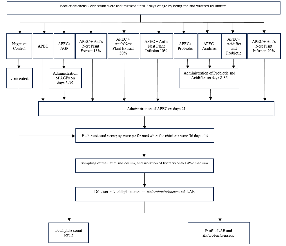
The experimental design of this study is illustrated in Figure 1. Broilers were randomly allocated into ten treatment groups (Table 1). All broilers were acclimatized until 7 days of age with ad libitum access to feed and drinking water. From day 8 to day 35, dietary treatments were administered according to group allocation, including antibiotic growth promoter (zinc bacitracin), ant’s nest plant (Myrmecodia sp.) extract, ant’s nest plant infusion, probiotics, acidifiers, or combinations thereof. On day 21, all groups except the negative control were challenged with an avian pathogenic E. coli (APEC) O-78 field isolate via drinking water.
Table 1: Group and sample treatment.
Group
Treatment
T1
Negative Control
T2
APEC
T3
APEC + AGP
T4
APEC + Ant’s Nest Plant Extract 15%
T5
APEC + Ant’s Nest Plant Extract 30%
T6
APEC + Ant’s Nest Plant Infusion 10%
T7
APEC + Probiotic
T8
APEC + Acidifier
T9
APEC + Acidifier and Probiotic
T10
APEC + Ant’s Nest Plant Infusion 20%
An avian pathogenic Escherichia coli (APEC) field isolate serotype O-78 was used for experimental infection. The bacterial culture was grown overnight and adjusted to a final concentration of approximately 1 × 10⁸ CFU/mL. The APEC suspension was administered through drinking water at an estimated dose of 1 × 10⁸ CFU per bird,based on average water consumption per broiler. Infection was confirmed phenotypically by re-isolation of Enterobacteriaceae from cecum samples collected at necropsy, followed by biochemical identification tests, as described in the microbiological analysis section.
For the ant’s nest plant (Myrmecodia sp.) extract and infusion were prepared prior to administration. For the infusion, dried ant’s nest plant material was ground into powder. A number of concentration (15; 20; 30) based on weight per volume (w/v) infusion. It was prepared by adding 15 or 20 or 30 g of powdered plant material to distilled water and adjusting the final volume to 100 mL. The mixture was heated at approximately 90–95°C for 15 minutes, allowed to cool, and filtered using sterile filter paper. The infusion was freshly prepared and administered via drinking water according to the treatment groups.
At 35 days of age, all broilers were humanely euthanized, followed by necropsy and collection of ileum and cecum samples for microbiological analysis. Enterobacteriaceae and LAB populations were subsequently quantified using the total plate count method.
Sample collection
At 35 days of age, all broiler chickens were humanely euthanized following standard animal welfare procedures. Necropsy was immediately performed, and ileum and cecum samples were aseptically collected from each bird. The samples were placed in sterile containers and transported under cool conditions to the Laboratory of Microbiology and Veterinary Immunology, Faculty of Veterinary Medicine, Universitas Brawijaya, Indonesia, for subsequent microbiological analysis.
Calculation of enterobacteriaceae and lactic acid bacteria (LAB) counts
For Enterobacteriaceae enumeration, cecum samples were aseptically collected, weighed, and homogenized in sterile Buffered Peptone Water (BPW). The homogenates were subjected to serial tenfold dilutions up to 10⁻⁷. From appropriate dilutions (10⁻⁵ to 10⁻⁷), 0.1 mL aliquots were spread-plated onto Violet Red Bile Agar (VRBA) and incubated aerobically at 37°C for 24 hours. For LAB enumeration, ileum samples were processed using the same homogenization and serial dilution procedures. Aliquots (0.1 mL) from appropriate dilutions were plated onto de Man, Rogosa, and Sharpe (MRS) agar and incubated anaerobically at 37°C for 48 hours using an anaerobic jar with a gas-generating kit.
After incubation, bacterial colonies were counted using the standard plate count method. The Total Plate Count (TPC) was calculated according to Azizah and Soesetyaningsih (2020). Plates with 30-300 colonies were counted. The number of viable bacteria per gram was calculated using the formula: CFU/g= (number of colonies counted) × (dilution factor). Plates with merged colonies were counted as a single colony, whereas plates exceeding the countable range were classified as too numerous to count (TNTC) . Bacterial counts were expressed as colony-forming units per gram (CFU/g) and reported as ×10⁷ CFU/g.
Isolation and identification of enterobacteriaceae and LAB biochemical tests
Isolation and identification in this study were limited to the genus level based on morphological and biochemical characteristics. Species-level identification using molecular methods (e.g., 16S rRNA gene sequencing) was beyond the scope of this study. Enterobacteriaceae isolates obtained from ileum samples were subjected to biochemical identification using standard differential and confirmatory tests. Colonies suspected as Enterobacteriaceae were first streaked onto Eosin Methylene Blue Agar (EMBA) to observe characteristic metallic green sheen formation. Further identification was performed using Sulfide–Indole–Motility (SIM) medium to assess sulfide production, indole formation, and motility. Methyl Red (MR) and Voges–Proskauer (VP) tests were conducted to evaluate glucose fermentation pathways, while citrate utilization was assessed using Simmons Citrate Agar (SCA).
Triple Sugar Iron Agar (TSIA) tests were performed to determine carbohydrate fermentation patterns (slant and butt reactions), gas production, and hydrogen sulfide (H₂S) formation. Urease activity was also evaluated using urease medium. Biochemical reaction profiles were interpreted by comparing test results with standard identification keys for Enterobacteriaceae, allowing presumptive identification of E. coli isolates.
LAB isolates obtained from cecum samples were identified based on colony morphology, microscopic characteristics, and biochemical tests. Colony morphology, including shape and color, was recorded following incubation on de Man, Rogosa, and Sharpe (MRS) agar. Gram staining was performed to determine Gram reaction and cell morphology. Catalase and coagulase tests were conducted to differentiate LAB from other Gram-positive bacteria. Isolates showing Gram-positive reaction, absence of catalase and coagulase activity, and characteristic rod-shaped or coccoid morphology were classified as LAB, consistent with the phenotypic characteristics of Lactobacillus spp. and Enterococcus spp., following established identification procedures ; ; .
Statistical analysis
Statistical analyses were performed using IBM SPSS Statistics software (version 22; IBM Corp., Armonk, NY, USA). Data distribution was assessed for normality using the Shapiro–Wilk test. As the data were not normally distributed and the sample size per group was small (n = 5), non-parametric statistical tests were applied. Differences in Enterobacteriaceae and LAB counts among treatment groups were analyzed using the Kruskal–Wallis test. When a significant overall effect was detected, pairwise comparisons were conducted using the Mann–Whitney U test with Holm–Bonferroni correction to adjust for multiple comparisons. Bacterial counts were expressed as colony-forming units per gram (CFU/g) and reported as ×10⁷ CFU/g. Data are presented as median (Q1–Q3) and mean rank. A p-value < 0.05 was considered statistically significant.
Results and Discussion
Characteristics of lactic acid bacteria isolates
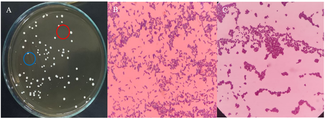
Isolation of LAB from ileum samples resulted in two distinct colony morphologies, designated as isolates A and B (Figure 2). Isolate A formed large, circular colonies with an entire margin and flat elevation, whereas isolate B produced smaller colonies with similar morphological characteristics. Both isolates displayed a milky white coloration on MRS agar. Isolation of LAB from ileum samples resulted in two distinct colony morphologies, designated as isolates A and B (Figure 3).
Gram staining revealed that both isolates were Gram-positive. Microscopic observation showed rod-shaped cells for isolate A and coccoid cells for isolate B. Biochemical testing demonstrated that both isolates were negative for catalase and coagulase activity, which is consistent with the phenotypic characteristics of LAB. Based on morphological and biochemical features, isolate A was presumptively identified as belonging to the genus Lactobacillus, while isolate B was consistent with Enterococcus spp., in agreement with previous descriptions of LAB isolated from poultry intestines ; . Species-level identification using molecular methods was beyond the scope of this study.
Identification of Enterobacteriaceae isolates
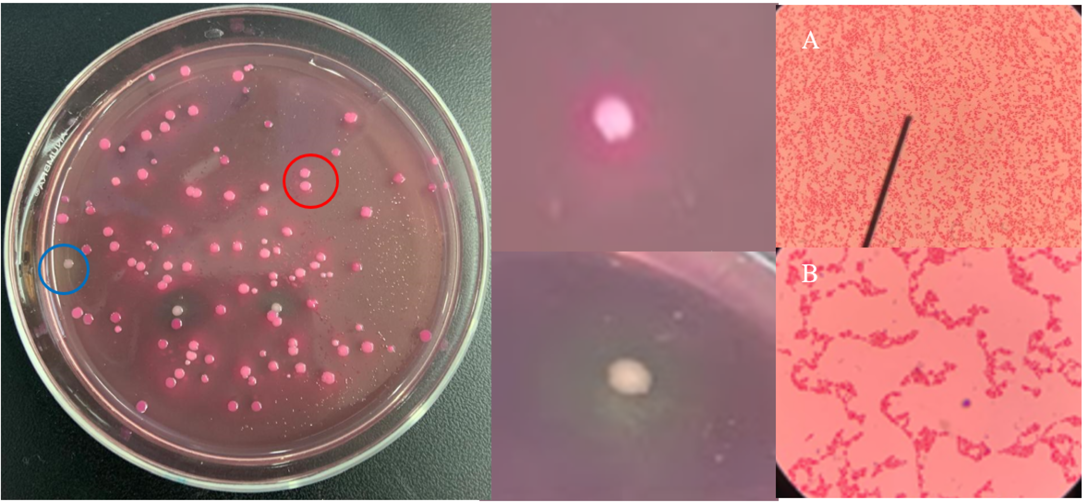
Isolation on VRBA yielded two dominant colony morphologies (Figure 2). Colonies exhibiting a purple coloration accompanied by a precipitation zone were indicative of lactose-fermenting Enterobacteriaceae, whereas pale or colorless colonies without precipitation suggested non-lactose fermenters. Gram staining confirmed that the isolates were Gram-negative bacilli, consistent with members of the Enterobacteriaceae family.
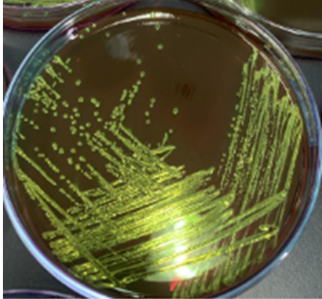
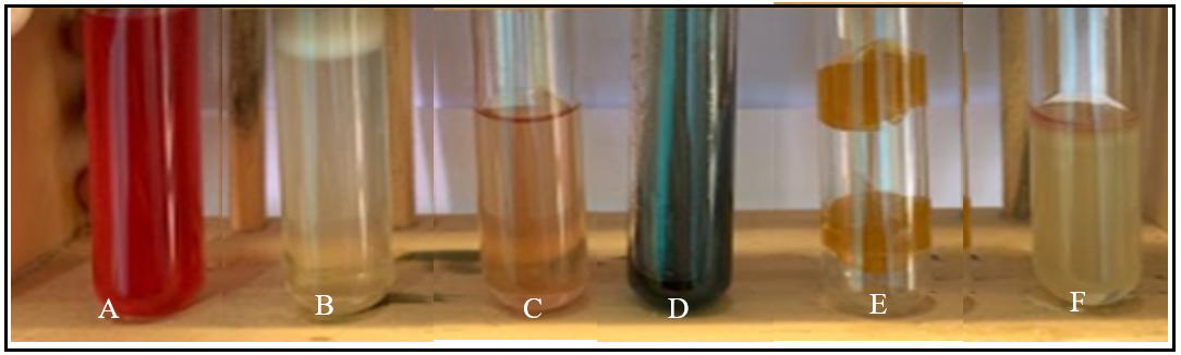
Further biochemical characterization using IMViC, TSIA, SCA, urease medium, and EMBA (Figures 4 and 5) demonstrated reaction patterns characteristic of E. coli. The presence of a metallic green sheen on EMBA, positive methyl red reaction, negative VP and citrate tests, and gas production on TSIA collectively supported the identification of E. coli as the predominant Enterobacteriaceae isolated from cecal samples. These findings are consistent with previous reports describing the biochemical profile of E. coli in broiler intestinal samples ; .
Effects of dietary treatments on Enterobacteriaceae and LAB counts
The distribution of Enterobacteriaceae and LAB counts across treatment groups is presented in Table 2. The Kruskal–Wallis test revealed a significant overall difference in Enterobacteriaceae counts among treatment groups (χ² = 40.809; df = 9; P < 0.001), indicating that dietary interventions significantly influenced the intestinal abundance of Enterobacteriaceae under avian pathogenic E. coli (APEC) challenge conditions. Broilers receiving probiotic (T7), acidifier (T8), and 20% ant’s nest plant infusion (T10) exhibited the lowest median Enterobacteriaceae counts, with median values of 9.0, 8.9, and 16.2 × 10⁷ CFU/g, respectively. These groups also showed the lowest mean rank values, reflecting a consistent
Table 2: Distribution of Enterobacteriaceae (10⁷ cfu/g) and lactic acid bacteria (LAB) counts (10⁷ cfu/g) by treatment.
Group
Treatment
n
Enterobactericeae (10⁷ cfu/g)*
LAB (10⁷ cfu/g)**
Median (Q1–Q3)
Mean rank
Median (Q1–Q3)
Mean rank
T1
Negative control
5
37.0 (26.9–47.2)
33.1
18.7 (5.2–32.3)
21.0
T2
APEC
5
32.7 (30.1–35.4)
35.5
27.2 (22.7–31.8)
27.0
T3
APEC + AGP
5
26.7 (23.7–29.8)
23.6
25.3 (21.8–29.1)
25.3
T4
APEC + Ant’s Nest Plant Extract 15%
5
42.9 (41.4–45.0)
48.0
17.5 (14.4–20.7)
13.2
T5
APEC + Ant’s Nest Plant Extract 30%
5
21.8 (19.9–23.7)
19.8
29.1 (26.8–31.5)
36.4
T6
APEC + Ant’s Nest Plant Infusion 10%
5
36.2 (34.3–38.1)
38.2
21.7 (19.5–25.1)
17.4
T7
APEC + Probiotic
5
9.0 (8.6–9.5)
6.7
30.1 (26.4–33.8)
36.6
T8
APEC + Acidifier
5
8.9 (8.6–9.2)
6.3
27.6 (24.5–30.8)
33.7
T9
APEC + Acidifier and Probiotic
5
31.0 (28.5–33.5)
29.8
10.6 (7.4–13.8)
5.7
T10
APEC + Ant’s Nest Plant Infusion 20%
5
16.2 (14.9–17.5)
14.0
44.6 (41.8–47.5)
48.0
* Data are presented as median (Q1–Q3). Kruskal–Wallis analysis indicated a significant overall difference among treatments (χ² = 40.809; df = 9; P < 0.001). Post hoc pairwise comparisons using the Mann–Whitney U test with Holm–Bonferroni correction did not reveal statistically significant differences between individual treatment pairs. ** Data are presented as median (Q1–Q3). Kruskal–Wallis analysis indicated a significant overall difference among treatments (χ² = 34.997; df = 9; P < 0.001). Post hoc pairwise comparisons using the Mann–Whitney U test with Holm–Bonferroni correction did not reveal statistically significant differences between individual treatment pairs.
reduction in potentially pathogenic bacteria compared with the APEC-only group (T2). In contrast, higher median Enterobacteriaceae counts were observed in the negative control (T1) and in treatments receiving lower concentrations of ant’s nest plant extract or infusion (T4 and T6), suggesting limited suppressive effects at these concentrations.
A significant overall difference in LAB counts was also detected among treatment groups (χ² = 34.997; df = 9; p < 0.001). The highest median LAB count was observed in the 20% ant’s nest plant infusion group (T10), reaching 44.6 × 10⁷ CFU/g, followed by probiotic supplementation (T7) and 30% ant’s nest plant extract (T5). These treatments also demonstrated the highest mean rank values, indicating a strong tendency toward enrichment of beneficial LAB populations in the intestinal tract.
Although post hoc pairwise comparisons using the Mann–Whitney U test with Holm–Bonferroni correction did not reveal statistically significant differences between individual treatment pairs (adjusted p > 0.05), consistent directional trends were observed in both median values and mean ranks. In particular, the 20% ant’s nest plant infusion consistently combined lower Enterobacteriaceae counts with higher LAB abundance, suggesting biologically meaningful modulation of gut microbial composition despite the absence of pairwise statistical significance after correction.
Implications for gut microbial modulation and AGP substitution
The observed reduction in Enterobacteriaceae counts in broilers receiving acidifier, probiotic, and ant’s nest plant infusion treatments may be attributed to alterations in intestinal pH and the presence of bioactive compounds with antibacterial properties. Acidifiers are known to lower intestinal pH, creating an unfavorable environment for pathogenic bacteria and inhibiting their metabolic activity and proliferation .
Ant’s nest plant (Myrmecodia sp.) contains bioactive phenolic compounds, including flavonoids and triterpenoids, which exhibit antibacterial activity by disrupting bacterial cell membranes and activating enzymes that will reduce the growth of pathogenic bacteria. Flavonoids also provide antioxidant effects by forming a defence for cells in the body so as to minimize damage that can be caused by free radicals ; ; . The superior performance of the 20% infusion compared with lower concentrations suggests a dose-dependent effects, whereby sufficient levels of bioactive compounds are required to effectively modulate the gut microbiota.
Notably, while probiotic supplementation alone increased LAB abundance, the combined acidifier–probiotic treatment (T9) resulted in comparatively lower LAB counts. This finding suggests a potential antagonistic interaction, possibly due to excessive acidification adversely affecting probiotic viability or colonization. This observation highlights the importance of formulation compatibility when combining gut-modulating agents.
Overall, the consistent association between the 20% ant’s nest plant infusion and a favorable microbial profile that is characterized by reduced Enterobacteriaceae and enhanced LAB populations supports its potential as a functional alternative to antibiotic growth promoters in broiler production systems under APEC challenge. It should be noted that conclusions regarding gut health are based on microbial indicators only, as physiological and immunological parameters were not assessed in this study.
Conclusions
This study characterizes the dominant intestinal bacteria in broiler chickens challenged with avian pathogenic Escherichia coli (APEC) and evaluates the effects of dietary interventions on gut microbial populations. Lactic acid bacteria isolated from ileum samples were presumptively identified as members of the genera Lactobacillus and Enterococcus based on morphological and biochemical characteristics, while Enterobacteriaceae isolated from cecum samples were predominantly identified as E. coli. Species-level identification using molecular methods was beyond the scope of this study. Dietary supplementation with ant’s nest plant (Myrmecodia sp.) infusion consistently resulted in lower Enterobacteriaceae counts and highest lactic acid bacteria populations compared with other treatment groups under APEC challenge conditions. These microbial patterns indicate a shift toward a more favorable intestinal bacterial composition.
Overall, the findings demonstrate that a 20% ant’s nest plant infusion shows strong potential as a non-antibiotic dietary strategy for reducing Enterobacteriaceae and enriching beneficial lactic acid bacteria in broiler chickens. Conclusions regarding gut health are based solely on microbiological indicators, as physiological, immunological, and performance parameters were not assessed in this study. Nevertheless, the observed microbial modulation supports the potential role of ant’s nest plant infusion as an alternative to antibiotic growth promoters in broiler production systems.
Acknowledgement
The authors would like to thank Penelitian Kolaboratif Pendanaan Internal DPP/SPP Fakultas Kedokteran Hewan, Universitas Brawijaya Tahun 2024, with grant number: 1733/UN10.F13/2024
Novelty Statement
The novelty of this study lies in the comparative evaluation of ant’s nest plant (Myrmecodia sp.) extract and infusion, probiotics, acidifiers, and their combination as non-antibiotic alternatives to antibiotic growth promoters in broilers challenged with avian pathogenic Escherichia coli (APEC). By assessing Enterobacteriaceae and lactic acid bacteria (LAB) as complementary microbial indicators, this study shows that 20% ant’s nest plant infusion produced a favorable microbial profile, with the highest LAB abundance and relatively low Enterobacteriaceae counts.
Author’s Contribution
FNAEPD: Supervision, methodology, writing-reviewing and editing. WPL: Conceptualization, formal analysis. EFL: Writing-reviewing and editing. EE: Writing-reviewing and editing. NIA: Sample collection, laboratory analysis, writing-reviewing and editing. MTI: Sample collection, laboratory analysis, writing-original draft. AHSy: Sample collection, laboratory analysis, writing-reviewing and editing. MAAS: Laboratory supervision, data curation, validation. RYOD: Data curation and writing-reviewing. RS: Data curation and validation, writing-reviewing and editing.
Generative AI and AI assisted technology statement
The authors declare that no generative AI and AI assisted technology was used in the creation of this manuscript.
Conflict of interest
The authors have declared no conflict of interest.
Figures
Figure 1:.
Experimental design schematic.
Figure 2:.
Bacterial colonies on VRBA’s media and Gram staining, A Colony A, B Colony B. Note: The red circle is colony A and the blue circle is colony B. Microscopic images were observed at 1000× magnification (oil immersion).
Figure 3:.
A: Bacterial colonies on MRSA media, B: Colony A, C: Colony B. Note: The red circle is colony A and the blue circle is colony B. Microscopic images were observed at 1000× magnification (oil immersion).
Figure 4:.
Results of isolation Eschericia coli on EMBA medium.
Figure 5:.
Identification results of Eschericia coli in the TSIA, IMViC, SCA and Urease. (A) MR (+), (B)VP (-), (C) Urease (-), (D) SCA (-), (E) TSIA, (F) SIM.
References
Arifin M, Pramono V (2014). The effect of sinbiotic supplement as replacement for antibiotic growth promotor on growth and size of intestiunal villi in broiler chicken. J. Sain Vet., 32(2): 205-217.
Assidiqi MH, Lisnanti MF, Sigit M (2021). Pengaruh pemberian ekstrak tanaman sarang semut (Myrmecodia sp.) terhadap produktivitas ayam petelur. J. Ilmiah Fillia Cendekia, 6(1).
Astuti LA, Yani S (2023). Uji Toksisitas akut, respon penyembuhan luka gingina dan uji iritasi sediaan gel sarang semut (Myrmecodia pendens) Kalimantan (Studi in vivo) Sebagai Bahan Dasar Terapi Poket Periodontal. AGMA, Sulawesi Selatan, Indonesia.
Azizah A, Soesetyaningsih E (2020). Akurasi Perhitungan bakteri pada daging sapi menggunakan metode hitung cawan. Berkala Sainstek. 8(3): 75-79. https://doi.org/10.19184/bst.v8i3.16828[CrossRef]
Dameanti FNAEP, Yanestria SM, Widodo A, Effendi MH, Plumeriastuti H, Tyasningsih, W, Ugbo EN, Sutrisno R, Akramsyah MA (2023). Prevalence of multidrug resistance and extended-spectrum beta-lactamase-producing Klebsiella pneumoniae from dairy cattle farm wastewater in East Java Province, Indonesia. Int. J. One Health, 9(2): 141–149. https://doi.org/10.14202/IJOH.2023.141-149[CrossRef]
Dameanti FNAEP, Yanestria SM, Widodo A, Effendi MH, Plumeriastuti H, Tyasningsih, W, Sutrisno R, Akramsyah MA (2023). Incidence of Escherichia coli producing Extended-spectrum beta-lactamase (ESBL) in wastewater of dairy farms in East Java, Indonesia. Biodiversitas, 24: 1143-1150. https://doi.org/10.13057/biodiv/d240254[CrossRef]
Daud M (2005). Performan ayam pedaging yang diberi probiotik dan prebiotik dalam ransum. J. Ilmu Ternak. 5(2): 75–79.
Fajrih N (2022). Pembangunan pertanian dan peternakan berkelanjutan. Deepublish, Yogyakarta, Indonesia.
Firdaus MA, Soeharsono S, Lokapirnasari WP, Hidanah S, Kusnoto K, Safitri E, Silfia HI, Lisnanti, EF (2024). Probiotic Lactobacillus sp. as a substitute for growth antibiotic (AGP): Its effects on production performance and contribution margin valuein broilers infected with Escherichia coli. J. Ilmu-Ilmu Petern., 34(3): 299-308. https://doi.org/10.21776/ub.jiip.2024.034.03.2[CrossRef]
Hamid H, Shi HQ, Ma GY, Fan Y, Li WX, Zhao LH, Zhang JY, Ji C, Ma QG (2018). Influence of acidified drinking water on growth performance and gastrointestinal function of broilers. Poult. Sci., 97(10): 3601–3609. https://doi.org/10.3382/ps/pey212[CrossRef]
Hestiyani F, Wahyuni A (2024). Isolasi dan identifikasi bakteri pada sekum ayam petelur (Gallus gallus domesticus) sehat sebagai kandidat probiotik. Resiliensi Industri Peternakan Tropis. 1: 85-94.
Hidayat K, Wibowo S, Sari L, Darmawan A (2018). Acidifier alami air perasaan jeruk nipis (Citrus Aurantiun) Sebagai Pengganti Antibiotik Growth Promotor Ayam Broiler. J. Ilmu Nutrisi dan Teknologi Pakan. 16(2): 27-33. https://doi.org/10.29244/jintp.16.2.27-33[CrossRef]
IBM Corp. (2013). IBM SPSS Statistics for Windows, Version 22.0. Armonk, NY: IBM Corp.
Kartikasari A, Hamid IS, Purnama MT, Damayanti R, Fikri F (2019). Isolasi dan identifikasi bakteri Eschericia coli kontaminan pada daging ayam broiler di rumah potong ayam kabupaten lamongan. J. Med. Vet., 2(1): 66-71. https://doi.org/10.20473/jmv.vol2.iss1.2019.66-71[CrossRef]
Koutsianos D, Athanasiou L, Mossialos D, Koutoulis KC (2020). Colibacillosis in poultry: A disease overview and the new perspectives for its control and prevention. J. Hellenic Vet. Med., 71(4): 2425-2436. https://doi.org/10.12681/jhvms.25915[CrossRef]
Kurniawan J, Tugiyanti E, Susanti E (2021). Pengaruh pemberian feed additive sebagai pengganti antibiotik terhadap konsumsi pakan dan pertambahan bobot badan ayam broiler. J. Anim. Sci. Technol., 3(2): 134-140.
Lima B, Aguero MB, Zygaldo J, Tapiai A, Solis C, De Arias AR, Yaluff G, Zacchino S, Feresin GE, Hirschman GS (2009). Antimicrobial activity of extracts, essential oils and metabolites obtained from Tagetes mendocina. J. Chil. Chem. Soc., 54: 68-72. https://doi.org/10.4067/S0717-97072009000100016[CrossRef]
Lisnanti EF, Lokapirnasari WP, Al-Arif MA, Hestianah EP, Hidanah S, Moses IB, Yuliani MGA, Baihaqi ZA, Dameanti FNAEP, Khairullah AR, Hendrawan VF, Rehman (2025). The ameliorative effects of Myrmecodia sp. as an antibiotic growth promoter in broiler chicken with avian pathogenic Escherichia coli infection. Open Vet. J., 15(11): 5858-5871. https://doi.org/10.5455/OVJ.2025.v15.i11.41[CrossRef]
Lisnanti EF, Lokapirnasari WP, Hestianah EP, Al-Arif MA, Baihaqi ZA, Yulianto AB (2024). Antibacterial alternatives using the potential of the ant nest plant (Myrmecodia spp.). Int. J. One Health, 10(1): 148-152. https://doi.org/10.14202/IJOH.2024.148-152[CrossRef]
Lokapirnasari WP, Hidayatik N, Hestianah EP, Silfia HI, Firdaus MA, Sherasiya A, Yulianto AB, Lamidi M, Al-Arif MA, Lisnanti EF, Baihaqi ZA, Marbun TD (2025). The potency of probiotics and acidifiers on body weight, feed efficiency, cholesterol, HDL, LDL in meat of broiler chickens infected with Escherichia coli. Adv. Anim. Vet. Sci., 13(1): 131-138. https://doi.org/10.17582/journal.aavs/2025/13.1.131.138[Full text][CrossRef]
Mubarak PR, Mahfudz LD, Sunarti D (2019). Pengaruh pemberian probiotik pada level protein pakan berbeda terhadap perlemakan ayam kampung. J. Sain Peternakan Indones., 13(4): 357-364. https://doi.org/10.31186/jspi.id.13.4.357-364[CrossRef]
Obioha PI, Ouoba LII, Anyogu A, Awamaria B, Atchia S, Ojimelukwe PC, Sutherland JP, Ghoddusi HB (2021). Identification and characterisation of the lactic acid bacteria associated with the traditional fermentation of dairy fermented product. Braz. J. Microbiol., 52(2): 869-881. https://doi.org/10.1007/s42770-021-00461-y[CrossRef]
Pio PO, Ardana IBK, Suastika P (2017). Efektivitas berbagai dosis asam organik dan anorganik sebagai acidifier terhadap histomorfometri duodenum ayam pedaging. Indones. Med. Vet., 6(1): 47-54.
Putri ALO, Kusdiyantini E (2018). Isolasi dan Identifikasi Bakteri Asam Laktat dari Pangan Fermentasi Berbasis Ikan (Inasua) yang Diperjualbelikan di Maluku-Indonesia. J. Biol. Trop., 1(2): 6-12. https://doi.org/10.14710/jbt.1.2.6-12[CrossRef]
Rahmatillah Z, Firdaus A (2025). Utilization of probiotics, fermented beverages, and Aspergillus niger in drinking water on performance and digestive quality of broilers (Gallus domesticus). J. Agripet., 25(2): 159-166. https://doi.org/10.17969/agripet.v25i2.43233[CrossRef]
Sari M, Abrar A, Merint (2013). Isolasi dan karakterisasi bakteri asam laktat pada usus ayam broiler. Agripet. 13(1): 43-48. https://doi.org/10.17969/agripet.v13i1.552[CrossRef]
Sinurat AP, Bahri S, Muahrsini S, Puastuti S, Priyanti A, Nurhayati IS, Priyono (2017). Kebijakan Pengendalian Penggunaan Antibiotic Growth Promoters dan Ractopamine dalam Mendukung Keamanan Pangan Nasional. Pusat Penelitian dan Pengembangan Peternakan, Bogor, Indonesia.
Syaifudin, Sigit M (2019). Pengaruh Penambahan Ekstrak Sarang Semut (Myrmecodia sp.) terhadap Performa Ayam Broiler Fase Finisher. J. Ilmiah Filia Cendekia. 4(2): 49-53.
Wardhani H, Lokapirnasari W, Soepronianondo K (2019). Penggunaan Probiotik Kombinasi Lactococcus Lactis Dan Lactobacillus acidophilus Sebagai Pengganti Antibiotika Pada Ayam Petelur Yang Diinfeksi Escherichia coli Terhadap Analisis Usaha. Buana Sains. 19(1): 61-68. https://doi.org/10.33366/bs.v19i1.1527[CrossRef]