A total of 34,776 animals were examined including 8405 cattle, 24554 sheep, and 1817 goats, at the Central slaughterhouse during the 2 years’ study period (January 2019 to November 2020) as presented in Table 1. The prevalence of the hydatid cyst in the year 2019 and 2020 was 6.58%, 5.23%, and 4.02% respectively for cattle, sheep and goat species examined, also, there was no significant difference between them as indicated in Table 2.
The overall infection rate in slaughtered local cattle was 11.63% while that of imported cattle was 4.77% in the year 2019. Similarly, the infection rate for slaughtered cattle in the year 2020 was 9.70% in the local cattle and 5.29% in the imported cattle as shown in Table 2.
From a total of 11323 slaughtered local and 500 imported sheeps examined in Dhamar abattoir in 2019, the infection rate in local and imported sheeps was 5.41% and 7.20% respectively, while in the year 2020, out of 11931 slaughtered local breed of sheep examined, 5.74% were infected. Similarly, for imported breed of sheep, the value was 800 heads and the rate of infection was 4.8%, as shown in Table 2.
The study also showed that the prevalence of hydatid cyst in 2019 in the local and imported breeds of goats were 6.08% and 5.93% respectively, whereas in 2020, the infection rate was 6.95% and 6.80% in local and imported goats respectively. There was no statistically significant association in the prevalence of hydatid cysts in the local and imported breeds of goat during study period as shown in Table 3.
The findings of the study revealed that the lung was most commonly infected organ in cattle, sheep and goats followed by the liver, while the heart and spleen were the least affected organs in all the carcasses examined. In addition, 51% (565) and 40.63% (449) of hydatid cyst were recorded in the lung and liver in the year 2019 respectively as presented in Table 3. While lesser numbers {37 (3.34%) and (54) 4.88% }were observed in the heart and spleen of imported animals in the year 2019 respectively as presented in Table 3. However, hydatid cysts were not found in the kidney in all species during the study period. Statistically significant variation in the prevalence of hydatid cysts in the organs of local and imported animal species was observed, with the lung and liver showing a significant increase among the local cattle and sheep compared to imported, while no significant variation in organ distribution in goats was recorded during the study period as displayed in Table 3. In the year 2020, a significant difference in organ distribution was recorded between the local and imported breeds of animals with a total of 582 (52.76%) and 428 (38.80%) cysts recorded in the lung and the liver of local and imported animals respectively. However, no significant difference in the cyst distribution in the heart and spleen was recorded between the local and imported animal. The record obtained from the heart was 25 (2.26%), while that of spleen was 68 (6.16%) as presented in Table 3 showed there was significant difference (P<0.05) between prevalence of infection and organs distribution in the current study as shown in Table 4.
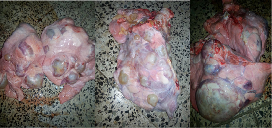
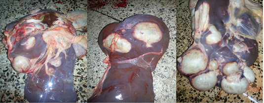
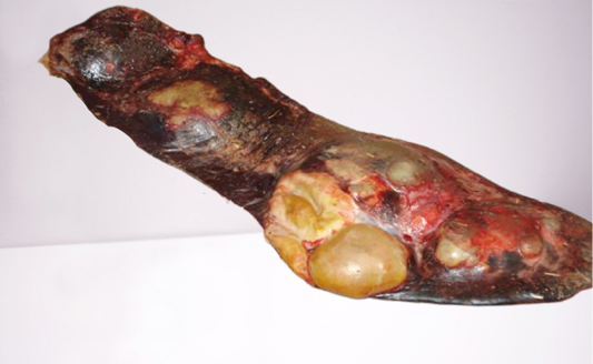
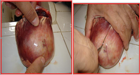
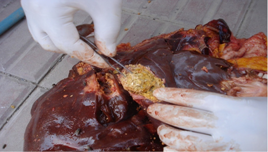
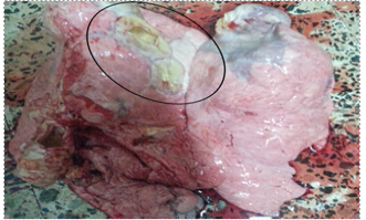
The gross lesion caused by hydatid cyst in the lungs in livestock revealed a solitary to multiple hydatid cysts of varying sizes as presented in Figure 1. Pathological lesions ranging from severe to moderate, usually table tennis ball shape but sometimes as big as a cricket ball were observed. The cysts were either fully immersed in the lung parenchyma or were partially immersed and visible on the lung surface. However, both the dorsal and ventral aspects of the lungs were affected single and multiple cysts of varying sizes on the parietal surface and visceral surface of the liver were observed as shown in (Figure 2). In addition, the cysts were soft and doughy to touch and were filled with clear to slightly turbid fluid. However, hydatid cysts (HCs) can be solitary or multiple as showed in (Figure 3) in spleen of cattle. The location of the cyst also determine the the cyst volume. The spleen and the heart show a decreased volume compared to the lung and liver which may be due to the structure of the spleen and heart (figure 3 and 4) that have small spaces between the tissue which hinders expansion of the cyst in spleen and heart compared to lung and heart.
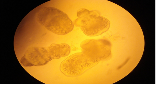
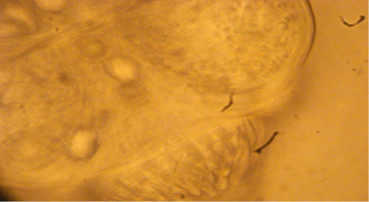
The study showed that sterile cysts constituted the highest percentage in the total samples examined followed by fertile cysts in comparison to other types of cysts (calcified, and encrusted (cheesy). Out of the 2208 cysts examined, 1236 (55.97%) were sterile, 515 (23.32%) fertile, 323 (14.62%) calcified, and 174 (7.88%) encrusted (cheesy) as shown in (Table 5). In addition, Figures (5) and (6) also show the encrusted cysts in the liver and lungs of infected cows. While Figure (7) refers to the external shape of the spines on the tapeworm heads taken from the liver of an infected cow, and Figure (8) shows the external shape of the spines on the tapeworm heads. The rates of fertile, sterile, calcified, and encrusted cysts were (20.7%), (27.1%), (14.1%) and (7.26%) in local cattle respectively, and (14.3%), (39.16%), (24.5%), and (13.28%) in imported cattle, respectively. The rate of fertile cyst for in local and imported sheeps was (23.37%), and (24.64%) respectively, while (65.77%) of the cyst in local sheeps and (39.61%) in imported sheeps were sterile, while (11.4%) and (20.7%) calcified cysts were found in local and imported breeds of sheep respectively. However, (7.29%) encrusted cysts were found in local breed of sheep and (8.21%) were found in imported breed of sheep Table 5 shows that statistically
Table 5: Fertility rate of hydatid cysts of all local and imported slaughtered livestock.
Table 6: Fertility rate of hydatid cysts of all infected organs in local and imported slaughtered livestock.
significant differences (P<0.05) exist between the different forms oc cyst (fertile, sterile, calcified and encrusted). The percentages of fertile, sterile, calcified, and encrusted cysts in local goats were (23.19%), (34.02%), (17.9%) and (9.78%) respectively.However, only two forms of cysts (fertile and sterile) were observed in imported goats and their percentages were (36.95%) fertile and (63.04%) sterile as presented in (Table 5). Higher percentage of sterile cysts followed by fertile cysts compared to calcified and encrusted cysts was discovered in cattle. It was observed that theorgan distribution of cysts was higher in local breeds compared to the imported breeds. The lungs and liver of local livestock have higher rates (45.06%) and (35.19) respectively, while the imported breeds appeared to have lower rates in the lungs and liver (6.88%) and (4.52%) respectively. Similarly, the heart and the spleenof local breeds have higher cysts rate (2.30% and 3.98%) in local breeds compared to (0.49% and 1.53%) found in the heart and spleen of imported breeds as presented in Table 6.