
Keloids and hypertrophic scars are an exaggeration of fibroplasia in dermal repair. Keloids are found solely in humankind, and not in animals. Thus far no practicable animal model of Keloid has been made. Recently a prototype rabbit ear keloid model has been produced by the authors by giving Transforming Growth Factor Beta 1 injection, and then skin punch excision on rabbit ear. In the present follow-up study consisting of eight refind techniques, best Keloid was produced by the technique where Transforming Growth Factor Beta 1 injection was followed by ventral skin, ventral perichondrium, cartilage, and dorsal perichondrium punch excision, leaving behind only dorsal skin.
Short Communication · Volume 49, Issue 1, 22 January 2017 · Open Access
A New Technique for Development of Rabbit Ear Keloid Model
· ·
Affiliations & Notes ⌄
- 1Department of Surgery, Postgraduate Medical Institute, Ameeruddin Medical College, Lahore General Hospital, Lahore, Pakistan
Article Info ⌄
- Journal:
- Pakistan Journal of Zoology
- Published:
- 22 January 2017

From the article
- In this study, the authors used eight different techniques to find out ideal method for production of rabbit ear keloid model.
- It is formed in greater amounts in keloids and keloid fibroblasts react to lesser amounts of TGFB1 than non-keloid fibroblasts.
- Best rabbit ear Keloid model can be produced by giving TGFB1 injection, followed by ventral skin, ventral perichondrium, cartilage, and dorsal perichondrium excision .
Selected from the article's own text.
Abstract
Material and methods
This was a quasi-experimental study done at Experimental Research Laboratory. Its duration was twelve months. Sample comprised of eight New Zealand White rabbits. Sampling technique was simple convenience.
Anaesthesia to rabbits was given with mixture comprising Ketamine and Xylazine. Study zone was cleaned using normal saline, and ethyl alcohol swab prior to creation of wound.
Eight rabbits were each given injection of Transforming Growth Factor Beta 1 (TGFB1) (Santa Cruz Biotechnology, 10410 Finnell Street Dallas, Texas 75220, USA; Tel +1-8004 57-3801; email: scbt@scbt.com; URL: www.scbt.com) 100 pg in 0.1 ml water, prepared in insulin syringe.Then one of the following eight different surgical techniques of punch excision was used: (a) ventral skin excision, (b) dorsal skin excision, (c) ventral skin and ventral perichondrium excision, (d) dorsal skin and dorsal perichondrium excision, (e) ventral skin, ventral perichondrium, and cartilage excision, (f) dorsal skin, dorsal perichondrium, and cartilage excision, (g) ventral skin, ventral perichondrium, cartilage, and dorsal perichondrium excision, and (h) dorsal skin, dorsal perichondrium, cartilage, and ventral perichondrium excision.
Skin punch excision was done using a 4 mm skin biopsy punch (Acu-Punch; Acuderm Inc., 5370 NW, 35 Terrace, Ft. Layderdale, FL 33309, USA; Tel +1-8003 27-0015; URL: www.acuderm.com). It is a pre-sterilized prepacked single-use skin punch biopsy gadget, which has a circular sharp edge acting as a knife. When pressure is applied at its back end, and circular movements are given, it cuts a disc of skin and underlying cartilage if required. Perichondrium is cut with a sharp pointed scissors. If there is bleeding, it is stopped with cotton gauze pressure for a short time.
Wounds were cleaned with sterilized gauze and dried with hot air dryer. Dressing was done with hydrocolloid sheet. Usual post-operative care was given. Standard wound care was given. Hydrocolloid sheet was permitted to drop off on its own. Results were recorded at twenty eight days.
Results and discussion
Rabbits were inspected at twenty eight days. Out of eight rabbits, best Keloid was created by technique (g) where injection of TGFB1 was given, followed by ventral skin, ventral perichondrium, cartilage, and dorsal perichondrium punch excision, leaving behind only dorsal skin. Gross height of this keloid was 1.355 mm. Scar elevation index (SEI) was calculated to be 3.45 (Table I).
Keloid was a clinically substantial. Its consistency was hard. It was dome-shaped, and colour was red. It had smooth surface, and there were no hair on it. Even after follow-up of one year, keloid was still present (Fig. 1A).
On gross examination, keloid specimen was hard, and was palpated as distinct nodule. Its colour was red, and surface smooth. On dissection, there was a gritty sensation. Colour of cut surface was pale. Microscopically, keloid depicted homogeneous, brilliantly eosinophilic, haphazardly dispersed, glassy, hyalinized collagen. Fibroblasts were scarce, and lied parallel to collagen (Fig. 1B).
In this study, the authors used eight different techniques to find out ideal method for production of rabbit ear keloid model. Out of eight techniques, best keloid was produced by the technique where TGFB1 injection 1 was followed by ventral skin, ventral perichondrium, cartilage, and dorsal perichondrium punch excision, leaving behind only dorsal skin.
TGFB1 is a strong chemotactic factor for fibroblasts and excites them to build main extracellular matrix constituents like collagen. It is manufactured by macrophages, and also by extracellular matrix itself. It is formed in greater amounts in keloids and keloid fibroblasts react to lesser amounts of TGFB1 than non-keloid fibroblasts. Adding exogenous TGFB1 results in stimulation of fibroblasts, which enhances secretion of collagen, and ultimately production of keloids.
Table I.- Size of keloids produced by different techniques.
| Technique Code | Tissues excised with 4 mm Skin Punch, after injection of 100 pg TGF Beta 1 | Gross Keloid Height (mm) | Ranking No |
| a | Ventral skin | 0.512 | 8 |
| b | Dorsal skin | 0.550 | 7 |
| c | Ventral skin and ventral perichondrium | 0.939 | 6 |
| d | Dorsal skin and dorsal perichondrium | 0.954 | 4 |
| e | Ventral skin, ventral perichondrium, and cartilage | 1.049 | 3 |
| f | Dorsal skin, dorsal perichondrium, and cartilage | 1.253 | 2 |
| g | Ventral skin, ventral perichondrium, cartilage, and dorsal perichondrium | 1.355 | 1 (Best) |
| h | Dorsal skin, dorsal perichondrium, cartilage, and ventral perichondrium | 0.918 | 5 |
Table II.- Comparison of techniques of production of rabbit ear keloid models.
| Author | Babar and Nagi, (2016) | Morris et al. (1997) | Li et al. (2001) | Kloeters et al. (2007) | Kryger et al. (2007) | Jia et al. (2011) | Diao et al. (2013) | Xiao and Xi (2013) | Wang et al. (2015) |
| Parameter | |||||||||
| Clinical | |||||||||
| Duration | 365+ days | 288 days | 262 days | NR | NR | NR | NR | 180 days | NR |
| Gross | |||||||||
| Scar elevation index (SEI) | 3.45 | 2.72 | NR | 1.70 | 1.67 | 2.10 | 2.07 | 1.73 | 2.93 |
| Texture | Hard | Palp able | NR | NR | NR | Protr usive | Palp able | NR | NR |
| Microscopic | |||||||||
| Hyalinized collagen | Abundant | Absent | NR | Absent | Absent | NR | Absent | Absent | Absent |
| Fibro asts | Scant | NR | Increa sed | NR | Decre ased | NR | NR | NR | NR |
NR, Not reported.
Our technique of production of rabbit ear keloid model is superior to previous techniques, as keloids produced by us had maximum scar elevation index, hard texture, long duration, abundant hyalinized collagen, and scant fibroblasts. Hyalinized collagen, which is pathognomonic, characteristic and hallmark of keloid was not reported by any of the previous authors. Morris et al, whose technique has been considered as gold standard so far, reported: ‘No hyalinized collagen such as seen in keloids was present.’ Comparison with previous keloid production techniques is given in Table II.
Conclusion
Best rabbit ear Keloid model can be produced by giving TGFB1 injection, followed by ventral skin, ventral perichondrium, cartilage, and dorsal perichondrium excision
Acknowledgment
This study was funded by Higher Education Commission, Govt. of Pakistan, Islamabad, vide Project No. 20-1427/R&D/09.
Conflict of interest statement
We declare that we have no conflict of interest.
Figures
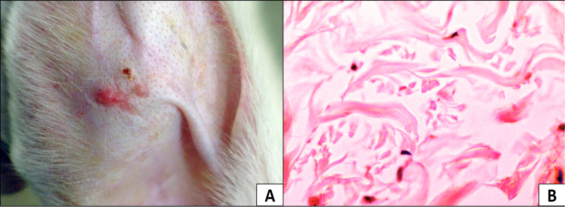
Rabbit ear keloid: A, gross view; B, microscopic view
References
- Babar, A.M., 2015a. Keloids and hypertrophic scars. University of Health Sciences, Lahore, Pakistan.
- Babar, A.M., 2015b. Keloids and hypertrophic scars: Treatment with intralesional interferon gamma versus triamcinolone acetonide. PhD thesis, University of Health Sciences, Lahore, Pakistan.
- Babar, A.M. and Nagi, A.H., 2016. Prof. med. J., 23: 907-912. http://dx.doi.org/10.17957/TPMJ/16.2941[CrossRef]
- Carmassi, M., Eraud, J., Gonelli, D., Magalon, G. and Andrac-Meyer, L., 2015. Ann. Pathol., 35: 148-153. http://dx.doi.org/10.1016/j.annpat.2014.10.007[CrossRef]
- Diao, J.S., Xia, W.S., Yi, C.G., Yang, Y., Zhang, X., Xia, W., Shu, M.G., Wang, Y.M., Gui, L. and Guo, S.Z., 2013. Plast. Reconstr. Surg., 132: 61e-69e. http://dx.doi.org/10.1097/01.prs.0000435930.88835.38[CrossRef]
- Jia, S., Xie, P., Hong, S.J., Galiano, R., Singer, A., Clark, R.A. and Mustoe, T.A., 2014. Wound Repair Regen., 22: 730-739. http://dx.doi.org/10.1111/wrr.12231[CrossRef]
- Jia, S., Zhao, Y. and Mustoe, T.A., 2011. J. Plast. Reconstr. Aesthet. Surg., 64: e332-e334. http://dx.doi.org/10.1016/j.bjps.2011.05.008[CrossRef]
- Kloeters, O., Tandara, A. and Mustoe, T.A., 2007. Wound Repair Regen., 15: S40-S45. http://dx.doi.org/10.1111/j.1524-475X.2007.00236.x[CrossRef]
- Kryger, Z.B., Sisco, M., Roy, N.K., Lu, L., Rosenberg, D. and Mustoe, T.A., 2007. J. Am. Coll. Surg., 205: 78-88. http://dx.doi.org/10.1016/j.jamcollsurg.2007.03.001[CrossRef]
- Lee, J.P., Jalili, R.B., Tredget, E.E., Demare, J.R. and Ghahary, A., 2005. J. Interf. Cytok. Res., 25: 627-631. http://dx.doi.org/10.1089/jir.2005.25.627[CrossRef]
- Li, H., Liu, J. and Xia, W., 2001. Chinese J. Plast. Surg., 17: 276-278.
- Morris, D.E., Wu, L., Zhao, L.L., Bolton, L., Roth, S.I., Ladin, D.A. and Mustoe, T.A., 1997. Plast Reconstr. Surg., 100: 674-681. http://dx.doi.org/10.1097/00006534-199709000-00021[CrossRef]
- Nagi, A.H. and Babar, A.M., 2015. Surgical adjuvant intralesional cytokines versus steroids for hypertrophic scars and keloids. Higher Education Commission, Islamabad, Pakistan.
- Qian, L.W., Fourcaudot, A.B., Yamane, K., You, T., Chan, R.K. and Leung, K.P., 2015. Wound Repair Regen., 24: 26-34. http://dx.doi.org/10.1111/wrr.12381[CrossRef]
- Rabello, F.B., Souza, C.D. and Farina Jr., J.A., 2014. Clinics, 69: 565-573. http://dx.doi.org/10.6061/clinics/2014(08)11[CrossRef]
- Ramos, M.L., Gragnani, A. and Ferreira, L.M., 2008. J. Burn Care Res., 29: 363-368. http://dx.doi.org/10.1097/BCR.0b013e31818b9e5c[CrossRef]
- Wang, H., Chen, Z., Li, X.J., Ma, L. and Tang, Y.L., 2015. Eur. J. Pharmacol., 751: 42-49. http://dx.doi.org/10.1016/j.ejphar.2015.01.040[CrossRef]
- Xiang, J., Wang, Z.Y., Jia, S.X., Jin, S.W., Lu, S.L. and Liao, Z.J., 2004. Chinese J. Burns, 20: 281-283.
- Xiao, Z. and Xi, C., 2013. Adv. Skin Wound Care, 26: 266-270. http://dx.doi.org/10.1097/01.ASW.0000429705.02588.f5[CrossRef]
- Yagmur, C., Guneren, E., Kefeli, M. and Ogawa, R., 2011. J. Plast. Reconstr. Aesthet. Surg., 64: 1359-1365. http://dx.doi.org/10.1016/j.bjps.2011.04.028[CrossRef]
- Zhang, Q., Liu, L.N., Yong, Q., Deng, J.C. and Cao, W.G., 2015. Stem Cell Res. Ther., 6: 145. http://dx.doi.org/10.1186/s13287-015-0249-0[CrossRef]
- ZhiYong, W., Fei, S., LianJu, X., Yingkai, L., Chun, Q., Shuliang, L. and XiQiao, W., 2012. Int. J. Low. Extrem. Wounds, 11: 271-276. http://dx.doi.org/10.1177/1534734612463698[CrossRef]
- Zhu, G.Y., Xu, B. and Cai, J.L., 2008. Chinese J. Plast. Surg., 24: 216-219.
Services
Article usage statistics
Loading…