
Despite the presence of vaccines, Newcastle disease (ND) and Infectious Bronchitis (IB) outbreaks are not uncommon in endemic countries. Therefore, the current study was conducted to investigate the efficacy of commercially available vaccines against each virus alone as well as co-infections with NDV and IBV in commercial poultry. For assessment of vaccine efficacy, commercial chickens were vaccinated with La Sota (against NDV) and Massachusetts (against IBV) vaccine preparations. After screening the protective titers in birds at 19 days post inoculation three week-old broilers were challenged with 10-5.76 and 10-6.03 EID50 dose of NDV and IBV, respectively. The hosts were divided into six groups of 35 birds each. These were groups A (NDV-challenged), B (IBV- challenged), C (NDV+IBV-challenged), D (IBV+NDV challenged), E (NDV and IBV- challenged) and F (negative control). Mild to moderate clinical presentations were observed in most of the co-infected birds of groups C, D and E. Tissue samples collected at 2nd, 4th and 6th days of post infection (dpi) showed histopathological signs of clinical infection. Overall, the severity of infection was higher in the co-infected birds as compared to host challenged with either of the viruses alone. Notably, moderate clinical infections raised concerns vaccine efficacies. The current study concluded that the commercially available vaccines may not provide enough protection particularly in the case of co-infection with NDV and IBV. Therefore, here is need to conduct further studies on production of vaccines with current prevailing strains in disease endemic countries such as Pakistan.
Research Article · Volume 52, Issue 5, 20 May 2020 · Open Access
A Comparative Assessment of Efficacy of Currently Applied Vaccines in Broiler Chicken against Individual and Co-Infection with Field Prevailing Newcastle Disease and Infectious Bronchitis Viruses
· · · ·
Affiliations & Notes ⌄
- 1Department of Pathology, University of Veterinary and Animal Sciences Lahore, 54000, Punjab, Pakistan 2Department of Microbiology, University of Veterinary and Animal Sciences, Lahore 3KBCMA, CVAS, Narowal Subcampus, University of Veterinary and Animal Sciences, Lahore , * Corresponding author: drasimaslamch@uvas.edu.pk
Article Info ⌄
- Journal:
- Pakistan Journal of Zoology
- Published:
- 20 May 2020

From the article
- While performing postmortems pathological lesions revealed the infection of NDV in 95 (32.2%) birds, IBV in 150 (50.85%) birds whereas 50 (16.95%) birds showed lesions suggesting co-infection of NDV and IBV.
- Therefore, the current study was conducted to investigate the commercially available vaccines efficacy against individual as well as co-infections with NDV and IBV in commercial poultry.
- The current study indicated that infection by one virus may increase host susceptibility to a secondary infection by another virus species having similar tissue tropism; co infection, in turn may reduce vaccine efficacy levels in disease host.
- Moreover, current vaccines against IBV/NDV have inherent limitation in strain cross.
Selected from the article's own text.
Abstract
INTRODUCTION
Respiratory diseases epidemics have been frequently observed in commercial poultry, causing economic losses in poultry production, particularly in diseases endemic regions such as Pakistan. Primary among avian respiratory diseases are the Newcastle disease viruses (NDVs) , Infectious bronchitis viruses (IBVs) and avian influenza viruses (AIVs). Among these, both NDV and IBV are particularly noteworthy due to their widespread circulation in commercial poultry ; . Both viruses have single stranded RNA genome but NDV is negative sense virus belongs to the genus Avulavirus in the Paramyxoviridae family, while IBV is positive sense virus belongs to gamma-Cornoavirus in the Cornoaviridae family . NDVs are primarily classified as velogenic (highly virulent), mesogenic (moderately virulent) and lentogenic (mildly virulent) strains whereas, IBVs are classified as classical and variant . Infections due to NDV and IBV have high morbidity and mortality rates with involvement of mainly respiratory system in IBV infections while digestive and nervous systems are the primary targets of NDV .
Pakistani poultry sector has experience significant growth recently; this has been accompanied by an increase of devastating economic losses to poultry production due to high rates of morbidity and mortality . In order to effectively control theses outbreaks, mass vaccination programs have been implemented in commercial poultry with limited success . The emergence of new NDV and IBV strains belongs to diverse genotypes has raised issues concerning limitations of the currently applied field vaccines. In recent years, infection due to NDV and IBV strains was observed in vaccinated flocks (Gowthaman et al., 2018; . The genotypes or serotypes of these circulatory viruses still remain unknown. Moreover, the immune pressure induced by irrational use of different vaccine types, may increase the occurrence of co-infection . The failure of vaccines, due to the co-infection may cause the disease outbreaks even in vaccinated poultry flocks. Therefore, the present study was designed to evaluate the efficacy of the currently used vaccines in commercial poultry birds upon single and cumulative challenges using prevailing viruses. The current study will establish the basis for understanding dynamics of NDV and IBV strains causing NVD/IBV outbreaks in vaccinated commercial poultry farms.
MATERIALS AND METHODS
Virus isolation and identification
During a 2016-2018 period, several NDV and IBV outbreaks were investigated from eleven poultry houses (PH) rearing commercial broiler birds in Lahore district. A total of 295 birds showing respiratory distress and neurological signs with the history of mortality were processed for diagnostic screening. Tissue samples (Trachea, lungs) were collected for the isolation and molecular identification of the viruses. The collected suspected samples were propagated individually in the 9-d-old embryonated chicken eggs and confirmed by Haemagglutination Inhibition (HI) assay using NDV- and IBV-specific antisera as per described previously . The harvested fluids from these eggs were later processed for the assessment of pathogenicity including Mean Infectious Dose (EID50) De Wit et al. (2011) and Mean death time (MDT) as per the procedures described previously . The viral genome extraction was carried out from the HI-positive harvested fluids using QIAamp Viral RNA extraction Mini Kit as per manufacturer’s instructions (Qiagen, Valencia city, CA, USA). The hypervariable regions at the fusion gene of NDV and Spike subunit S1 gene of IBV were amplified for molecular identification using one-step reverse transcriptase polymerase chain reaction (RT-PCR) .
Experiment design
A total of one hundred and twenty (n = 120) day-old broiler chicks were procured from local well reputed ISO certified hatchery. Standard management conditions were provided for birds with ad libitum feeding and watering to avoid cross contamination. All birds were screened for the presence of maternal antibodies at 0 day. At 6th day of age, all birds (n = 120) were divided into six different groups (35 birds per group) and vaccinated with commercially available NDV (LaSota) and IBV (Massachusetts) vaccines via eye drop method or combination accordingly as group A (LaSota-inoculation), B (Massachusetts- inoculation), C (first LaSota then Massachusettsinoculation after 24 hrs), D (first Massachusetts then LaSota inoculation after 24 hrs), E (LaSota and Massachusettsinoculation simultaneously) and F (negative control). On day 19, serological titers were assessed from vaccinates for both of ND and IB using haemagglutination inhibition assay. On day 21, the birds in each group were challenged with NDV (EID5010-5.76/ml) and IBV (EID5010-6.03/ml) accordingly; the birds in group A were challenged with NDV alone, birds in Group B were challenged with IBV alone, birds in group C were challenged with NDV first followed by exposure to IBV, birds in group D were challenged with IBV first followed by an exposure to NDV, whereas, birds in group F were kept as such and were administered sterile PBS. Birds were monitored for clinical signs twice a day up to 6 dpi with special monitoring of respiratory and nervous disorders. A scoring system for evaluation of degree of severity of infection was used by following scales: as no sign (0), slight or mild signs (1), moderate signs (2) and severe signs (3). The mean score of clinical signs was measured as sum of clinical scores for each sign divided by the number of birds showing signs in each group as previously described .
Necropsy findings and histopathological examination
From each group, a total of 05 chickens each were randomly slaughtered at day 0 pi and thereafter, 10 chickens from each group (A, B, C, D, E and F) were sacrificed at 2nd, 4th and 6th days post infection. Necropsy was performed during the post-mortem of birds. The presence of pathologic lesions of trachea, lung, proventriculus, kidney and cecal tonsils were observed and scored as follows: no lesion (0), mild lesions (1), moderate lesions (2) and severe lesions (3). The sum of lesions score in one group was used for statistical comparison to investigate the severity of infection between groups. Approximately 5 mm3 tissue samples were collected from each aforementioned organ. For this purpose, tissue sections of corresponding organ preserved in 10% formalin were fixed on microscopic glass and stained with hematoxylin and eosin (HE) as per method described previously. The paraffin-embedded tissues were sectioned, mounted, stained with and examined under light microscope for histological changes under 100X magnification and 3X view field repetition. During histopathological examination, lesions or any pathological change was graded as follows no lesion (-), slight or mild lesions (+), moderate lesions (++) and marked or severe lesions (+++) as described previously .
RESULTS
While performing postmortems pathological lesions revealed the infection of NDV in 95 (32.2%) birds, IBV in 150 (50.85%) birds whereas 50 (16.95%) birds showed lesions suggesting co-infection of NDV and IBV. The mean death time (MDT) was found with the range of 49.2 to 56.8 h and embryo infective dose rate (EID50) was found between the range of 105.76 and 106.03. The haemagglutination assay on harvested allantoic fluid samples showed a high titer (log29/50μl) and revealed an inhibition of haemagglutination (HI titer) with NDV and IBV-specific antisera containing antibodies against NDV and IBV. The harvested allantoic fluid showing HA and HI positivity were used for the molecular identification of viruses. After extraction of viral RNA, the NDV and IBV viruses were confirmed using specific primers in reverse transcriptase polymerase chain reaction (RT-PCR) a 375 bp band size indicated NDV whereas a 636 bp band size showed the presence of IBV.
Estimation of antibodies titer post vaccination
To investigate the antibodies titer against NDV in group A, C, D and E and, IBV in group B, C, D and E, HI was performed using specific antigens by collecting blood at 0 and 19 post vaccination days. The antibody titer in 96.9% of the serum collected at 19 days were found at/or above protective levels (1:7) for NDV, whereas, 93.5% of these were found at/or above protective levels (1:8) for IBV. The mean antibody titer for NDV was ranges between 7.29-8.57. The highest mean titer (8.57±0.80) was found in the birds of group E, followed by 8.31±0.63 in group D, 8.27±0.88 in group C and 7.29±0.72 in group A. Similarly, mean antibody titer for IBV ranged between 7.33-8.89. The highest mean titer (8.89±0.59) in the birds of group B following the 8.45±0.77 in group E, 8.27±0.88 in group C and 7.33±0.61 in group D (Table I).
Clinical presentation of experimental infection in vaccinated broiler chickens with field isolates of ND and IB virus
Generally, varying degrees of clinical signs were observed from low to moderate infection in all infected birds of group A, B, C, D and E. For respiratory signs, gasping (24%), sneezing (8%) and coughing (4%) were less frequent in infectious hosts. Nervous signs were relatively frequent in group E (32.1%), C (23.7%), D (19.4%), and A (18.9%) as compared to group B (0%). There were high rate of respiratory signs (26.6%) in group E birds, followed by 13.33% in group C and D, 6.67% in group B and 4% in the group A (Fig. 1). The group E showed the highest score for respiratory and nervous signs as compared to other infected groups. 2nd dpi in group E; these signs increased to moderate level. Clinical signs became severe at about 6th dpi. There was no clinical sign in the control birds (group F).
Gross pathological and histopathological findings
Pathological lesions on the submucosa of organs/tissues were mild to moderate as were haemorrhagic tracheitis, mucosal congestion and catarrhal exudates in the trachea in the birds. Many of the infected hosts showed mild and moderate haemorrhaging in the trachea and in lungs collected from groups B, C and E birds. Such evidences were truly the indication of partial or less infection with both ND and IB viruses. The proventriculus was found swollen in many birds of group A, C and E with the presence of haemorrhages in submucosa and mild haemorrhagic papillae of the proventriculus. Severe haemorrhaging in the internal mucosa of caecum was observed in group E birds. Severely congested and haemorrhagic lesions were seen in the spleen of group E birds. The kidneys collected from the birds of groups A, D and E showed mild to severe congestion and mild to moderate haemorrhaging in the kidneys along with inflammation. Likewise, mild hemorrhaging was observed in the cecal tonsils collected from the group A and E.
At necropsy most of the birds from group A-E showed variable intensity (mild to moderate) pathological lesions suggestive of NDV and IBV infection. Lesions on the parenchymatous organs/tissues particularly trachea, proventriculus, lungs, kidneys and cecal tonsils were
Table I. Serum antibody responses in chickens following respective commercially available vaccination detected by haemagglutination inhibition assays (HI).
| Group | Vaccine | HI GMT ± S.D | |
| Day 0 | Day 19 | ||
| A | LaSota | 2.55 ± 0.51 | 7.29 ± 0.72 |
| B | Massachusetts | 2.48 ± 0.52a | 8.89 ± 0.59 a |
| C | LaSota +Massachusetts | 2.55 ± 0.51, 2.14 ± 0.49 a | 7.31 ± 0.91, 8.27 ± 0.88 a |
| D | Massachusetts+LaSota | 2.48 ± 0.52 a, 2.50 ± 0.53 | 7.33 ± 0.61 a, 8.31 ± 0.63 |
| E | LaSota and Massachusetts | 1.48 ± 0.52, 1.46 ± 0.52 a | 8.57 ± 0.80, 8.45 ± 0.77 a |
| F | Control | 1.55 ± 0.51 | ND |
recorded. Generally, haemorrhagic tracheitis in trachea, hyperaemia in lungs, enlargement of kidneys, haemorrhagic and swollen proventriculus and cecal tonsils were observed. Haemorrhagic tracheitis, mucosal congestion and catarrhal exudates in the trachea were observed in all birds in groups A, B, C, D and E. Necropsy showed mild and moderate congestion and hemorrhaging in the trachea and lungs collected from host in groups C, D and E. Proventriculus was found swollen in several hosts in groups A, C, D and E with the presence of hemorrhaging in submucosa and mildly hemorrhagic papillae. The kidneys and cecal tonsils collected from the birds of group B showed mild congestion and hemorrhaging along with inflammation.
Histopathological examination showed sign of mild to moderate pathogenesis of NDV and IBV infection in the trachea, lung, proventriculus, kidney and caecal tonsil samples collected at 0, 2nd, 4th and 6th dpi. Infiltration of lymphocytes, prominent blood vessels, hemorrhaging of blood vessels with oedematous fluid in surrounding area were observed in the proventriculus, kidneys and caecal tonsils tissues collected from group A. Similar types of lesions was also observed in the trachea and lung tissues collected from group B. These findings were also recorded in trachea, kidneys, proventriculus and caecal tonsils of groups C and D. Comparably, these all histopathological features with sloughing of ciliary cells in the trachea were observed in all tissues collected from group E. As compared to all groups, no histopathological changes were observed in any tissues collected from group F (Fig. 2).
DISCUSSION
Newcastle disease (ND) and Infectious Bronchitis (IB) are most economical significant diseases of poultry industry worldwide, especially in particular endemic countries such as Pakistan . Vaccination programs rely mainly on the use of LaSota and Massachusetts strains for NDV and IBV, respectively, however, existence of disease in vaccinates was commonly observed (Gowthaman et al., 2018; . Therefore, investigations on protection afforded by currently applied vaccines against field prevailing strains are crucial. Current study clearly shows that commercial available vaccines are not providing enough protection against infection . The emergence of NDV and IBV co infection has recently complicated efforts to combat both of these pathogens. Therefore, the current study was conducted to investigate the commercially available vaccines efficacy against individual as well as co-infections with NDV and IBV in commercial poultry. Subsequent to viral infection, the birds of group E showed high degree of infection as compared to birds of other groups such as A, B, C and D. These findings are in agreement with previous study where high clinical infection and mortality were seen in co-infected vaccinated commercial chickens . However, promising findings concerning vaccine efficacy were observed after challenged with NDV and IBV as compared to the vaccinates infected with individual virus ; . The immune stress in result of vaccination or viral infection may facilitate host susceptibility to secondary bacterial or viral infection. Conversely, infections caused by one virus in a host may likely provoke similar infections caused by a second virus . In birds, an initial viral infection appears to significantly increase the probability of another viral infection having similar tissue tropisms such NDV, IBV and AIV .
Overall, the infected birds displayed mild to moderate clinical signs including sneezing, coughing, tracheal rales and head shaking, sign observed following co-infection in poultry . Histopathologically, microscopic findings following the infection of NDV and IBV were similar to those found in vaccinated commercial poultry ; . Subsequent to NDV and IBV infection in groups C, D and E, severe haemorrhaging in tracheal submucosa were seen to progress to congestion and tracheitis, accompanied with gasping behavior. Such findings further support the hypothesis that IBV play a role in increasing severity of clinical sings in respiratory infection, possibly via impairment of clearance for other bacterial pathogens in the air way tract of host . Additionally, the congestion seen in different tissues indicates deciliation and leukocytic infiltration in the trachea . Remarkably, the lungs, kidneys and spleens of groups A, B, C, D and E displayed congestion followed by atrophy as compared to tissues of group F control birds. Such findings indicate that virus infected immune cells aids viral dissemination throughout the body via blood or lymph vessels .
Experimental infection modeling revealed a peak of disease severity in co-infected birds at 6th dpi which is suggestive of partial protection provided by vaccines against field prevailing NDVs and IBVs (Belkasmi et al., 2017). However, it has been reported that vaccination may reduce the severity of clinical sign birds, but not hinder the multiplication and shedding of the virus leading to the environmental contamination and pathogen peristence in the field ; . Moreover, vaccination does not protect the lymphoid organs in the survivors due to immunosuppression ; . Few studies have observed full protection provided by NDV and IBV vaccines in terms of significant reduction in disease outcomes : . Neither vaccine appears to offer full cross protection against all pathogenic strains of NDV and IBV. . Nonetheless, in current study, only a single dose of vaccine was used without a follow up booster. Therefore, it is probably of more practical in terms of control strategies to perform vaccination protection studies using prevalent field strains in commercial poultry; additionally, research efforts on IBV/NDV vaccine efficacy may focus on inoculations with preparations made from multiple strains to these respective pathogens.
CONCLUSION
The current study indicated that infection by one virus may increase host susceptibility to a secondary infection by another virus species having similar tissue tropism; co infection, in turn may reduce vaccine efficacy levels in disease host. Moreover, current vaccines against IBV/NDV have inherent limitation in strain cross.
Statement of conflict of interest
All authors declared no competing interest regarding this article
Figures
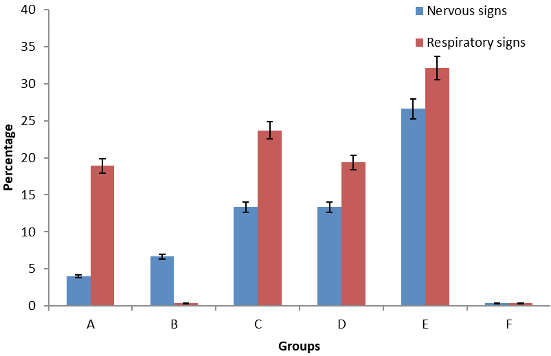
Clinical presentation showing the involvement of respiratory and nervous system in the experimental infection for different vaccinates birds (group A-E) and non-vaccinated control birds (group F).
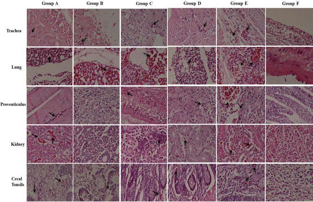
The distribution of NDV and IBV antigens in different tissues collected from vaccinated birds of groups (A-E) and non-vaccinated control birds (group F).
References
- Alexander, D. and Senne, D., 2008. Newcastle disease, other avian paramyxoviruses, and pneumovirus infections, Diseases of poultry, 12th ed. Blackwell Publishing, Ames, I.A., pp. 75–98
- Beato, M.S., De Battisti, C., Terregino, C., Drago, C., Capua, A. and Ortali, I.G., 2005. Evidence of circulation of a Chinese strain of infectious bronchitis virus (QXIBV) in Italy. Vet. Rec., 156: 720-26. https://doi.org/10.1136/vr.156.22.720[CrossRef]
- Belkasmi, S.F., Fellahi, S., Umar, S., Delpont, M., Delverdier, M., Lucas, M.N., Bleuart, C., Kichou, F., Nassik, S., Guerinand, J.L. and Fihri, O.F., 2017. Vaccines against infectious bronchitis Moroccan-Italy 02 virus in specific-pathogen-free chickens and commercial broilers. Avian Dis., 61: 466-471. https://doi.org/10.1637/11686-060817-Reg.1[CrossRef]
- Bwala, D.G., Clift, S., Duncan, N.M., Bisschop, S.P. and Oludayo, F.F., 2012. Determination of the distribution of lentogenic vaccine and virulent Newcastle disease virus antigen in the oviduct of SPF and commercial hen using immunohistochemistry. Res. Vet. Sci., 93: 520-528. https://doi.org/10.1016/j.rvsc.2011.06.023[CrossRef]
- Chhabra, R., Ball, C., Chantrey, J. and Ganapathy, K., 2018. Differential innate immune responses induced by classical and variant infectious bronchitis viruses in specific pathogen free chicks. Develop. comp. Immunol., 87: 16-23. https://doi.org/10.1016/j.dci.2018.04.026[CrossRef]
- Cook, J.K., Jackwood, M. and Jones, R.C., 2012. The long view: 40 years of infectious bronchitis research. Avian Pathol., 41: 239-250. https://doi.org/10.1080/03079457.2012.680432[CrossRef]
- De Wit, J.J., Cook, J.K. and Van Der Heijden, H.M., 2011. Infectious bronchitis virus variants: a review of the history, current situation and control measures. Avian Pathol., 40:223-235. https://doi.org/10.1080/03079457.2011.566260[CrossRef]
- Dolz, R., Vergara-Alert, J., Pérez, M., Pujols, J. and Majó, N., 2012. New insights on infectious bronchitis virus pathogenesis: characterization of Italy 02 serotype in chicks and adult hens. Vet. Microbiol., 156: 256-264. https://doi.org/10.1016/j.vetmic.2011.11.001[CrossRef]
- Naggar, R.F., Rohaim, M.A., Bazid, A.H., Ahmed, K.A., Hussein, H.A. and Munir, M., 2018. Biological characterization of wild-bird-origin avian avulavirus 1 and efficacy of currently applied vaccines against potential infection in commercial poultry. Arch. Virol., 163:2743-2755. https://doi.org/10.1007/s00705-018-3916-5[CrossRef]
- Ezema, W.S., Eze, D.C., Shoyinka, S.V.O. and Okoye, J.O.A., 2016. Atrophy of the lymphoid organs and suppression of antibody response caused by velogenic Newcastle disease virus infection in chickens. Trop. Anim. Hlth. Prod., 48:1703-1709. https://doi.org/10.1007/s11250-016-1147-x[CrossRef]
- Gowthaman, V., Ganesan, V., Gopala Krishna Murthy, T.R., Nair, S., Yegavinti, N., Saraswathy, P.V., Suresh Kumar, G., Udhayavel, S., Senthilvel, K. and Subbiah, M., 2019. Molecular phylogenetics of Newcastle disease viruses isolated from vaccinated flocks during outbreaks in Southern India reveals circulation of a novel sub-genotype. Transbound. Emerg. Dis., 66: 363-372. https://doi.org/10.1111/tbed.13030[CrossRef]
- Haghighat-Jahromi, M., Asasi, K., Nili, H., Dadras, H. and Shooshtari, A.H., 2008. Coinfection of avian influenza virus (H9N2 subtype) with infectious bronchitis live vaccine. Arch. Virol., 153: 651-655. https://doi.org/10.1007/s00705-008-0033-x[CrossRef]
- Jirjis, F.F., Noll, S.L., Halvorson, D.A., Nagaraja, K.V., Martin, F. and Shaw, D.P., 2004. Effects of bacterial coinfection on the pathogenesis of avian pneumovirus infection in turkeys. Avian Dis., 48: 34-49. https://doi.org/10.1637/7017[CrossRef]
- Keeler Jr, C.L., Reed, K.L., Nix, W.A. and Gelb Jr, J., 1998. Serotype identification of avian infectious bronchitis virus by RT-PCR of the peplomer (S-1) gene. Avian Dis., 1: 275-284. https://doi.org/10.2307/1592477[CrossRef]
- Kimura, Y., Norrby, E., Nagata, I., Ito, Y., Shimokata, K. and Nishiyama, Y., 1976. Homologous interference induced by a temperature-sensitive mutant derived from an HVJ (Sendai virus) carrier culture. J. Gen. Virol., 33: 333-343. https://doi.org/10.1099/0022-1317-33-2-333[CrossRef]
- Kiss, I., Mató, T., Homonnay, Z., Tatár-Kis, T. and Palya, V., 2016. Successive occurrence of recombinant infectious bronchitis virus strains in restricted area of Middle East. Virus Evol., 2: 1-2. https://doi.org/10.1093/ve/vew021[CrossRef]
- Kouakou, A.V., Kouakou, V., Kouakou, C., Godji, P., Kouassi, A.L., Krou, H.A., Langeois, Q., Webby, R.J., Ducatez, M.F. and Couacy-Hymann, E., 2015. Prevalence of Newcastle disease virus and infectious bronchitis virus in avian influenza negative birds from live bird markets and backyard and commercial farms in Ivory-Coast. Res. Vet. Sci., 102: 83-88. https://doi.org/10.1016/j.rvsc.2015.07.015[CrossRef]
- Kwon, J.S., Lee, H.J., Lee, D.H., Lee, Y.J., Mo, I.P., Nahm, S.S., Kim, M.J., Lee, J.B., Park, S.Y., Choi, I.S. and Song, C.S., 2008. Immune responses and pathogenesis in immunocompromised chickens in response to infection with the H9N2 low pathogenic avian influenza virus. Virus Res., 133: 187-194. https://doi.org/10.1016/j.virusres.2007.12.019[CrossRef]
- Lounas, A., Oumouna-Benachour, K., Medkour, H. and Oumouna, M., 2018. The first evidence of a new genotype of nephropathogenic infectious bronchitis virus circulating in vaccinated and unvaccinated broiler flocks in Algeria. Vet. World, 11: 1630-1636. https://doi.org/10.14202/vetworld.2018.1630-1636[CrossRef]
- Mayo, M., 2002. A summary of taxonomic changes recently approved by ICTV. Arch. Virol., 147: 1655-1656. https://doi.org/10.1007/s007050200039[CrossRef]
- Naguib, M.M., El-Kady, M.F., Lüschow, D., Hassan, K.E., Arafa, A.S., El-Zanaty, A., Hassan, M.K., Hafez, H.M., Grund, C. and Harder, T.C., 2017. New real time and conventional RT-PCRs for updated molecular diagnosis of infectious bronchitis virus infection (IBV) in chickens in Egypt associated with frequent co-infections with avian influenza and Newcastle disease viruses. J. Virol. Meth., 245: 19-27. https://doi.org/10.1016/j.jviromet.2017.02.018[CrossRef]
- Nili, H. and Asasi, K., 2002. Natural cases and an experimental study of H9N2 avian influenza in commercial broiler chickens of Iran. Avian Pathol, 31: 247-252. https://doi.org/10.1080/03079450220136567[CrossRef]
- Okwor, E.C., Eze, D.C., Echeonwu, G.O.N., Ibu, J.O., Eze, P.C. and Okoye, J.O.A., 2016. Comparative studies on the effects of La Sota and Komarov vaccine antibodies on organ distribution, persistence and shedding of Kudu 113 virus in chickens. J. Anim. Pl. Sci., 26: 1226-1235.
- Roohani, K., Tan, S.W., Yeap, S.K., Ideris, A., Bejo, M.H. and Omar, A.R., 2015. Characterisation of genotype VII Newcastle disease virus (NDV) isolated from NDV vaccinated chickens, and the efficacy of LaSota and recombinant genotype VII vaccines against challenge with velogenic NDV. J. Vet. Sci., 16: 447-457. https://doi.org/10.4142/jvs.2015.16.4.447[CrossRef]
- Shabbir, M.Z., Zohari, S., Yaqub, T., Nazir, J., Shabbir, M.A.B., Mukhtar, N., Shafee, M., Sajid, M., Anees, M., Abbas, M. and Khan, M.T., 2013. Genetic diversity of Newcastle disease virus in Pakistan: a countrywide perspective. Virol. J., 10: 170-171. https://doi.org/10.1186/1743-422X-10-170[CrossRef]
- Shirvani, E., Paldurai, A., Manoharan, V.K., Varghese, B.P. and Samal, S.K., 2018. A recombinant Newcastle disease virus (NDV) expressing S protein of infectious bronchitis virus (IBV) protects chickens against IBV and NDV. Sci. Rep., 8: 1951-1954. https://doi.org/10.1038/s41598-018-30356-2[CrossRef]
- Su, Q., Li, Y., Zhang, Y., Zhang, Z., Meng, F., Cui, Z., Chang, S. and Zhao, P., 2018. Newcastle disease virus-attenuated vaccine LaSota played a key role in the pathogenicity of contaminated exogenous virus. Vet. Res., 49: 80-84. https://doi.org/10.1186/s13567-018-0577-z[CrossRef]
- Susan, S., Ekram, S. and Amal, A., 2012. Efficacy of some living classical and variant infectious bronchitis vaccines against local variant isolated from Egypt. Nat. Sci., 10: 292-299.
- Swayne D.E., 1998. Laboratory manual for the isolation and identification of avian pathogens. American Association of Avian Pathologists, University of Pennsylvania.
- Wajid, A., Dimitrov, K.M., Wasim, M., Rehmani, S.F., Basharat, A., Bibi, T., Arif, S., Yaqub, T., Tayyab, M., Ababneh, M. and Sharma, P., 2017. Repeated isolation of virulent Newcastle disease viruses in poultry and captive non-poultry avian species in Pakistan from 2011 to 2016. Prevent. Vet. Med., 142: 1-6. https://doi.org/10.1016/j.prevetmed.2017.04.010[CrossRef]
Services
Article usage statistics
Loading…