
The Journal of Advances in Parasitology


Research Article
The Journal of Advances in Parasitology 1 (4): 49 – 53Prevalence and Associated Risk Factors of Cryptosporidium and Giardia species in Cattle within Mandalay Region, Myanmar
Saw Bawm1*, Sandar Kyi2, Khin Khin Lay3, Lat Lat Htun1, Tin Tin Myaing4
- Department of Pharmacology and Parasitology, University of Veterinary Science, Yezin, Nay Pyi Taw, Myanmar
- Livestock Breeding and Veterinary Department, Myingyan Townships, Myanmar
- Department of Animal Science, University of Veterinary Science, Yezin, Nay Pyi Taw, Myanmar
- Myanmar Veterinary Association, Myanmar
*Corresponding author:bestshadow@gmail.com; sawvet@uvsyezin.edu.mm
ARTICLE CITATION:
Bawm S, Kyi S, Lay KK, Htun LL, Myaing TT (2014). Prevalence and associated risk factors of Cryptosporidium and Giardia species in cattle within Mandalay region, Myanmar. J. Adv. Parasitol. 1 (4): 49 – 53.
Received: 2014–11–15, Revised: 2014–12–01, Accepted: 2014–12–04
The electronic version of this article is the complete one and can be found online at
(
http://dx.doi.org/10.14737/journal.jap/2014/1.4.49.53
)
which permits unrestricted use, distribution, and reproduction in any medium, provided the original work is properly cited
ABSTRACT
The information on prevalence of highly infectious protozoan parasites Cryptosporidium and Giardia capable of causing gastrointestinal illness in humans and many other species of mammals in the dairy calves of Myanmar is very limited. The overall prevalence of Cryptosporidium and Giardia spp. were 56% (224/400) and 22.5% (90/400), respectively. Calves with under 6 months of age had significantly higher prevalence than older animals (OR = 2.27, CI = 1.11–4.66, P = 0.02) on Cryptosporidium and Giardia infection. Management of calves was associated with a higher risk of exposure to Cryptosporidium spp. infection if they grazed (OR = 3.78, CI = 2.35–6.06, P = < 0.001) and drunk water from river or pond (OR = 0.25, CI = 0.16–0.37, P = < 0.001). Our observed information may useful in designing prevention plans in cattle to minimize economic losses due to Cryptosporidium and Giardia and potential hazards to public health.
INTRODUCTION
The highly infectious protozoan parasites belonging to Cryptosporidium and Giardia spp. are capable of causing gastrointestinal illness in humans and many other species of mammals. They are the cause of serious diarrhoea in calves in tropical region (Xiao et al., 2004; Bhat et al., 2012; Randhawa et al., 2012). In cattle, direct transmission through the contamination of surroundings by infected animals seems to be the principal mode of infection. Farm animals play significant role in contributing parasite cysts/oocysts in large proportion because of their high abundance on farms (Hunter and Thompson, 2005; O’Handley and Olson, 2006; Xiao and Fayer, 2008) and can be act as the causal agents of human Giardiasis and cryptosporidiosis. Giardia and Cryptosporidium have emerged as important parasites of dairy cattle because of their proven pathogenecity, economic losses and the potential public health significance of zoonotic transmission (Olson et al., 2004; Bhat et al., 2013).
There has also been considerable interest and discussed regarding potential of zoonotic transmission of these pathogens, particularly from livestock. Transmission of this type may occur through either direct contact in the case of farmers, veterinarians, and petting zoos, or through indirect routes such as contaminated surface water or foods (Dixon, 2009). A high prevalence of both Giardia and Cryptosporidium has been reported worldwide in dairy and beef cattle. Moreover, cattle can harbor a number of protozoan parasites and plays important role in the causes of human Giardiasis and cryptosporidiosis. Giardia and Cryptosporidium have emerged as important parasites of dairy cattle because of their proven pathogenecity, economic losses and the potential public health significance of zoonotic transmission (de Graaf et al., 1999; Olson et al., 2004).
Control of cryptosporidiosis and Giardiasis in cattle has become important, not only to reduce the risk of disease in cattle, but also to reduce the risk of infection in humans especially in immunosuppressed individuals (Garber et al., 1994). The information on prevalence of Cryptosporidium and Giardia species in dairy calves of Myanmar is scaring (Lay et al., 2008). The present study was therefore conducted to investigate the regional prevalence of Cryptosporidium and Giardia species infections in cattle in Yamethin and Pyawbwe townships, Mandalay Region of Myanmar.
MATERIALS AND METHODS
Study Area, Population and Fecal Sample Collection
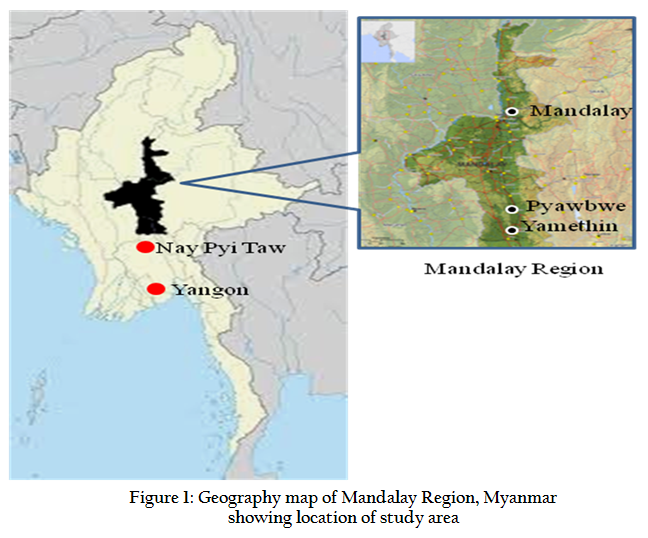
A cross–sectional study was carried out in twelve villages within Yamethin Township (located within 20 15′ 0″ N and 96 15′ 0″ E) and Pyawbwe Township (located within 20 35′ 0″ N and 96 4′ 0″ E) (Figure 1). This study was conducted between July and December 2011, mainly on small holder dairy cow with local breed (Zebu). The sample size was calculated according to the method described by Thrusfield (2007) based on the estimated cattle population of 132,000 and 126,000 in Yamethin and Pyawbwe Towships, respectively (Source: Township Veterinary Officer, Yamethin and Pyawbwe Townships, Livestock Breeding and Veterinary Department).The selection of farms was done using the stratified random sampling method.
The cattle were kept in a wooden building with bamboo roofing. They were provided with concentrate in feeding troughs and drinking water at morning and evening. At daytime, they were grazed in pasture. As a routine, cattle were fed roughages and concentrate (mixed of sesame cake, pea, bean bran and grain husks) twice a day. A total of 400 faecal samples were collected as 191 (47.75% of total samples collected) and 209 (52.25% of total samples collected) samples from Yamethin and Pyawbwe Townships, respectively. Twenty to twenty five grams of faecal samples per cattle was collected either directly from rectum or from the ground immediately after defecation. For each animal sampled, a questionnaire was completed by the owner at the time of sampling. Recorded questionnaires included information about age, sex, breed, anthelmintic treatment, sources of water, feeding and cleaning routines for cattle and presence of gastrointestinal illness.
Laboratory Analysis of Fecal Samples
In this study, standard laboratory techniques were used and all laboratory works were followed standard procedures. Direct identification of Cryptosporidium oocysts was conducted by using modified Ziehl–Neelsen acid fast staining method (Clarke and McIntyre, 2001). Briefly, a thin faecal smear was made, left it to the air to dry and fixed it in methanol for 2–3 minutes. The smear was stained with cold carbol–fuchsin for 5–10 minutes and differentiated in 1% hydrochloric acid–ethanol until colour ceases to flood out. It was rinsed in tap–water and counterstained with 0.25% malachite green (or methylene blue) for 30 seconds. Then, rinsed in tap–water again, blotted or drained dry. The stained smear was examined by microscope, Cryptosporidium oocysts appeared as bright rose–pink spherical on a pale green background.
Identification of Giardia cysts was performed by using Zinc Sulfate flotation method (Margaret et al., 1994). Briefly, approximately 2–3 g of faeces was mixed with 6–7 ml of water. The mixture was strained through a tea strainer and poured into a 15–ml centrifuge tube and then, centrifuged for about 5 minutes at approximately 2500 rpm. The centrifuge tube was filled with flotation solution until a reverse meniscus was presented and a cover slip was then added. The cover slip was removed from the top of the tube, placed on a slide, and examined.
Oocysts/cysts density was determined according to the oocysts/cysts scoring system of Dagnall Teaching Laboratory (1998), as follows: rare (+) for ≤ 5 oocysts per slide; few to moderate (++) for 1–10 oocysts per field of view; and numerous (+++) for 11 or more oocysts per field of view.
Statistical Analysis
Data files of questionnaires and laboratory results were prepared in Epi–Info version 7.1.4 (CDC, USA) to determine prevalence and odds ratios. Logistic regression analysis was performed in SPSS version 16 for Windows at 95% Confidence Interval (CI).
RESULTS AND DISCUSSION
In this study, a total of 191 animals from 6 villages from Yamethin Township and 209 animals from 6 villages of Pyawbwe Township were sampled. Prevalence amongst the locations in the different age groups and herd management, such as grazing and sources of water and their respective 95% CI are shown in Table 1.
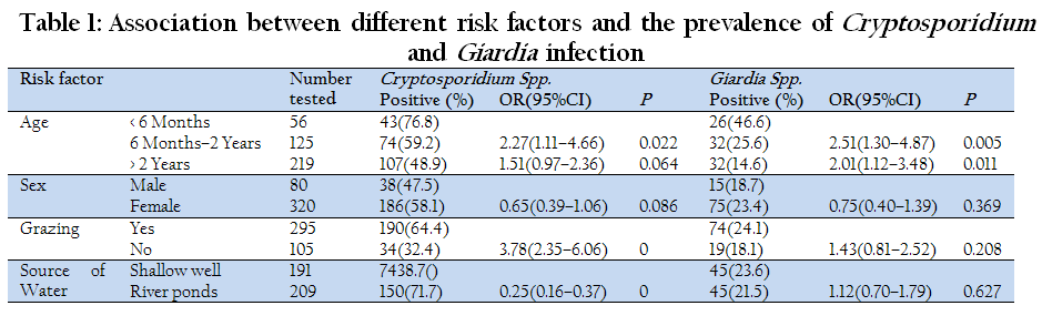
Table 1: Association between different risk factors and the prevalence of Cryptosporidium and Giardia infection
The result of the present study showed that the overall prevalence of Cryptosporidium was 56% (224/400) in Yamethin and Pyawbwe Townships, Mandalay region. Similar results have been reported from farms around Mandalay City, Mandalay region of Myanmar by Lay et al. (2008) who demonstrated that the prevalence of Cryptosporidium was 57.3% in 1 to 17 weeks old calves. Although Cryptosporidium and Giardia infections have been reported for cattle in many parts of the world, prevalence data have often varied markedly. The present detection rate of Cryptosporidium infection based on microscopic examination is much higher compared to the obtained prevalence in former studies in Thailand, Argentina, Ethiopia, England, India and Wales with the prevalence of 0.6, 19.35, 7.8, 12.50 and 10.2 per cent, respectively (Jittapalapong et al., 2006; Tiranti et al., 2011; Wegayehu et al., 2013; Singla et al., 2013; Smith et al., 2014). Similar results with this study, high prevalence rates were also reported in Canada, Germany, Nigeria, Tanzania and Brazil with the prevalence rates of 32, 41.3, 33.0, 35 and 45 per cent, respectively (Budu–Amoako et al., 2012; Gillhuber et al., 2014; Faleke et al., 2014; Swai and Schoonman, 2010; Almeida et al., 2010).
The detected prevalence of Giardia infection in the cattle (22.5%, 90/400), was lower than that of Cryptosporidium (56%). Similar results with microscopic based prevalence were also reported elsewhere (Bjorkman et al., 2003). The prevalence of Giardia infection in cattle in this study is relatively higher than in Canada, Germany, Ethiopia and Turkey with 14, 7.2, 2.3 and 3.7 per cent, respectively (Budu–Amoako et al., 2012; Gillhuber et al., 2014; Faleke et al., 2014; Degerli et al., 2005). Nevertheless, it was lower than the 34.5 percent prevalence in calves reported in Argentina (Tiranti et al., 2011).
Calves younger than six months of age were almost 2 times more likely to be infected with Cryptosporidium and Giardia than those older than six months of age (odds ratio [OR] = 2.27, CI = 1.11–4.66, P = 0.02) (Table 1 and Figure 2). Adult cattle are generally considered refractory to heavy infections by Cryptosporidium and associated clinical diseases because of a strong immune response (Ralston et al., 2003). The young calves can also be protected from Giardia infection by consumption of colostrums (Olson et al., 2000).

Sex of the calf was not associated with oocyst/cyst shedding of Cryptosporidium and Giardia species and this is in agreement with the report of Swai et al. (2007) and Almeida et al. (2010). Although, grazing in pasture (OR = 3.78, CI = 2.35–6.06, P = < 0.001) and sources of water (OR = 0.25, CI = 0.16–0.37, P = < 0.001) were highly associated with Cryptosporidium, there was no evident influence on the occurrence of infection by Giardia spp. Cattle grazed in pasture were more likely to be infected with Cryptosporidium than those kept intensive in the barn with cut and carry feeding. Cows grazing outside are more likely to contaminate the environment (including feed and water) with oocysts compared with cattle kept intensively (Table 1). Similar findings were also reported by Budu–Amoako et al. (2012). Moreover, the animals grazed on pasture near streams and ponds were often contaminated with slurry as well as other manure.
Furthermore, almost all studied farms had feed and water troughs and the presence of water troughs was probably potential reservoir of Cryptosporidium, since the oocysts can survive in water for a long period of time (Simmons et al., 2001; Almeida et al., 2010) and probably there is constant fecal contamination of that water by animals and even by humans. Thus, hygiene methods to clean and disinfect water troughs should be taken into account as important preventive strategies. The high percentage of Cryptosporidium positive samples (56% of analyzed animals) showed the high occurrence of the parasite and possible highly contaminated by the infected animals. Since the study period was rainy season, high number of animals crowded in the same pasture could favour to close contact each other. This may be the possibility of direct transmission of pathogens from one animal to another. Moreover, oocysts and cysts survive longer at moderate temperature than at higher temperature.
In this finding, most samples showed moderate or mild infection, especially in adult cattle, may act as a healthy carrier and it can be a source of infection for younger animals, especially during the periparturient period (Xiao et al., 1994). They can also play as a potential role and reservoirs for humans those infected with immunodepressive diseases. The variation of prevalence among geographical localities may be due to differences in the levels of calf management practices employed at farm level, housing–related factors, and calf–related factors at a time of sampling, nature of the study, and fecal screening technique used (Swai and Schoonman, 2010).
In this study, there was evidence that, in and around Mandalay Region, calves are infected by Cryptosporidium spp., indicating that bovine cryptosporidiosis is endemic and locally widespread. In order to better understand the zoonotic potential of Cryptosporidium and Giardia infections in cattle within these areas, it has been important to determine whether humans and other animals may be susceptible to infection with these parasites. Although this study did not identify the molecular characterization of the parasites due to lack of facilities in our laboratory, the results are still important because there is only limited information can be available in Myanmar. Further studies relevant to the genetic identification of these protozoa and risk factors associated with human infection from public health aspects are necessary to conduct.
COMPETING INTERESTS
The authors declare that they have no competing interests.
ACKNOWLEDGMENTS
The authors thank all the stockmen who participated and the townships veterinary officers and staff for their very considerable support and assistance. This study is partly supported by postgraduate research fund of the University of Veterinary Science, Yezin, Nay Pyi Taw, Myanmar.
REFERENCES
Almeida AJ, Oliveira FCR, Flores VMQ, Lopes CWG (2010). Risk factors associated with the occurrence of Cryptosporidium parvum infection in calves. Arq. Bras. Med. Vet. Zootec. 62 (6):1325 – 1330.
http://dx.doi.org/10.1590/S0102-09352010000600005
Bhat SA, Juyal PD, Singla LD (2012). Prevalence of cryptosporidiosis in neonatal buffalo calves in Ludhiana district of Punjab, India. Asian J. Anim. Vet. Adv. 7 (6): 512 – 520.
http://dx.doi.org/10.3923/ajava.2012.512.520
Bhat SA, Juyal PD, Singla LD (2013). Bovine cryptosporidiosis: brief review of its distribution in india . Trends Parasitol. Res. 2 (2): 5 – 13.
Bjorkman C, Svensson C, Christensson B, Verdier K (2003). Cryptosporidium parvum and Giardia intestinalis in calf diarrhea in Sweden. Acta. Vet. Scand. 44 (3–4): 3 – 4.
Budu–Amoako E, Greenwood SJ, Dixon BR, Barkema HW, McClure JT (2012). Giardia and Cryptosporidium on dairy farms and the role these farms may play in contaminating water sources in Prince Edward Island, Canada. J. Vet. Intern. Med. 26 (3): 668 – 673.
http://dx.doi.org/10.1111/j.1939-1676.2012.00930.x
PMid:22489682
Clarke SC, McIntyre M (2001). Acid–fast bodies in faecal smears stained by the modified Ziehl–Neelsen technique. Br. J. Biomed. Sci. 58 (1): 7 – 10.
PMid:11284227
Dagnall Teaching Laboratory (1998). Liverpool School of Tropical Medicine, Pembroke Place, Liverpool, UK. 106.
de Graaf DC, Vanopdenbosch E, Ortega–Mora LM, Abbassi H, Peetere JE (1999). A review of the importance of cryptosporidiosis in farm animals. Int. J. Parasitol. 29 (8): 1269 – 1287.
http://dx.doi.org/10.1016/S0020-7519(99)00076-4
Degerli S, Celiksoz A, Kalkan K, Ozcelik S (2005). Prevalence of Cryptosporidium spp. and Giardia spp. in cows and calves in Sivas. Turk. J. Vet. Anim. Sci. 29: 995 – 999.
Dixon BR (2009). The role of livestock in the food borne transmission of Giardia duodenalis and Cryptosporidium spp. to humans In: Ortega–Pierres MG, Caccio` SM, Fayer R, Smith H (eds.), Giardia and Cryptosporidium: from molecules to disease. CAB International, Wallingford, UK. 107 – 122.
http://dx.doi.org/10.1079/9781845933913.0107
PMid:20024140
Faleke OO, Yabo YA, Olaleye AO, Dabai YU, Ibitoye EB (2014). Point prevalence of Cryptosporidium oocyst in calves grazing along River Rima bank in Sokoto, Nigeria. Pak. J. Biol. Sci. 17 (3): 443 – 446.
http://dx.doi.org/10.3923/pjbs.2014.443.446
PMid:24897803
Garber LP, Salman MD, Hurd HS, Keefe T, Schlater JL (1994). Potential risk factors for Cryptosporidium infection in dairy calves. J. Am. Vet. Med. Assoc. 205: 86 – 91.
PMid:7928557
Gillhuber J, Rügamer D, Pfister K, Scheuerle MC (2014). Giardiosis and other enteropathogenic infections: a study on diarrhoeic calves in Southern Germany. BMC Res. Notes. 7: 112.
http://dx.doi.org/10.1186/1756-0500-7-112
PMid:24568139 PMCid:PMC3941484
Hunter PR, Thompson RCA (2005). The zoonotic transmission of Giardia and Cryptosporidium. Int. J. Parasitol. 35 (1–2): 1181 – 1190.
http://dx.doi.org/10.1016/j.ijpara.2005.07.009
PMid:16159658
Jittapalapong S, Pinyopanuwat N, Chimnoi W, Siripanth C, Stich RW (2006). Prevalence of Cryptosporidium among dairy cows in Thailand. Ann. NY. Acad. Sci. 1081: 328 – 335.
http://dx.doi.org/10.1196/annals.1373.045
PMid:17135534
Lay KK, Hoerchner HCF, Morakote N, Kreausukon K (2008). Prevalence of Cryptosporidium, Giardia and Other Gastrointestinal Parasites in Dairy Calves in Mandalay, Myanmar. In: The 15th Congress of FAVA. OIE Joint Symposium on Emerging Diseases, Bangkok, Thailand, 27–30 October 2008, 273.
Margaret WS, Russell LK, Anne MZ (1994). Faecal Examination. In Veterinary Clinical Parasitology, 6th edition, Iowa State University Press, USA.
PMid:7927545
O'Handley RM, Olson ME (2006). Giardiasis and cryptosporidiosis in ruminants. Vet. Clin. North. Am. Food. Anim. Pract. 22 (3): 623 – 643.
http://dx.doi.org/10.1016/j.cvfa.2006.07.002
PMid:17071357
Olson ME, Ceri H, Morck DW (2000). Giardia vaccination. Parasitol. Today. 16 (5): 213 – 217.
http://dx.doi.org/10.1016/S0169-4758(99)01623-3
Olson ME, O' Handley RM, Ralston BJ, McAllister TA, Thompson RCA (2004). Update on Cryptosporidium and Giardia infections in cattle. Trends. Parasitol. 20 (4): 185 – 191.
http://dx.doi.org/10.1016/j.pt.2004.01.015
PMid:15099558
Randhawa SS, Zahid UN, Randhawa SS, Juyal PD, Singla LD, Uppal SK (2012). Therapeutic management of cryptosporidiosis in cross bred dairy calves. Indian Vet. J. 89 (2): 17 – 19.
Ralston BJ, McAllister TA, Olson ME (2003). Prevalence and infection pattern of naturally acquired Giardiasis and cryptosporidiosis in range best calves and their dams. Vet. Parasitol. 114 (2): 113 – 122.
http://dx.doi.org/10.1016/S0304-4017(03)00134-1
Simmons OD, Sobsey MD, Heaney CD, Schaefer FW, Francy DS (2001). Concentration and detection of Cryptosporidium oocysts in surface water samples by method 1622 using ultrafiltration and capsule filtration. Appl. Environ. Microbiol. 67 (3): 1123 – 1127.
http://dx.doi.org/10.1128/AEM.67.3.1123-1127.2001
PMid:11229901 PMCid:PMC92704
Singla LD, Gupta MP, Singh H, Singh ST, Kaur P, Juyal PD (2013). Antigen based diagnosis of Cryptosporidium parvum infection in cattle and buffalo faeces. Indian J. Anim. Sci. 83 (1): 37 – 39.
Smith RP, Clifton–Hadley FA, Cheney T, Giles M (2014). Prevalence and molecular typing of Cryptosporidium in dairy cattle in England and Wales and examination of potential on–farm transmission routes. Vet. Parasitol. 204 (3–4): 111 – 119.
http://dx.doi.org/10.1016/j.vetpar.2014.05.022
PMid:24909077
Swai ES, Schoonman L (2010). Investigation into the prevalence of Cryptosporidium infection in calves among small–holder dairy and traditional herds in Tanzania. Vet. Med. Int. 676451.
PMid:21234370 PMCid:PMC3014725
Swai ES, French NP, Karimuribo ED, Fitzpatrick JL, Bryant MJ, Kambarage DM, Ogden NH (2007). Prevalence and determinants of Cryptosporidium spp. infection in smallholder dairy cattle in Iringa and Tanga Regions of Tanzania Onderstepoort J. Vet. Res. 74 (1): 23 – 29.
Thrusfield M (2007). Veterinary Epidemiology, 3rd edition. Blackwell Science, UK. 238 – 239.
Tiranti K, Larriestra A, Vissio C, Picco N, Alustiza F, Degioanni A, Vivas A (2011). Prevalence of Cryptosporidium spp. and Giardia spp., spatial clustering and patterns of shedding in dairy calves from Córdoba, Argentina. Rev. Bras. Parasitol. Vet. 20 (2): 140 – 147.
http://dx.doi.org/10.1590/S1984-29612011000200009
PMid:21722489
Wegayehu T, Adamu H, Petros B (2013). Prevalence of Giardia duodenalis and Cryptosporidium species infections among children and cattle in North Shewa Zone, Ethiopia. BMC Infect. Dis. 13: 419.
http://dx.doi.org/10.1186/1471-2334-13-419
PMid:24010794 PMCid:PMC3849630
Xiao L, Fayer R (2008). Molecular characterisation of species and genotypes of Cryptosporidium and Giardia and assessment of zoonotic transmission. Int. J. Parasitol. 38 (1): 1239 – 1255.
http://dx.doi.org/10.1016/j.ijpara.2008.03.006
PMid:18479685
Xiao L, Fayer R, Ryan U, Upton SJ (2004). Cryptosporidium Taxonomy: Recent Advances and Implications for public health. Clin. Microbiol. Rev. 17 (1): 72 – 97.
http://dx.doi.org/10.1128/CMR.17.1.72-97.2004
PMid:14726456 PMCid:PMC321466
Xiao L, Herd RP, McClure KE (1994). Periparturient rise in the excretion of Giardia spp. cysts and Cryptosporidium parvum oocysts as a source of infection for lambs. J. Parasitol. 80 (1): 55 – 59.
http://dx.doi.org/10.2307/3283345
PMid:8308659

