
The Journal of Advances in Parasitology


Research Article
The Journal of Advances in Parasitology 1 (1): 6 – 8Advances in Therapeutic Management of Complicated Demodicosis in Canines
Muhammad Iqbal Yatoo*, Ricky Jhambh, Deepa Padinjaree Malepad, Pankaj Kumar, Umesh Dimri,
*Corresponding author: iqbalyatoo@gmail.com
ARTICLE CITATION:
Yatoo MI, Jhambh R, Deepa PM, Kumar P and Dimri U (2014). Advances in therapeutic management of complicated demodicosis in canines. J. Adv. Parasitol. 1 (1): 6 – 8.
Received: 2013–10–13, Revised: 2014–01–03, Accepted: 2014–01–07
The electronic version of this article is the complete one and can be found online at
(http://dx.doi.org/10.14737/journal.jap/2014/1.1.6.8)
which permits unrestricted use, distribution, and reproduction in any medium, provided the original work is properly cited
ABSTRACT
Mixed infection by ectoparasites and bacteria are common among dogs. Such cases need efficiently devised therapeutic approach as mere treatment of one of the pathogens will not be much beneficial. In one such case of German Shephered presented with signs of alopecia, erythema and hyperpigmentation, comedones, crust formation, itching, oozing of serum and pus formation at certain points, proper diagnostic and therapeutic approach lead to improvement. Skin scraping for parasitic examination and swab for bacterial culture was taken. After skin scraping and bacterial culture it was diagnosed as a mixed infection of demodicoses and pyoderma. As the case was complicated, both antiparasitic and antibacterial medication was started in a proper way along with antiinflammatory and antiallergic agents. Animal showed improvement and treatment was continued with some modifications. Finally the animal recovered. Advancement in therapeutic management of complicated demodicosis in canines is becoming a need of the hour as such cases are on rise among pets.
INTRODUCTION
Nowadays concurrent occurrence of mixed infection by mites and bacteria are becoming common among pet dogs (Mueller, 2004; Mueller et al., 2012). This may be due to the unhygienic management and complications post demodicosis. Due to itching and scratching by mite infestation, skin is predisposed to bacterial invasion and hence development of pyoderma. Such cases are usually diagnosed based on history, clinical examination along with skin scraping test and bacterial culture (Muller et al., 2012). Treatment involves medication against both mites and bacteria besides preventing inflammation and allergy (Taylor, 2001; Ettinger and Feldman, 2010). Demodex canis is common cause of demodicosis or red mange in dogs. It is a tiny parasitic mite living in or near hair follicles and invades deep in dermis. Infection is acquired either from infected animal or objects or following immunosuppressive conditions or treatments, or may be related to a genetic immune deficiency (Greve and Gaafar, 1966). Dalmatian, the American Bulldog and the American Pit Bull Terrier appear to be more susceptible (Urquhart, 1996). Most common sites lesions are the face, muzzle, forelimbs and periorbital regions. Usually two types of manifestions are there. These include squamous and pustular forms. Squamous form causes dry alopecia and thickening of the skin and pustular form which is the more severe form, causing secondary bacterial infection resulting in the characteristic red, numerous pustules and wrinkling of the skin (Mueller, 2004; Mueller et al., 2012). Usually demodicosis is complicated by Staphylococcal infection (especially Staphylococcus epidermidis) (Mueller, 2004) but other bacteria like streptococcus can also occur. This results in pustule formation and systemic reactions like fever and septicemia.
MATERIALS AND METHODS
A one and half year German Shepherd dog was presented with signs of alopecia, erythema and hyperpigmentation, comedones, crust formation, itching, oozing of serum and pus formation at certain points on body. Clinical examination was done and clinical parameters like temperature, respiration rate and pulse rate, were taken. Skin scrapings were taken for detection of mites and pus soaked swab for bacterial culture. Scrapings were taken from edge of lesion by a sharp blade smeared with glycerol in a petri dish until bleeding occurred. Skin scrapings were digested in warm 10% KOH and smear was prepared. Swab was cultured in nutrient broth and slides were stained with Gram's stain for bacterial examination
RRESULT
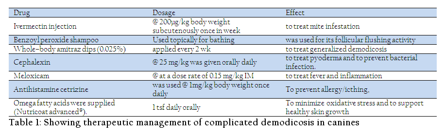
On clinical examination alopecia, crusting of skin and pustules were noticed around ventral abdomen, face, neck and back. Animal had itching and scratching lead to oozing of serum like fluid. Pus formation was noticed around lesions. Temperature was slightly elevated (103.8oF), respiration rate (28/minute) and heart rate (72/minute). were within normal range. Mucous membranes were congested. Animal was dull, depressed and anorectic. Pinna–pedal reflex showed jerking of foot on pinching pinna. Skin scraping revealed presence of small mites under microscopic examination. These were categorized as Demodex canis by parasitological examination. Bacterial culture showed presence of Gram positive cocco–bacillary bacteria mostly Staphylococci. Treatment was started with ivermectin injection @ 200µg/kg body weight subcutenously once in week to treat mite infestation. Topically benzoyl peroxide shampoo was used for its follicular flushing activity. Whole–body amitraz dips (0.025%) were applied every 2 wk to treat generalized demodicosis. Higher concentrations (0.05%) and shorter treatment intervals (1 wk) may be more efficient. At times hair clipping and body cleansing, especially with benzoyl peroxide shampoo was used. Cephalexin @ 25 mg/kg was given orally daily to treat pyoderma and to prevent bacterial infection. Meloxicam @ at a dose rate of 0.15 mg/kg was given to treat fever and inflammation. To prevent allergy/icthing, antihistamine cetrizine was used @ 1mg/kg body weight once daily. To minimize oxidative stress and to support healthy skin growth omega fatty acids were supplied (Nutricoat advanced@). Animal responded well to treatment and same treatment was continued for one week. After this ivermectin was stopped, topical preparations were continued along with omega fatty acids. Besides hepatoprotectants like vitamin B complex and antioxidants like selenium and vitamin E was also provided in follow up treatments.
DISCUSSION
Treatment of canine generalized demodicosis is multimodal (Mueller et al., 2012). Taylor et al. (2001) has reviewed recent developments in ectoparasitcides. Mueller (2008) has used different topical agents for demodicosis. Benzoyl peroxide (2–3%) and chlorhexidine–based shampoos (3–4%) are commonly recommended for dogs with demodicosis. They also have antimicrobial action. Amitraz is widely used in canine demodicosis. The recommended concentration varies from 0.025 to 0.06%, with a frequency of once weekly to every 2 weeks (Mueller, 2004). It has many advantages as summarized by Scott et al. (2001) and Mueller et al. (2012). Recently efficacy of metaflumizone plus amitraz for the treatment of juvenile and adult onset demodicosis in dogs was studied by Rosenkrantz (2009). Use of macrocyclic lactones in demodicosis is well established. Milbemycin oxime (Holm, 2003; Mueller, 2004), ivermectin (Delayte et al. 2006; Mueller, 2004) and doramectin (Murayama et al., 2010) are the common ones used. Recent ones include selamectin (Schnabl et al., 2010). Also use of amitraz collars, closantel, deltamethrin, vitamin and Se, PUFAs (Behera et al., 2011; Singh et al., 2011), herbal and homeopathic preparations (Dimri et al., 2008), muramyl dipeptide and phoxime has been recommended in skin affections (Mueller, 2004; Gorte, 2006). Demodicosis complicated by bacterial infection presents a challenge for veterinary practitioners. Therapeutic regime requires a strategic approach that overcomes both effects of mite and bacteria and the damage they cause in the animal in general and in the skin in particular. Besides proper timing, frequency and duration of treatment will determine the outcome of therapy.
ACKNOWLEDGEMENT
Authors are thankful to Director IVRI for allowing this research.
CONFLICT OF INTEREST
Author's declare that they do not have any conflict of interest.
REFERENCES
Behera SK, Dimri U, Singh SK, Mohanta RK (2011). The curative and antioxidative efficiency of ivermectin and ivermectin + vitamin E–selenium treatment on canine Sarcoptes scabiei infestation. Vet Res Commun. 35(4):237–44.
http://dx.doi.org/10.1007/s11259-011-9468-8
PMid:21336571
Delayte EH, Otsuka M, Larsson CE Castro RCC (2006). Efficacy of systemic macrocyclic lactones (ivermectin and moxidectin) for the treatment of generalized canine demodicosis. Arquivo Brasileiro de Medicina Veterinária e Zootecnia. 58: 31–38.
http://dx.doi.org/10.1590/S0102-09352006000100006
Dimri U, Ranjan R, Kumar N, Sharma MC, Swarup D, Sharma B, Kataria M (2008). Changes in oxidative stress indices, zinc and copper concentrations in blood in canine demodicosis. Vet Parasitol. 2008 Jun 14: 154(1–2): 98–102.
http://dx.doi.org/10.1016/j.vetpar.2008.03.001
PMid:18440148
Ettinger SJ, and Feldman EC (2010). Textbook of Veterinary Internal Medicine. Philadelphia, W.B. Saunders. 368–371.
Urquhart GM (1996). Veterinary Parasitology (2nd ed.). Blackwell Publishing.
Gortel K (2006). Update on canine demodicosis. Vet Clin North Am Small Anim Pract. 36(1):229-41.
http://dx.doi.org/10.1016/j.cvsm.2005.09.003
PMid:16364787
Greve JH and Gaafar SM (1966). Natural transmission of Demodex canis in dogs. Journal of the American Veterinary Medical Association. 148: 1043–1045.
PMid:5949148
Holm BR (2003). Efficacy of milbemycin oxime in the treatment of canine generalized demodicosis: a retrospective study of 99 dogs (1995–2000). Veterinary Dermatology. 14: 189–195.
http://dx.doi.org/10.1046/j.1365-3164.2003.00339.x
PMid:12895223
Mueller RS (2008). Topical dermatological therapy. In: Maddison JE, Page SW, Church DB, eds. Small Animal Pharmacology. Philadelphia, W.B. Saunders. 546–556.
http://dx.doi.org/10.1016/B978-070202858-8.50026-9
PMid:18063367
Mueller RS (2004) Treatment protocols for demodicosis: an evidence–based review. Veterinary Dermatology. 15: 75–89.
http://dx.doi.org/10.1111/j.1365-3164.2004.00344.x
PMid:15030556
Mueller RS, Bensignor E, Ferrer L, Holm B, Lemarie S, Paradis M, Shipstone MA (2012). Treatment of demodicosis in dogs: 2011 clinical practice guidelines. Veterinary Dermatology. 23(2): 86–e21.
http://dx.doi.org/10.1111/j.1365-3164.2011.01026.x
PMid:22329600
Murayama N, Shibata K and Nagata M (2010) Efficacy of weekly oral doramectin treatment in canine demodicosis. Veterinary Record. 167: 63–64.
http://dx.doi.org/10.1136/vr.b4885
PMid:20622206
Rosenkrantz W (2009) Efficacy of metaflumizone plus amitraz for the treatment of juvenile and adult onset demodicosis in dogs: pilot study of 24 dogs. Veterinary Dermatology. 20: 227 (Abstract).
Schnabl B, Bettenay S, Glos N Linek, M. Loewenstein, C. and Mueller, R S. (2010). Oral selamectin in the treatment of canine generalised demodicosis. Veterinary Record. 166: 710–714.
http://dx.doi.org/10.1136/vr.4850
PMid:20525946
Scott DW, Miller WH Jr and Griffin CE (2001). Canine demodicosis. Muller & Kirk's Small Animal Dermatology. Philadelphia, W.B. Saunders, 457–474.
PMCid:PMC1718796
Singh SK, Dimri U, Sharma MC, Swarup D, Sharma B, Pandey HO, Kumari P. (2011). The role of apoptosis in immunosuppression of dogs with demodicosis. Vet Immunol Immunopathol. 144(3–4):487–92.
http://dx.doi.org/10.1016/j.vetimm.2011.08.008
PMid:21890219
Taylor MA (2001). Recent Developments in Ectoparasiticides. The Veterinary Journal. 161(3): 253–268.
http://dx.doi.org/10.1053/tvjl.2000.0549
PMid:11352483

