Advances in Animal and Veterinary Sciences
Research Article
Influence of Worm Infection on Immunopathological Effect of Mycoplasma Capricolum Capripneumoniae Pathogen in Goats
Noah Maritim1*, Moses Ngeiywa2, Donald Siamba3, Simion Mining4, Hezron Wesonga5
1Department of Animal Sciences, Pwani University Kenya; 2Department of Biological Sciences, University of Eldoret, Kenya; 3Department of Agriculture and Veterinary Sciences, Kibabii University, Kenya; 4Department of immunology, Moi University, Kenya; 5Veterinary Sciences and Research Institute, KARLO Muguga North, Kenya.
Abstract | Contagious caprine pleuropneumonia, caused by Mycoplasma capricolum capripneumoniae (Mccp), is a highly contagious disease of goats usually with a high morbidity and mortality in naïve goats and a major threat to food security. Twenty four goats were used to investigate the influence of nematode infection on immunopathological responses to live Mccp antigens. Twelve goats in two groups of six (Group E1 without and E2with detectable helminthes), were inoculated intratracheally with Mccp organisms. The remaining 12 goats, six in each group, (F1 without and F2 with detectable helminthes) were used for contact transmission investigation. Clinical observations and records were done daily at 8.30 AM, blood for sera analysis was collected weekly, while pathological data was collected at postmortem. Analysis of variance and Tukey Honest Significant Difference, a post hoc test, were performed using R statistical packages (Rx64 3.2.4 revised). The results showed that red and gray lung consolidations were significantly different statistically (p< 0.05) thus corresponding to the observed clinical signs, fibrin deposition along with pleural effusions in nematode infected groups. There was a high morbidity in group E2 (4/6) compared to E1 (1/6) that necessitated euthanasia for welfare reasons. Fibrous adhesion, a sign of chronic disease was more pronounced in none helminthes infected groups though not statistically significant (p> 0.05). Evidence from this study indicates that worm infection impacts negatively on immune response to microbial infection and the resultant pathological picture. Thus we recommend that deworming exercise should be carried out, where feasible, prior to planned vaccination campaigns.
Keywords | Mycoplasma, Nematode, Consolidation, Fibrous adhesions, Goats
Editor | Kuldeep Dhama, Indian Veterinary Research Institute, Uttar Pradesh, India.
Received | March 15, 2018; Accepted | May 07, 2018; Published | June 21, 2018
*Correspondence | Maritim K, Department of Animal Sciences, Pwani University Kenya; Email: maritimkipkoech@gmail.com
Citation | Maritim N, Ngeiywa M, Siamba D, Mining S, Wesonga H (2018). Influence of worm infection on immunopathological effect of mycoplasma capricolum capripneumoniae pathogen in goats. Adv. Anim. Vet. Sci. 6(6): 234-241.
DOI | http://dx.doi.org/10.17582/journal.aavs/2018/6.6.234.241
ISSN (Online) | 2307-8316; ISSN (Print) | 2309-3331
Copyright © 2018 Maritim et al. This is an open access article distributed under the Creative Commons Attribution License, which permits unrestricted use, distribution, and reproduction in any medium, provided the original work is properly cited.
Introduction
Contagious Caprine Pleuropneumonia (CCPP) caused by Mycoplasma capricolum subsp. capripneumoniae (Mccp) listed as notifiable by the Office International des Epizootics (OIE, 2017), is a highly contagious disease, usually with a morbidity and mortality of up to 100% and 80% respectively in naive goats or none vaccinated, dyspnoea, cough on exertion, mucopurulent nasal discharge, fibrinous pleuropneumonia (OIE, 2017; MacVey et al., 2013). The disease has been reported in nearly40 countries of Africa, Asia and Eastern Europe though the etiological agent has been isolated in about 20 countries (Nicholas and Churchward, 2012; Centikaya et al., 2009; Ozdemir et al., 2005). Outbreaks have been reported in Saudi Arabia (El-Deeb et al., 2017), Greece 2006, Iran 2006-2007, Ethiopia 2007, Oman in 2008-2009, Tajikistan in 2009, China in 2006 and Yemen (Nicholas and Churchward, 2012), Mauritius 2009 (Srivastara et al., 2010) Pakistan 2008 (Awan et al., 2010). Strict host-specificity of Mccp stands challenged by isolation of the organism from sheep (Centikaya et al., 2009) and wild ruminants’ samples (Nicholas et al., 2008, Arif et al., 2007). Kipronoh et al. (2016) reported a high sero-prevalance of Mycoplasma capricolum among goats found in the regions along the international borders and game reserves teeming with wild small ruminants and these may act as disease reservoirs.
Upon infection with Mccp, cells of the host’s innate immune system react to bacterial components such as modulins (Chambaud et al., 1999) by release of pro inflammatory cytokines, oxygen radicals, nitric oxide and interferon gamma which in turn can lead to shock and death. The toxic oxidant (peroxynitrite) accumulation and the hydrolytic enzymes produced by Mycoplasma also contribute to tissue damage (Darzi et al., 1998).
In order for Mycoplasma or any other pathogen to survive within its host tissues, evasion of the host immune system is of paramount importance and consequently they have devised diverse stratagem to circumvent host immune mechanisms (Citti et al., 2010; Sanchez-Vargas et al., 2008; Shlomo, 2003) and thus ensuring their survival.
Helminthes and Co-infection
Helminthes are group of multi-cellular organisms of veterinary, medical and economic importance as they infect food and game animals and humans provoking either fatal or often chronic disease or poor growth (Dabasa et al., 2017, Moreau and Chauvin, 2010).
Helminth infections induce regulatory T cells secreting IL-10 and transforming growth factor (TGF-β) (Doetze et al., 2000) as well as CD4+CD25+Treg expressing the Foxp3 transcription factor in the host (Cervi et al., 2009; Pacífico et al., 2009) that may alter the course of inflammatory disorders (Correale and Farez, 2007). This scenario may also represent a potential explanation regarding how exposure to a parasite could alter immune reactivity to unrelated antigens.
Helminthes secretory-excretory products have been reported to able to impair functions of antigen-presenting cells (Dondji et al., 2008); possesanti proliferative properties (Mylonas et al., 2000), suppress pro- inflammatory immune responses (Johnston et al., 2016), activate alternatively activated macrophages (AAMΦ) (Herbert et al., 2004).
Pathogens may interact with each other to modify transmission, virulence and resulting pathology (Maizels and McSorley, 2016). Previous work done in Senegal and Ethiopia, reported that a high prevalence of coinfection with helminthes was associated with the severity of malaria attacks, higher densities of malaria parasites and lower haemoglobin concentrations (Degarege et al.,2012; Le Hesran et al., 2004). However, there are contrasting reports of protection from acute malaria (Lyke et al., 2005), decreased severity of renal pathology and jaundice (Nacher, 2001) and diminished severity of cerebral malaria (Nacher, 2000). Helminth coinfection has been shown to alter the cytokine response to malarial antigens and extracts (Hartgers et al., 2009; Diallo, et al., 2010).
Helminthes also stimulate potent regulatory cell populations of the innate and adaptive systems that differ from the effects mediated by cytokines of the Th2 subset of helper T cells (McSorley and Maizels, 2012). In contrast, pathogenic micro-organisms typically trigger a type 1 immune response which results in elevation in IL-12, IL-23 and IFN gamma and IL-17 (Anthony et al., 2007). Helminth excretory-secretory products can directly influence antigen presentation suppressing differentiation of Th1 and Th17 cells that are pro-inflammatory to support the development of the generally anti-inflammatory Th2 and T regulatory cells (Hewitson et al., 2009; Lightowlers and Rickard 1988; White and Artavanis-Tsakonas, 2012). Through immune modulation, infections can increase host susceptibility to other parasites (Abu-Raddad et al., 2006), enhance the intensity of other pathogens infections and increase disease duration.
Working with mice, O’Neill et al. (2001), showed that Fasciola hepatica inhibits the Th1-like response induced by Bordetella pertussis and similarly, Flynn et al. (2007) showed that Fasciola hepatica was able to change predictive value of the bovine tuberculosis diagnosis by modifying the immune response against Mycobacterium bovis.
MATERIALS AND METHODS
Study Design
Twenty-four Mccp antigen naïve goats, aged between 9-12 months, were used for the study. The goats were housed in well ventilated pens at Veterinary Sciences Research Institute- Muguga North. They were randomly placed into four groups each comprising of six goats namely E1, E2, F1 and F2.Groups E1 and F1were dewormed with Albendazole (ValbazenRNorvatis) at 10mg/Kg body weight. Groups E2 and F2 were not dewormed and were under observation for four weeks.
GroupE1 consisting of 6 goats without detectable helminthes were inoculated intratracheally with 30 ml of inoculum containing Mccp organisms passaged to level 3, then 15 ml of agar followed by 10 ml of phosphate buffered saline as described by Wesonga et al. (2004). Group E2 made up of 6 goats with worms was treated as group E1described above. Group F1withoutdetectable helminthes and Group F2with detectable worms were introduced to the inoculated goats for contact transmission and to study the effect of helminthes on the clinical, antibody response and pathology to Mccp infection.
The course of infection was monitored through clinical examination for vital parameters recorded at 8.30-10.00 AM for 35 days. Weekly collection of blood for serology was done. Fever, severe clinical signs for 5 days or off-feed for 2 consecutive days respectively necessitated euthanasia of the experimental animals.
Faecal Egg Counts
Modified McMaster Technique (Zajac and Conboy, 2012) was used for faecal egg counts on faecal specimens collected directly from the rectum of the experimental goats.
Post Mortem
Goats that were euthanized on ethical reasons or at the end of trial were carefully dissected and complete necropsy performed. Pleural fluid was collected in sterile falcon tubes, thoracic cavity examined in natural light and lung pathology assessed noting any fibrin depositions, consolidation and adhesions and lung samples being taken aseptically. The abomasums and the entire intestinal tract were taken out for worm recovery, count and identification.
Lesion Scoring
Lesions were observed under natural light. Consolidation, adhesions and deposition were scored on a scale of 0-4 as follows: No lesion (0), Mild (1), moderate (2), severe (3) and extremely severe and death (4). The above scoring method was used in combination with the method used by Wesonga et al. (2004).
Blood for Complete Count Analysis and cELISA
Blood samples were collected for complete cell count and investigated for serum late antibodies to M. capricolum capri pneumoniae antigen on experimental infection as described by Wesonga et al. (2004) using competitive ELISA (c-ELISA).
Statistical Analysis
R statistical package (Rx64 3.2.4 Revised) was used to perform data analyses. Analysis of variance (ANOVA) was performed to compare the differences among and between the groups means of antibody response to Mycoplasma bacterium antigen in goats with or without helminthes. Tukey honest significant difference (HSD) multiple comparisons of means giving 95% family-wise confidence level was performed on all sets of data to establish groups that differed from the others.
Ethical Statement
This research was approved by the Institutional Animal Care and Use Committee of KARLO-Veterinary Science Research Institute, Muguga North upon compliance with all provisions vetted under and coded: KALRO-VSRI/ IACUC012/22092017.
RESULTS
After the initiation of the study, a total of 9/12 of tracheal inoculated goats showed clinical signs of fever. 3/6 intubates among the non- dewormed and 1/6 intubates among the dewormed together with 2/6 of contact transmission goats were euthanized due to severe clinical disease. 3/6 group F1 showed clinical signs of fever transiently. 1/6 group F2 contact transmission goat died. Upon termination of study the overall analysis of post mortal finding across showed 14/24 had severe lesions, 9/24 had mild lesions and 1/24 had normal lung texture.
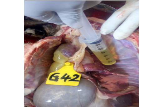
The pathological findings included pleural effusions that were straw coloured and became gel-like when left standing in the open air. Acute fibrinous pneumonia was also observed in some cases, some with extensive red and grey hepatization, including overlying fibrinous pleuritis. Serious cases showed severe pleural adhesions with unilateral or bilateral lung involvement.
Fibrous Adhesions
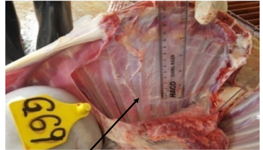
3/6 of the intratracheally inoculated goats (group E1 without detectable worm infection) had fibrous adhesions 1/6 severe and given a score of 3 in a scale of 4 (Goat 18E1). All the six goats belonging to group E2 (intratracheally inoculated goats with detectable worm infection) had fibrous adhesions of varying severity, 3/6 with most extensive lesions where one of them was awarded a score of 4 (Goat 69 group E2 Figure 2). The contact transmission groups, F1 (without worms) and F2 (with detectable worm infection), also had fibrous adhesion lesions of varying intensity ranging from 0-4. One particular case, goat 63 from group F2 (Figure 3), had extensive lung fibrous adhesion to thoracic cavity, a sign of chronic disease and red hepatization a sign of acute disease process. This suggests that there was a relapse to acute phase of the disease.
One-way analysis of fibrous adhesions group means yielded no statistically significant difference between the groups p-value = 0.7577.
Lung Consolidation
More than 50% of the experimental goats’ had lung consolidations varying from red to grey hepatization. The lesion sizes also varied with the most extensive lesions observed in both inoculated and in contact goats with worms. The overall analysis of post mortal finding across showed 14/24 goats had severe lesions, 9/24 goats had mild lesions and 1/24with normal lung texture. Consolidations were unilateral or bilateral with the diaphragmatic lobes more often affected.
From one-way analysis of lung consolidation group means the p-value was equal to 0.0003056 which is less than 0.05. There was a significant difference between the groups with and without nematode infection. To establish which of the group differed from the others, a Post Hoc Test, Tukey HSD multiple comparisons of means giving 95% family-wise confidence level was performed and this revealed that the means of lung consolidations in groups E2-E1, E2-F1 and F2-E2 (p-values 0.0019574, 0.0043982 and 0.0003869 respectively) were statistically significantly different.
DISCUSSIONS
Fibrous Adhesion
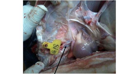
Fibrous adhesion was observed on post mortem across the groups with some having focal lesions while others had extensive lung fibrous adhesion to the thoracic cavity and unilateral lung collapse and this agreed with the works of Wesonga et al. (2004). Generally, group E1 had lower scores of fibrous adhesion with respect to surface area. This group was intratracheally inoculated and had no detectable worm infection at the time of experimental infection. Figure 1 (goat 18 group E1) shows extensive left lung adherence to the thoracic rib cage. All the six goats belonging to group E2 (intratracheally inoculated goats with detectable worm infection) had fibrous adhesions of varying severity, 3 out of 6 had the most extensive lesions with one of them the awarded a score of 4. The contact transmission groups, F1 (without worms) and F2 (with detectable worm infection), also had fibrous adhesion lesions of varying intensity ranging from 0-4. One particular case, goat 63 from group F2 (Figure 3), had lung fibrous adhesion to thoracic cavity a sign of chronic disease and red hepatization a sign of acute disease process. The same scenario was also observed with goat 69 (Figure 2) belonging to group E2 in which the whole right lung had fibrous adhesion to the thoracic cavity with restricted function. In spite of this, group F2 goats had less pathology with respect to fibrous adhesions compared with group F1. It appears that innate and adaptive immune response to Mycoplasma observed as fibrous adhesions was compromised in worm infected goats as evidenced by less adhesions.
Despite the above post mortal observation , a one-way analysis of fibrous adhesions group mean scores showed that there was no statistically significant difference between the experimental groups (p-value = 0.7577) though clinical and pathology suggested slight individual and group differences.
Lung Consolidation
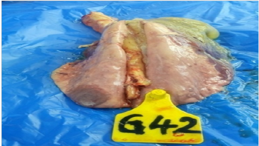
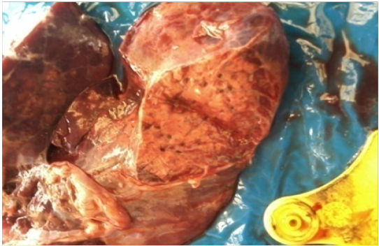
Several of the experimental goats showed bilateral or unilateral lung consolidation that ranged from red to grey hepatization on post mortem. The lesions were confined to the thoracic cavity and these findings were in agreement with observations of previous studies on classical contagious caprine pleuro pneumonia (Wesonga et al., (2004), Arif et al., 2007, MacVey, et al., 2013). Host susceptibility to Mccp infection is determined by the vigour of the immune response. Too strong an inflammatory response induces production of free radicals leading to acute tissue damage and consolidation whereas an inadequate immune response allows for chronic disease development and widespread fibrous deposition. On post mortem, several goats (4 out of 6 from E2 inoculates with helminthes infection and 5 out of 12 contact transmission goats) showed grey and red areas of hepatization (consolidation) in their lungs, in some cases pathological lesions were found affecting more than one lobe and animals showed marked pleuritis and pleural effusion depicting the adverse reaction to the infecting organism. It was found out that majority of the goats with severe pathology (extensive gray and red hepatization (Figure 5 and Figure 7) with an accompanying fibrin deposition (Figure 4) and increased pleural fluids on post mortem belonged to group E2. Six goats had pleural effusion, the pleural fluid being thick and straw coloured, with two of the goats each giving a yield of more than 200 mls. The cut surface of some affected lungs (Refer to Figure 6) revealed a fine granular texture with grey hepatization. One-way analysis of lung consolidation group means showed a statistically significant difference in hepatization between the groups with and without nematode infection (p-value equal to 0.0003056). Even so, this finding called for further analysis to segregate inequality of scores between the groups. Posthoc test, Tukey HSD multiple comparisons of means revealed that E2-E1, E2-F1 and F2-E2 (p-values 0.0019574, 0.0043982 and 0.0003869 respectively) were statistically significant. The clinical signs with pathological findings and statistical results for groups E2-E1 showed there was a negative impact of helminthes infection on host immune response. The finding also proved Koch’s postulate in that a similar pattern of clinical and pathological findings were reproduced after isolation of putative causal organism followed by experimental infection of the research goats.
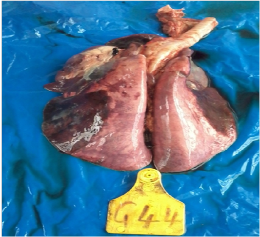
Figure 7: Lung consolidation G44 E2
CONCLUSIONS AND RECOMMENDATIONS
The above findings indicate that worm infection has a negative influence on immune response to bacterial infection. The clinical signs showed that the disease was more severe in worm infected individuals in both tracheal inoculated and contact transmission goats. Antibody titres in response to live Mycoplasma antigens were lower in worm infected goats but none statistically significant though there was a significant pathology.
Fibrous adhesions, a sign of chronic disease was more pronounced in none helminthes infected group. These groups had more fibrous adhesions according to scoring method adopted and visual assessment of the lesion. Classical clinical and pathological findings in contagious caprine pleuro pneumonia such as fibrin deposition, pleural effusions, red and gray hepatization, were frequent in the helminthes infected groups with some worst episodes observed (p-value equal to 0.0003056). Mycoplasma infection in the presence of helminthes co-infection would lead to high mortality (p-values 0.0019574) and acute disease (4 out of 6 from group E2 goats). Conversely, helminthiasis control may compromise the capability of the host to tolerate microbial infections as evidenced more fibrous adhesions in chronic cases that were more prevalent in the none worm infected groups. Though resistance to those microbes may be enhanced, exacerbation of the associated harmful inflammation, read,hepatization, pleural effusions fibrin deposits and fibrous adhesion in the absence of parasitic worm-induced immunoregulatory networks, could potentially contribute to increased disease severity. Given that the nature of antigen determines the category of immune response, the interplay between the effects of parasitic coinfection on host microbial resistance and forbearance will differ with the particular antigen involved and with the gravity of the metazoan or microbial infection. We therefore recommend that livestock be dewormed before vaccination is carried out.
Further studies are required to elucidate how helminthes intrinsically reprogram adaptive immune response cells, namely, antigen specific T and B cells reactive to microbial pathogens. Another point to be looked into is how Mycoplasma induces the severe pathology, read, consolidation observed in the lungs of some of the infected goats and which of the cells of innate and adaptive immune response are involved.
ACKNOWLEDGEMENTS
To the National Commission for Science and Innovation for the financial support they gave towards completion of this work. To the Director of Veterinary Sciences Research Institute (VSRI), Kenya Agricultural & Livestock Research Organization(KALRO), Muguga North for acceptance and space to work in the institution and to the personnel for their assistance.To Professor P. P. Semenye, NUFFIC-NICHE KEN 212 for the support the project accorded me towards completion of this study.
CONFLICT OF INTEREST
Authors declare that they have no conflict of interest.
AUTHORS CONTRIBUTION
Moses Ngeiywa: Parasitology, PhD project supervisor and course work instructor.
Donald Siamba: Parasitology, PhD supervisor.
Simion Mining: Immunology, PhD supervisor.
Hezron Wesonga: Head of microbiology in Division Veterinary Sciences and Research Institute, KARLO gave guidance during isolation and culture of Mccp agent for experimental inoculation in goats.
REFERENCES


