
Advances in Animal and Veterinary Sciences


Research Article
Advances in Animal and Veterinary Sciences 1 (3): 80 – 83Detection of Antibiotic Residues and Determination of Microbial quality of Raw Milk from Milk Collection Centres
Nameirakpam Dinki1, Endale Balcha2*
- Assam Agricultural University, College of Veterinary Science, Guwahati–22, India
- Mekelle University, College of Veterinary Medicine, Mekelle, Ethiopia P.O.Box: 2084
*Corresponding author:endalebalcha@yahoo.com
ARTICLE CITATION:
Dinki N, Balcha E (2013). Detection of antibiotic residues and determination of microbial quality of raw milk from milk collection centres. Adv. Anim. Vet. Sci. 1 (3): 80 – 83
Received: 2013–07–10, Revised: 2013–07–19, Accepted: 2013–07–20
The electronic version of this article is the complete one and can be found online at
(
http://www.nexusacademicpublishers.com/table_contents_detail/4/57/html
)
which permits unrestricted use, distribution, and reproduction in any medium, provided the original work is properly cited
ABSTRACT
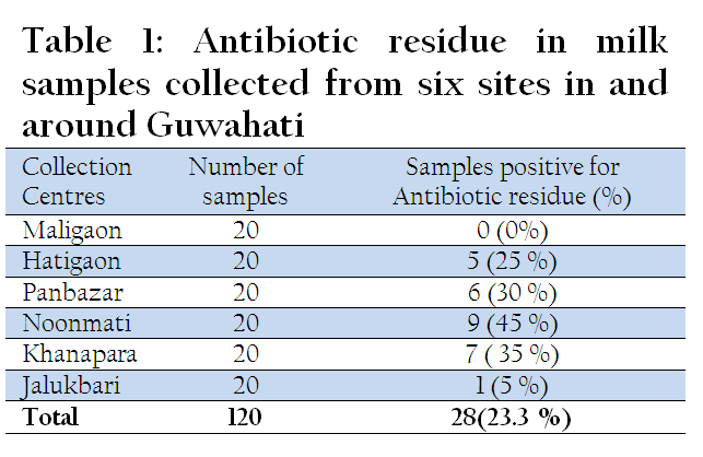
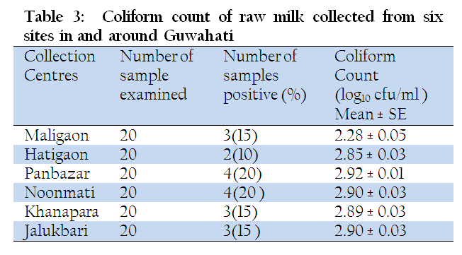
An investigation was carried out from September 2010 to January 2011 to detect antibiotic residues and assess microbial load of milk samples of cattle collected from six different consumers’ collection centres of Guwahati city, India viz. Maligaon, Hatigaon, Panbazar, Noonmati, Khanapara and Jalukbari. A total of 120 milk samples were aseptically collected from randomly selected milk cans. Antibiotic residues were found in 28 samples with 23.3 % detection rate. The mean standard plate count of raw milk of Maligaon, Hatigaon, Panbazar, Noonmati, Khanapara and Jalukbari were recorded as 6.38 ± 0.02, 6.31 ± 0.02, 6.33 ± 0.02, 6.34 ± 0.03, 6.32 ± 0.02 and 6.29 ± 0.02 log10 cfu per ml, respectively. Whereas the mean coliform count of raw milk of the six centres were recorded as 2.28 ± 0.05, 2.85 ± 0.03, 2.92 ± 0.01, 2.90 ± 0.03, 2.89 ± 0.03 and 2.90 ± 0.03 log10 cfu per ml, respectively. The results of the current study indicate that the milk produced and distributed in the study area can be considered as of fair quality from microbial load point of view. Since the presence of antibiotic residues could pose human health risk, awareness should be created on the judicious use of antibiotic and adherence to drug withdrawal period.
INTRODUCTION
Milk is a highly nutritious food, ideal for microbial growth and the fresh milk easily deteriorates to become unsuitable for processing and human consumption (FAO, 2001; Lingathurai and Vellathurai, 2010; Mubarack et al., 2010; Ali and Abdelgadir, 2011). Presence of bacteria in raw milk reduces the keeping quality of milk and certain bacteria with their associated enzymes and toxins may even survive pasteurization creating health hazards (Salman and Hamad, 2011). High bacterial counts in raw milk are indicator of poor production hygiene (Harding, 1999).
The safety of milk with respect to food–borne diseases is of great concern around the world. This is especially true in developing countries where production of milk and various milk products takes place under unsanitary conditions and poor production practices (Mogessie, 1990).
Milk is sterile during secretion from healthy animals but the components that are foreign to it enter the milk in the udder or during or after milking as well as any changes occurring in the milk are often detrimental to its quality (Waltsra et al., 1999). Once, the milk comes outside the udder, microbial contamination may occur due to normal handling procedures and in between milking, teats may become soiled with dung, mud and bedding materials. Number and type of microorganisms vary according to type and amount of soil materials (Gierl and Putz, 1992).
Coliforms particularly Escherichia coli are frequently used in the microbiological analysis of food as an indicator of poor hygienic condition. Microbiological examination of milk is essential to find the degree of contamination with detection and enumeration of indicator organisms. The Coliforms are defined as the indicator (faecal coliform) of suitability of milk for drinking (SMCWWA, 1981).
The hygienic quality problems of milk may arise from raw milk of diseased animals (Murphy and Boor, 2000). The presence of antimicrobial substances in raw milk could have serious toxicological and technical consequences (Kang et al., 2005).
Antibiotic residues are of concern due to their possible adverse effects on people allergic to antibiotics, potential build up of antibiotic–resistant organism in humans and inhibition of starter cultures used to produce cultured milk products such as yogurt and cheeses (Jones and Seymou, 1988).
Understanding the microbial load of raw milk needs to measure the hygienic quality of milk. High population of bacteria in aseptically drawn milk samples or detection antibiotic residues is an evidence of the public health risk of milk (Abrahamsen et al., 2007).
Although milk and milk products represent an important place in the nutrition of consumers as well as nutrition and income of producers, there is limited work so far undertaken regarding detection of antibiotic residues and assessing microbial load of raw milk in Guwahati, India. Therefore the present investigation was undertaken to analyse the milk samples collected from various pockets in and around Guwahati with the objective of determining the microbial load and detection of antibiotic residues.
MATERIALS AND METHODS
Milk Sample Collection
A total of 120 samples were collected from six different consumer’s collection centres of Guwahati city: Maligaon, Hatigaon, Panbazar, Noonmati, Khanapara and Jalukbari. From each collection centre 20 milk samples of morning milk were collected at week interval for a total of 4 weeks. The samples were collected in 250 ml capacity sterilized container and each sample measured 200 ml of milk. The samples were taken from bulk cans and all aseptic precautionary measures were taken to avoid external contamination till the laboratory examinations were over in the laboratory. Ice– boxes were used to carry the samples from the collection centres to the laboratory. Samples were stored at oC and subjected to bacteriological examination within 3 to 4 hours of collection.
Detection of Antibiotic Residues
The presence of antibiotic residues in milk was detected as per the method described by (Mitchew et al., 1998). Petri plates were prepared with each plate containing about 15–20 ml of nutrient agar media. The test organism used for the study was Bacillus subtilis. Spore suspension of organism in nutrient broth was used. The media in Petri plates were rubbed gently by a sterile cotton swab dipped in spore suspension so as to cover the entire agar surface. Sterile discs (HiMedia) were used. For each sample a separate disc was prepared by dipping the disc in small amount of milk sample for a little while and then shaking off the excess milk. The test disc prepared fresh and still wet were then placed on agar surface. Only 4 discs were placed on single plate. Plates were then incubated overnight at 37o C. After incubation discs surrounded by a distinct zone of growth inhibition around them were regarded as positive.
Assessment of Microbial load
The samples were examined for Total Bacterial Counts (TBC) and coliform counts by the pour plate technique described by (Collins et al., 1989). Milk samples were thoroughly mixed by shaking the sample bottle for at least 25 times and a serial ten–fold dilution was made in normal saline solution. After through mixing, 0.1 ml from each dilution of the sample was inoculated into sterile petri plates in duplicate, maintaining all aseptic precautions. Care was taken to transfer the last drop of sample into the petri plates. Then after, 15–20 ml of nutrient agar media previously melted and cooled to 45º C, was poured into each pairs of Petri dishes for total bacterial count and MacConkey’s agar was used to determine coliform counts. The inoculum and the media were thoroughly mixed by rotating and tilting. Petri dishes were allowed to cool down and set by keeping them on a level surface. Control samples were prepared in similar manner using sterile water in place of market milk samples to ascertain sterility of apparatus and the medium used.
Petri dishes were incubated at 37 ± 0.5º C for overnight in inverted position. After incubation plates having more that 30 but less than 300 separated colonies of bacteria were counted with the help of bacteriological colony counter. The number of colonies and dilution factors were recorded. Then, the mean from the two plates were calculated and multiplied with the specific dilution factor and lastly multiplied by ten and thus the total number of viable bacteria per ml of milk was determined and recorded.
Statistical Analysis
All the data were entered into Microsoft Excel. Descriptive statistic was used to analyse the data for antibiotic residues as percentage of samples tested positive for presence of antibiotic residues. Total bacterial counts and coliform counts of the different collection centres were transformed in to log values then data were analyzed.
RESULTS
Antibiotic Residue
Out of 20 milk samples collected from each zone, 5( 25 %) samples from Hatigaon, 6 ( 30 %) samples from Panbazar, 10 (45%) samples from Noonmati, 7 ( 35%) samples from Khanapara and 1( 5%) sample from Jalukbari were found positive for antibiotic residue. No samples were found to have antibiotic residue in Maligaon (Table 1).
Standard Plate Count
The mean standard plate count of raw milk (cfu/ml) was highest in Maligaon (6.38 ± 0.02) followed by Noonmati (6.34 ± 0.03), Panbazar ( 6.33 ± 0.02), Khanapara (6.32 ± 0.02), Hatigaon (6.31 ± 0.02) and Jalukbari (6.29 ± 0.02) (Table 2).

Coliform Count
The mean coliform count of raw milk (cfu/ml) was highest in Panbazar (2.92 ± 0.01) followed by Noonmati and Jalukbari (2.90 ± 0.03), Khanapara (2.89 ± 0.03), Hatigaon (2.85 ± 0.03) and Maligaon (2.28 ± 0.051) (Table 3).
DISCUSSION
In the present study a total of 120 milk samples collected from six different sites of Guwahati were screened for presence of antibiotic residue. Out of these 28 (23.3 %) samples were found to be positive for presence of residue. This somehow agrees with the result of Amonsin et al.(1996) who reported 27 % detection rate. However, Psomas et al. (1994) detected antibiotic residues in 46 % of samples whereas Movassagh and Karami (2010) reported 5% of raw milk samples to be positive for antibiotic residues. The variation might be due to the variation in the drug regiment used in the study areas and also variation in the drug withdrawal of the antibiotics used. This is demonstrated by the work of Zeng et al.(1996) where withholding time of milk for penicillin G was 72 hours and cephalosprin was 120 hours. So it can be inferred the milk delivered to milk collection centres could lead to the development of drug resistance as they contain antibiotic residues in them. This could be due to excessive and injudicious use of antimicrobials, prophylactic or therapeutic treatment of dairy animals which led to the excretion of the residues of drugs in milk.
The milk samples collected from six zones: Maligaon, Hatigaon, Panbazar, Noonmati, Khanapara and Jalukbari showed the average standard plate count log 10 cfu per ml of milk to be highest at Maligaon (6.38 ± 0.02) followed by Noonmati (6.34 ± 0.03), Panbazar (6.33 ± 0.02), Khanapara (6.32 ± 0.02), Hatigaon (6.31 ± 0.02) and Jalukbari (6.29 ± 0.02). Similar observations were also made from Karnal (Sakkarvarthi and Mathur,1990), Guwahati ( Rahman et al., 1992) , Srinagar (Hussain et al., 2005), Tabriz ( Ganzi et al., 2005) and Bangladesh (Khan et al., 2008) where total plate count were found to 6.14 –7.70 , 4.18–7.56, 6.08 , 6.13 and 5.920 – 6.075 log 10 cfu per ml, respectively. Pant et al. (2013) reported total bacterial viable count (log 10 cfu per ml) ranging from 5.17– 8.81.
The present findings revealed that the average total bacteria present in one millilitre of milk collected from different places of Guwahati city was well within the range of bacteriological standard of fair grade of milk as per ISI (1977).
Comparatively higher bacterial counts per ml of milk in Guwahati city might be due to the improper handling, use of unclean utensils, and unhygienic condition of the byre, long holding period of milk due to long distance of transportation from the source to the marketing place. Nevertheless, all the raw milk samples were found to be well within the maximum permissible limit of standard plate count per ml of milk according to ISI (1977) and hence could safely be accepted for consumption or further processing.
The mean ± SE coliform count cfu/ml of the six zones Maligaon, Hatigaon, Panbazar, Noonmati, Khanapara and Jalukbari were recorded as 2.28 ± 0.05 log10 cfu/ml, 2.85 ± 0.03 log10 cfu/ml, 2.92 ± 0.01 log10 cfu/ml, 2.90 ± 0.03 log10 cfu/ml, 2.89 ± 0.03 log10 cfu/ml and 2.90 ± 0.03 log10 cfu/ml, respectively. The results of this investigation are in agreement with the findings of Mutukumira et al.(1996). Saitanu et al.(1996) examined and found the total coliform count to be < 3 log10 cfu/ml. Kalilur and Khan(2002) also obtained the mean coliform count of 3.38 log10 cfu per ml from raw milk collected from sligarh city. The present findings are in good agreement with the findings of Nanu et al.(2006) who found the coliform count to be between 2.97 ± 0.05 to 3.20 ± 0.06 log10 cfu per ml .Similarly in a study conducted by Prejit et al.(2007) the coliform count were between 1.83 ± 0.22 to 3.24 ± 0.19 log 10 cfu per ml. In contrast to the present finding Edward and Inya (2013) reported coliform count ranging from 6.73 – 6.98 log 10 cfu per ml. This was explained to be due to the closeness of the udder to the anus of the animal since they are normal flora of the intestine. Also there is the tendency of the udder and the teat to be contaminated or dirtied by the animal dung when they lie down on it.
The present finding revealed that the mean coliform count of few of the milk samples collected from different places of Guwahati city was not within the range of bacteriological standard of milk as per ISI (1977). This might indicate that the milk might be adulterated with water which was contaminated with faeces and the unhygienic practices in some farms.
CONCLUSION
The mean standard plate count of the milk samples collected from the consumer’s collection centre were well within the maximum permissible limit of standard plate count per ml of milk according to hence Indian standards. However, the mean coliform count of few of the milk samples collected from different places of Guwahati city was not within the range of bacteriological standard of milk. According to the study 28 milk samples out of 120 have been detected for antibiotic residues which might suggest that the drug withdrawal period was not adhered in some farms. It can be inferred from the findings that there is lack of awareness of the milk producers on hygienic milk production and the impact of antibiotic residues on the health of human beings. Therefore creating awareness for milk producers by providing training and experience sharing forum may help improve to improve milk quality and safety in the city.
AKNOWLEDGEMENTS
The study was conducted in the Veterinary Public Health laboratory of Assam Agricultural University, College of Veterinary Science. The authors would like to thank all persons who contributed in this work.
CONFLICT OF INTEREST
There is no conflict of interest between the authors.
REFERENCES
Abrahamsen RK, Borg GI, Harstad OM, Haug A and Wetlessen A (2007). Milk Quality–Future Approach from a Researcher's Point of View. Norwegian food research institute (Matforsk), Osloveien, Norway. J. Animal & Feed Sci.16(1): 209–226.
Ali AA and Abdelgadir WS (2011). Incidence of Escherichia coli in raw cow's milk in Khartoum state. Br. J. Dairy. Sci. 2(1):23–26.
Amonsin A, Saitanu K and Teeverapanya S (1996). Asian Australasian J. Anim. Sci. 9:27.
Collins CH, Lyne PM and Grange JM (1989). Counting Microorganisms. In: Microbiological Methods, 6th edition, Butterworth/Heinemann.Pp. 127–140.
Edward KC and Inya IM(2013). The Microbial Quality of Raw Milk from four locations in Abia State, Nigeria. J. Pharmacy and Biological Sci.5(3): 30–33.
FAO (2001). The lactoperoxidase system of milk preservation. Regional Lactoperoxidase Workshop in West Africa. Burkina Faso. 17–19.
Gazani MH, Karami AR, Dolgharisharf J, Khajeh M and Najafian K (2008).Microbial Safety of Raw Milk in Tabriz Iran. J. Animal &Vet. Advances. 7 (7): 863–865.
Gierl H and Putz ZM (1992).Bacterial counts below 100,000 one achievable. Bayerisch Lanesanstalt Fur Tuerzucht Grub. Germany, Tierzuchter. 44(3): 34–37.
Harding F (1999). Milk Quality. A Chapman and Hall Food Science Book. An Aspen Publication. First edition.
Harrigan WF and McClane ME(1976). Laboratory Methods in Food and Dairy Microbiology. Academic Press, London.
Hussain SA, Rashid R, Willayat MM and Munshi ZH (2005). Prevalence of Food Borne Micro– organisms in Milk and Milk Products. J.Vet. Pub. Health. 3(1): 75–77.
ISI (1977). Part III. Bacteriological analysis of milk IS: 1479, Indian Standard Institute, Manak Bhavan, Bahadur Shah Zafar Marg, New Delhi–2 .
Jones GM and Seymour EH (1988). Cowside antibiotic residue testing. J. Dairy Sci. 71 (6): 1691.
http://dx.doi.org/10.3168/jds.S0022-0302(88)79734-9
Kalilur M, Khan R and Malik A (2002). Microbiological quality of milk, vegetable and fruit juice. J. Food & Food Sci. Technol.39:120–123.
Kang HJ, Jin HJ and Kondo F (2005).False–positive outcome and drug residue in milk samples over withdrawal times. J. Dairy Sci. 88: 908–913
http://dx.doi.org/10.3168/jds.S0022-0302(05)72757-0
Khan MTG, Zinnah MA, Siddique MP, Rashid MHA, Islam MA and Choudhury KA(2008). Physical and microbial qualities of raw milk collected from Bangladesh Agricultural University. Dairy Farm and the surrounding villages. Bangl. J.Vet. Med. 6 (2): 217–221.
Lingathurai S and Vellathurai P (2010). Bacteriological quality and safety of raw cow milk in Madurai, South India. Webmed Central. Microbiol. 1(10):1–10.
Mitchell JM, Griffiths MW, McEwen SA, McNab WB and Yee AJ (1998). Antimicrobial drug residues in milk and meat: Causes, concerns, prevalence, regulations, tests and test performance. J. Food. Prot.61: 742–756.
PMid:9709262
Mogessie A (1990). Microbiological quality of Ayib, a traditional Ethiopian cottage cheese. Int. J. Food Microbiol.10 : 263–268.
http://dx.doi.org/10.1016/0168-1605(90)90074-F
Movassagh MH and Karami AR (2010). Determination of Antibiotic Residues in Bovine Milk in Tabriz, Iran. Global Veterinaria. 5 (3): 195–197.
Mubarack MH, Doss A, Dhanabalan R and Balachander S (2010). Microbial quality of raw milk samples collected from different villages of Coimbatore district, Tamilnadu, South India. Ind. J. Sci. Technol. 3(1):61–63.
Murphy SC and Boor KJ (2000). Trouble–shooting sources and causes of high bacteria counts in raw milk. Dairy, Food & Environmental Sanitation.20: 606–611.
Mutukumira AN, Feresu SB, Narbhus JA and Abrahamsen RK(1996). Chemical and Microbiological Quality of raw milk produced by small holder farmers in Zimbabwe. J.Food Protection. 59 (9): 984–987.
Nanu E, Sunil B, Pretjit, Thomas M and Latha C (2006). Consumer Perception on Quality of Liquid Milk in Kerala. J. Vet. Pub. Health. 5 (1):49–51.
Pant R, Nirwal S and Rai N (2013). Prevalence of Antibiotic Resistant Bacteria and Analysis Of Microbial Quality Of Raw Milk Samples Collected From Regions Of Dehradun. Int.J. Pharm.Tech. Research. 5(2): 804–810.
Prejit, Nanu E and Latha C (2007).Microbial Quality Assurance of milk during Production, Processing and Marketing. American J. Food Technol. 2(3): 136–144.
Psomas IE, Fletoures DL and Papaqeorqiou DC (1994). Mantes AI. Null Hellenic Vet. Med. Soc. 45:295 Rahman H, Nath NC and Boro BR(1992). Bacterial flora and insecticidal residues in raw milk marketed in Guwahati city, Assam. Indian J. Comp. Microbiol. Immunol. Infect. Diseases. 13: 105–108.
Saitanu IA, Chuanchuen KR, Nuanuarsuwan S, Koowatananukul C and Rugkhaw V(1996). Microbiological quality of raw cow milk. Thai J. Vet. Med.26(3): 193–214.
Sakkarvarthi S, Mathur DK and Malik RK (1990). Bacteriological quality of buffalo milk in relation to processing at high temperatures. Indian J. Dairy Sci.43: 207–212.
Salman Adil MA and Hamad IM (2011). Enumeration and identification of coliform bacteria from raw milk in Khartoum state, Sudan. J. Cell Anim. Biol. 5(7):121–128.
SMCWWA (1981). Standard Method Committee for water and waste water analysis. J.Am. Public Health Association, Washington DC.
Walstra P, Geurts TJ, Noomen A, Jellema A and VanBookel MAS (1999).Composition, structure and properties. In: Dairy Technology: Principles of milk properties and processes. Marcel Dekker, New York.
Zeng SS, Escobar EN and Crowder B (1996). Evaluation of screening tests for detection of antibiotic residues in goat milk. Small Ruminant Research. 21:155–160
http://dx.doi.org/10.1016/0921-4488(95)00822-5


