
Advances in Animal and Veterinary Sciences


Research Article
Advances in Animal and Veterinary Sciences 2 (5S): 17 – 21Special Issue – 5 (2014) (Listeriosis and its public health concerns)
Seropositivity of Field Veterinarians for Listeric Infection by Indirect ELISAs Employing Recombinant and Wild–Type Listeriolysin O
Rahul Diliprao Suryawanshi1*, Bhushan Jayarao2, Sukhadeo Baliram Barbuddhe3, Sandeep Prabhakar Chaudhari4, Deepak Bhiwa Rawool1, , Vysakh Mohan1, Jess Vergis1, Mamta Negi1, Satya Veer Singh Malik1*
- Division of Veterinary Public Health, Indian Veterinary Research Institute, Izatnagar, Uttar Pradesh, India. Pin– 243 122
- Department of Veterinary and Biomedical Sciences, Pennsylvania State University, PA, U.S.A. Pin–16801
- National Institute of Biotic Stress Management, Raipur, Chhatisgarh, India. Pin– 492 012
- Department of Veterinary Public Health, Nagpur Veterinary College, Nagpur, Maharashtra, India. Pin–440 006
*Corresponding author:svsmalik@gmail.com
ARTICLE CITATION:
Suryawanshi RD, Jayarao B, Barbuddhe SB, Chaudhari SP, Rawool DB, Mohan V, Vergis J, Negi M, Malik SVS (2014). Seropositivity of field veterinarians for listeric infection by indirect ELISAs employing recombinant and wild–type listeriolysin O. Adv. Anim. Vet. Sci. 2 (5S): 17 – 21.
Received: 2014–07–12, Revised: 2014–07–28, Accepted: 2014–07–29
The electronic version of this article is the complete one and can be found online at
(
http://dx.doi.org/10.14737/journal.aavs/2014/2.5s.17.21
)
which permits unrestricted use, distribution, and reproduction in any medium, provided the original work is properly cited
ABSTRACT
Listeriolysin O (LLO) being an essential virulence marker produced by all the pathogenic strains of Listeria monocytogenes has been reported to be an immunodominant antigen for serodiagnosis of listeric infections. The present study explores the serodiagnostic potential of recombinant listeriolysin O (rLLO) vis–a–vis wild type LLO (wLLO) employed in an indirect plate ELISA for screening sera of 221 field veterinarians from Maharashtra, India. A higher seropositivity (73.30%) for antibodies against LLO (ALLO) was observed amongst field veterinarians in wLLO–based ELISA compared to 37.56% in rLLO–based ELISA. Further, adsorption of sera with streptolysin–O (SLO) resulted in more than three–fold reduction in the seropositivity for ALLO, which was observed to be 14.93% in wLLO–based ELISA and 13.57% in rLLO–based ELISA. The rLLO–based ELISA having advantage in terms of lesser cross–reactivity and ease of production of the employed antigen, appears to be a better option for serodiagnostic purposes than wLLO–based ELISA, which is classically employed as widely accepted reliable serodiagnostic test, especially on SLO adsorbed sera. However, rLLO based–ELISA needs to be further evaluated on the sera from known clinical cases of listeriosis, especially in the high risk groups of humans, for ascertaining its efficacy as rapid and reliable serodiagnostic test for mass screening. This study seems to be the first report on comparative diagnostic potential of rLLO and wLLO in plate ELISA.
INTRODUCTION
Listeriosis is an important bacterial infection caused by the pathogenic species of the genus Listeria, namely, Listeria monocytogenes and L. ivanovii (Barbuddhe et al., 2012). L. monocytogenes is of major concern as it accounts for about 98% of human and 85% of animal listeriosis cases (Liu, 2006). Categorized under List C of OIE, the disease in general exhibits neural, visceral and reproductive disorders particularly in various species of animals as well as humans who are immunocompromised or those that are in contact with animals (Barbuddhe and Chakraborty, 2008).
Globally, listeriosis has been reported to occur either in sporadic and epidemic form, however, there are certain Asian countries where the disease has been under reported due to lack of active surveillance systems (Tirumalai, 2013). In India, comprehensive review of reported sporadic cases of human and animal listeriosis suggested that the available epidemiological data is not sufficient to evaluate the extent of true infection in humans and animals; moreover, the disease remains mostly undiagnosed and under–reported due to unavailability of a suitable diagnostic assay (Malik et al., 2002; Barbuddhe et al., 2012). The most authentic diagnosis of listeriosis is made by isolation of the pathogen. However, it requires 2–3 days to provide presumptive positive results and additional 2–4 days for confirming suspected colonies by biochemical tests (Frece et al., 2010; Jadhav et al., 2012). On the other hand, serological tests have the advantage of large number of mass screening, and are economical, easy–to–perform and interpret. Ideally, such test must have sufficient sensitivity, specificity to detect the humoral response directed against the immunogenic component of the agent, preferably those linked to its virulence (Shoukat et al., 2013a). Many serodiagnostic assays have been developed for screening animal and human listeriosis cases either by employing the somatic (O), flagellar (H), cold–extracted or sonicated listerial antigens or outer membrane protein (OMP) of Listeria spp. (Chen and Chang, 1996). However, these conventional serological assays cannot be relied upon owing to their poor specificity and sensitivity (Berche et al., 1990; Chen and Chang, 1996). Moreover, these assays fail to discriminate between pathogenic and non–pathogenic Listeria strains. Therefore, it is important to have serodiagnostic tests based on virulence–linked antigens which may help in identifying true listeriosis cases. In this regard, there has been continuous search for virulence markers and/or the candidate protein antigens of Listeria species capable of eliciting the antibody response during listeric infection. Such virulence markers/antigens include listeriolysin–O (LLO), the hly gene encoded haemolysin produced by L. monocytogenes (Berche et al.,1990); internalins (InlA, InlB, InlC, InlC2, InlJ etc) (Das et al., 2013), the leucine rich repeat (LRR) proteins of Listeria spp. produced by virulence–linked family of genes (Boerlin et al., 2003); the actA gene encoded actin or Act A protein associated with cell–to–cell spread of the agent (Ellin Doyle, 2001), two phospholipases C namely the PI–PLC encoded by plcA gene and the PC–PLC encoded by plcB (Chaudhari et al., 2004a, 2004b); the autolysin p60 protein (Hess et al., 1996).
Among all the virulence associated proteins, LLO which is produced by only virulent strains of L. monocytogenes, has been extensively used as an antigen in development of Enzyme– linked immunosorbent assay (ELISA) for serodiagnosis of listeric infection in sheep (Low et al., 1992; Lhopital et al., 1993; Barbuddhe et al., 2000; Shoukat et al., 2013b), goats (Miettinen et al., 1990; Miettinen and Husu, 1991; Bhanu Rekha et al., 2006), buffaloes (Chaudhari et al., 2001) and cattle (Thakur, 2000) as well as in humans (Berche et al., 1990; Kaur et al., 2006). However, the cross–reactivity of antibodies against LLO (ALLO) with those produced against streptolysin O (SLO), a haemolysin produced by Streptococcus spp. remains a major limitation of this assay, which calls for adsorption of test sera with SLO prior to its testing by this assay (Berche et al., 1990; Kaur et al., 2006; Shivaramu, 2008; Shoukat et al., 2013a). Therefore, recombinant forms of LLO (rLLO) have been explored as an alternative to wild type LLO (wLLO) as a diagnostic antigen in Western blot assays (OIE, 2008).
In this context, the present study was undertaken to compare diagnostic potential of recombinant listeriolysin O (rLLO) with wild type LLO (wLLO) in indirect ELISA employed for screening the human serum samples of field veterinarians. The developed assays were also evaluated after adsorption of the test sera with SLO.
MATERIALS AND METHODS
Sample Collection
A total of 221 blood samples were collected aseptically from field veterinarians of Maharashtra State, India during September, 2013–March, 2014. All the samples were transported to the laboratory under chilled conditions. The sera from all the blood samples were separated and stored at –20 ºC for its further use in the developed assays. Before collection of the samples, informed consent was taken from all the persons.
Purification of Wild Type LLO (wLLO)
The wLLO was extracted and purified from the cell free supernatant of 18h–old Listeria monocytogenes (MTCC 1143, IMTECH, Chandigarh, India) growth in brain–heart infusion (BHI) broth (Himedia, India) at 37 ºC by ion–exchange chromatography technique as described by Lhopital et al. (1993).The eluted fractions having wLLO were pooled together and the protein concentration was estimated using BCA™ Protein Assay kit (Pierce, USA, Catalog No. 23225). Purity of the wLLO was checked by SDS–PAGE confirming it to be a homogenous 58.0 kDa protein. The pooled wLLO was stored at –80ºC til further use.
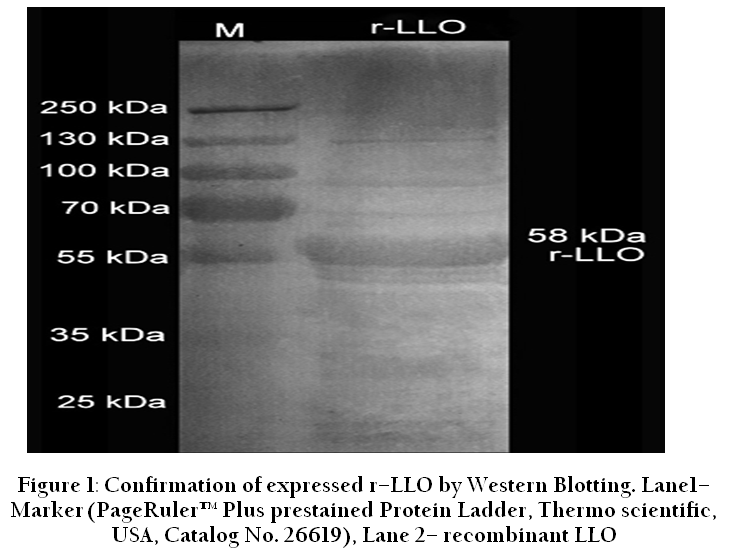
Figure 1: Confirmation of expressed r–LLO by Western Blotting. Lane1– Marker (PageRuler™ Plus prestained Protein Ladder, Thermo scientific, USA, Catalog No. 26619), Lane 2– recombinant LLO
Production of Recombinant LLO (rLLO)
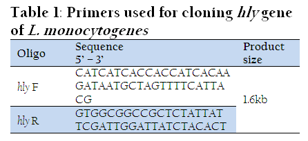
A precise directional cloning of hly gene of L. monocytogenes was performed using Expresso T7 Cloning and Expression System (Lucigen, USA, Catalog No. 49001–1). Primers were designed as per the manufacture’s kit protocol (Table 1). The expression of the 58 kDa target protein was confirmed by Western blot analysis (Figure 1) using anti–histidine antibodies (Abcam, USA, Catalog No. ab6442) and Goat anti–Rabbit IgG Fc (HRP) secondary antibodies (Abcam, USA, Catalog No. ab97200). The rLLO was purified using Ni–NTA Fast Start Kit (Qiagen, USA, Catalog No. 30600). Finally, the protein concentration of rLLO was measured using BCA™ Protein Assay kit (Pierce, USA, Catalog No. 23225) and stored at –80ºC until further use.
Indirect ELISA
The indirect plate ELISA was performed as described by Low et al. (1992). The ELISA was standardized by using checker–board titration method. A serum sample at a dilution of 1:200 with a positive to negative (P/N) ratio of more than 2 was considered as positive for listeriosis in a standardized ELISA by employing either wLLO (1µg/well) and rLLO (0.125µg/well) as an antigen and rabbit anti–human HRPO conjugate (1:2000, Sigma–Aldrich, India, Product No. A8792). These standardized indirect ELISA tests were then employed for screening ALLO before and after adsorption of the test sera with SLO (Sigma Aldrich, USA, Product No. S5265) as per the protocol described by Berche et al. (1990). All the test sera were evaluated three times independently by both the ELISAs.
Statistics
The data obtained from the present study was analyzed using paired student‘t’ test.
RESULTS
Detection of ALLO Antibodies by wLLO
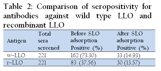
Screening of sera from field veterinarians by indirect ELISA employing wLLO revealed seropositivity for ALLO in 73.30% (162/221) cases (Table 2). On retesting of the sera showing positivity against wLLO following their adsorption with SLO, the seropositivity was considerably reduced to the level of 14.93% (33/221) (Table 2).
Detection of ALLO by rLLO–based ELISA
Screening of sera samples by rLLO–based ELISA revealed a positivity of 37.56% (83/221) for antibodies against rLLO. On retesting of the positive sera following their adsorption with SLO, the seropositivity was reduced to 13.57% (30/221) (Table 2).
Comparison of Seropositivity for Listeric Infection by wLLO and rLLO–based ELISAs
On overall basis, a high seropositivity (73.30%) was observed among the field veterinarians in wLLO–based indirect ELISA. However, subsequent to adsorption with SLO, the seropositivity reduced significantly (p<0.05) to 14.93% (Table 2). On comparison of these observations with that of rLLO–based ELISA, significantly lower seropositivity (p<0.05) was observed in case of unadsorbed sera i.e., 37.56% and SLO adsorbed sera, i.e., 13.57 % (Table 2).
DISCUSSION
L. monocytogenes is a zoonotic agent and its presence in all the critical stages of the food production and distribution chain, including the epidemiological surveillance of livestock plays a decisive role in the prevention of food–borne listeriosis in humans (Swaminathan and Gerner–Smidt, 2007). Thus, a sensitive and specific test to identify L. monocytogenes infected animals is of great importance in carrying out epidemiological surveys to develop appropriate control strategies. LLO is an important virulence marker of L. monocytogenes and a known dominant antigen target of anti–listerial immunity (Bouwer et al., 1992) which induces T–cell recognition during the course of an acute listeric infection (Berche et al., 1987). Antibodies to LLO (ALLO) have been detected soon after the clinical onset of disease in man (Morandi et al., 1981; Berche et al., 1990; Aljicevic et al., 2006) and their detectable levels have been found to persist for several months. Even the low dose of experimental infection in lambs has been reported to elicit a detectable ALLO response just like the high dose of infection (Lhopital et al., 1993). Therefore, LLO, which is produced in vivo during the process of intracellular multiplication of pathogenic Listeria spp., seems to be a good virulence– associated marker in clinical infection. Moreover, LLO has been reported to avoid the necessity of using multiple serotype antigens in immunoassays (Low and Donachie, 1997). However, LLO has been reported to share common antigenic domains with other haemolysins namely pneumolysin from Streptococcus pneumoniae, perfringolysin O from Clostridium perfringens, cereolysin O from Bacillus cereus, alveolysin from Bacillus alvei (Geoffroy et al., 1987).
In order to avoid the level of cross–reactivity in seropositivity observed against native or wild type of LLO (wLLO) we evaluated the serodiagnostic potential of rLLO as an antigen in indirect ELISA and compared with that of wLLO in terms of reduction caused, if any, in the cross–reactivity so as to have a more reliable serodiagnosis of listeric infection in field veterinarians. As expected, in wLLO–based ELISA a very high seropositivity (73.30%) for listeric infection was observed in field veterinarian which significantly reduced to 14.93% following sera adsorption with SLO. In rLLO–based ELISA, a lower seropositivity was observed (37.56%) which further reduced to 13.57% following adsorption of test sera with SLO.
The results of the present study are in agreement with earlier studies wherein a very high seropositivity for ALLO was observed in case of spontaneous abortions (48%) and in abattoir personnel (49.2%) when tested against purified LLO employed as antigen in indirect plate ELISA (Kaur et al., 2006; Barbuddhe et al, 1999). The observations are also comparable with earlier reports on seropositivity for listeric infection in case of animals ranging from 77% (Osebold and Aalund, 1968), 37% (Nass and Ortel, 1977), 53% (Husu, 1990; Lida et al., 1991), 53.33% (Morandi et al., 1981) as well as in case of human subjects 60% (Larsen and Jones, 1972) and 60% in women in reproductive age and some with spontaneous abortions (Aljicevic et al., 2006).
LLO is antigenically related to a number of cytolysins, including SLO from Streptococcus pyogenes, pneumolysin from Streptococcus pneumoniae and perfringolysin from Clostridium perfringens. Adsorption of the test sera with SLO has been found to cause 3–fold reduction in ALLO titers on account of eliminating the marked cross–reactivity in human clinical case of encephalitis (Berche et al., 1990) and abortions (Kaur et al., 2006) as well as animal listeriosis cases in cattle (Shivaramu, 2008) and sheep (Shoukat et al., 2013a).
Summarily, the rLLO–based ELISA turned out to be a superior serodiagnostic assay for human listeriosis than the conventional wLLO–based ELISA as the former assay showed less cross–reactivity. The exact reason for less cross–reactivity of rLLO in comparison to wLLO observed in the ELISA is not known, however, the differences in the antigenicity of both the antigens might be a probable cause. On comparison of seropositivity data, the rLLO–based ELISA employed on SLO adsorbed sera exhibited significantly superior serodiagnostic efficacy in terms of less cross–reactivity than that observed with rLLO–based ELISA employed on unadsorbed sera. Moreover, the rLLO employed as a diagnostic antigen in ELISA was having additional advantages, as it was easy to produce with assured consistent quality in each batch and cost effective in terms of sample analysis compared to the wLLO, which was tedious, time–consuming, cumbersome and costly to produce besides having the likelihood of batch–to–batch variations in the quality and quantity produced, owing to its vulnerability to proteolytic enzymes during its in vitro production and purification.
In conclusion, the rLLO–based ELISA employed on SLO adsorbed human sera can be used for reliable serodiagnosis of listeriosis in humans, especially in high risk groups. However, the assay needs to be validated on clinically confirmed listeriosis cases, with and without isolation of the pathogen from such subjects, before making final recommendation for its utilization as the diagnostic tool of choice for reliable serodiagnosis of listeriosis in human population.
ACKNOWLEDGEMENT
The authors thank Director, Indian Veterinary Research Institute, Izatnagar, UP, India and Head Division Department of Veterinary and Biomedical Sciences, Pennsylvania State University, PA, USA for providing necessary facilities to undertake the research. The financial support in the form of the grants from IUSSTF (Indo–US Science and Technology Forum) and PHFI (Public Health Foundation of India), New Delhi, Department of Biotechnology, Government of India. (NO.BT/01/CEIB/11/VI/13) for carrying out this research work also duly acknowledged.
REFERENCES
Aljicevic M, Beslagic E, Zvizdic S, Hamzic S, Mahmutovic S (2006). Agglutination as screening test in routine diagnostic of listeriosis. Med. Arch. 60: 93 – 95.
PMid:16528925
Barbuddhe S., Chakraborty T (2008). Biotechnological applications of Listeria's sophisticated infection strategies. Microbial Biotechnology. 1: 361 – 372.
http://dx.doi.org/10.1111/j.1751-7915.2008.00037.x
PMid:21261856 PMCid:PMC3815243
Barbuddhe SB, Malik SVS, Kumar P (1999). High seropositivity against listeriolysin O in humans. Annals. Trop. Med. Parasitol. 93: 537 – 539.
http://dx.doi.org/10.1080/00034989958294
PMid:10690251
Barbuddhe SB, Malik SVS, Bhilegaonkar KN, Kumar P, Gupta LK (2000). Isolation of Listeria monocytogenes and anti–listeriolysin–O detection in sheep and goats. Small Rum. Res. 38: 151 – 155.
http://dx.doi.org/10.1016/S0921-4488(00)00155-3
Barbuddhe SB, Malik SVS, Kumar AJ, Kalorey DR, Chakraborty T (2012). Epidemiology and risk management of listeriosis in India. Int. J Food Microbiol. 154: 113 – 118.
http://dx.doi.org/10.1016/j.ijfoodmicro.2011.08.030
PMid:21955732
Berche P, Gaillard JL, Geoffroy C, Alout JE (1987). T–cell recognition of listeriolysin – O is induced during infection with L. monocytogenes. J. Immunol. 139: 3813 – 3821.
PMid:3119720
Berche P, Reich KA, Bomichan M, Beretti JL, Geoffroy C, Raveneau J, Cossart R, Gaillard JL, Geslin P, Kreis H, Veron M (1990) Detection of anti–listeriolysin O for serodiagnosis of human listeriosis. Lancet. 335: 624 – 627.
http://dx.doi.org/10.1016/0140-6736(90)90411-W
Bhanu R, Malik SVS, Chaudhari SP, Barbuddhe SB (2006). Listeriolysin O–based diagnosis of Listeria monocytogenes infection in experimentally and naturally infected goats. Small Rum. Res. 66: 70 – 75.
http://dx.doi.org/10.1016/j.smallrumres.2005.07.021
Boerlin P, Boerlin–Petzold F, Jemmi T (2003). Use of Listeriolysin O and Internalin A in a Seroepidemiological Study of Listeriosis in Swiss Dairy Cows. J. Clin. Microbiol. 41: 1055 – 1061.
http://dx.doi.org/10.1128/JCM.41.3.1055-1061.2003
PMid:12624029 PMCid:PMC150307
Bouwer HGA, Nelson CS, Gibbins BL, Portnoy DA, Hinrichs DJ (1992). Listeriolysin–O is a target of the immune response to Listeria monocytogenes. J. Exptl. Med. 175:1467 – 1471.
http://dx.doi.org/10.1084/jem.175.6.1467
PMid:1588276
Chaudhari SP, Malik SVS, Barbuddhe SB (2004a). Humoral and delayed type hypersensitive responses against Listeria monocytogenes phosphatidylinositol–specific phospholipase C in experimentally infected buffaloes. Vet. Res. Commun. 28: 569 – 579.
http://dx.doi.org/10.1023/B:VERC.0000042864.98892.06
PMid:15563104
Chaudhari SP, Malik SVS, Banu Rekha G, Barbuddhe SB (2001). Detection of anti–listeriolysin O and Listeria monocytogenes in experimentally infected buffaloes (Bubalus bubalis). Trop. Anim. Hlth. Prod. 33: 285 – 293.
http://dx.doi.org/10.1023/A:1010579701464
PMid:11474862
Chaudhari SP, Malik SVS, Chatlod LR, Barbuddhe SB (2004b). Isolation of pathogenic Listeria monocytogenes and detection of antibodies against phosphatidylinositol–specific phospholipase C in buffaloes. Comp. Immunol. Microbiol. Infect. Dis. 27: 141 – 148.
http://dx.doi.org/10.1016/j.cimid.2003.08.002
PMid:14690723
Chen SC, Chang TC (1996). Identification of Listeria monocytogenes based on the detection of a 68–Kilodalton cell–surface antigen. J. Food Prot. 59(11): 1176 – 1181.
Das S, Malik SVS, Shrivastava S, Gandhale P, Kumar S, Shoukat S, Das DP, Barbuddhe SB, Rawool DB (2013). Comparative efficacy of Internalin C–based peptide and listeriolysin O–based enzyme linked immunosorbent assays for serodiagnosis of listeric infection in goats. Afr. J. Microbiol. Res. 7(48): 5471 – 5478.
Ellin DM (2001). Virulence characteristics of Listeria monocytogenes. FRI Briefings. Available at: http://fri.wisc.edu/docs/pdf/virulencelmono.pdf (Accessed on 24th June 2014).
Frece J, Markov K, Cvek D, Kolarec K, Delas F (2010). Comparison of conventional and molecular methods for the routine confirmation of Listeria monocytogenes in milk products produced domestically in Croatia. J. Dairy Res. 77: 112 – 116.
http://dx.doi.org/10.1017/S0022029909990379
PMid:19930757
Gellin BG, Broome CV (1989). Listeriosis. J. A. Med. Assoc. 261, 1313 – 20.
http://dx.doi.org/10.1001/jama.1989.03420090077035
http://dx.doi.org/10.1001/jama.261.9.1313
Hess J, Gentschev I, Miko D, Welzel M, Ladel C, Goebel W, Kaufmann SH (1996). Superior efficacy of secreted over somatic antigen display in recombinant Salmonella vaccine induced protection against listeriosis. Proc. Natl. Acad. Sci. 93: 1458 – 1463.
http://dx.doi.org/10.1073/pnas.93.4.1458
PMid:8643654 PMCid:PMC1079202
Husu JR (1990). Epidemiological studies on the occurrence of Listeria monocytogenes in the faeces of dairy cattle. J. Vet. Med. B. 37: 276 – 282.
http://dx.doi.org/10.1111/j.1439-0450.1990.tb01059.x
Jadhav S, Bhave M, Palombo EA (2012). Methods used for the detection and subtyping of Listeria monocytogenes. J. Microbiol. Methods. 88: 327 – 341.
http://dx.doi.org/10.1016/j.mimet.2012.01.002
PMid:22261140
Kaur S, Malik SVS, Vaidya VM, Kaur G (2006). Serological diagnosis of Listeria monocytogenes infection in women in spontaneous abortions. Indian J. Comp. Microbiol. Immunol. Infect. Dis. 27(1): 23 – 25.
Larsen S, Jones W (1972). Evaluation and standardization of an agglutination test for human listeriosis. Appl. Microbiol. 24(1): 101 – 107.
PMid:4626902 PMCid:PMC380554
Lhopital S, Marly J, Pardon P, Berche P (1993). Kinetics of antibody production against listeriolysin–O in sheep with Listeriosis. J. Clin. Microbiol. 31: 1537 – 1540.
PMid:8314995 PMCid:PMC265574
Lida T, Kanzaki M, Maruyama T, Inoue S, Kaneuchi C (1991). Prevalance of Listeria monocytogenes in intestinal contents of healthy animals in Japan. J. Vet. Med. Sci. 53: 873 – 875.
http://dx.doi.org/10.1292/jvms.53.873
Liu D (2006). Identification, subtyping and virulence determination of Listeria monocytogenes, an important foodborne pathogen. J. Clin. Microbiol. 55: 646 – 659.
Low JC, Donachie W (1997). A review of Listeria monocytogenes and listeriosis. The Vet. J. 153: 9 – 29.
http://dx.doi.org/10.1016/S1090-0233(97)80005-6
Low JC, Davies RC, Donachie W (1992). Purification of listeriolysin–O and development of an immunoassay for diagnosis of listeric infections in sheep. J. Clin. Microbiol. 30: 2705 – 2708.
PMid:1400971 PMCid:PMC270502
Malik SVS, Barbuddhe SB, Chaudhari SP (2002). Listeric infections in man and animals in Indian subcontinent: A Review. Trop. Anim. Hlth. Prod. 34: 359 – 381.
http://dx.doi.org/10.1023/A:1020051807594
PMid:12379055
Miettinen A, Husu A (1991). Antibodies to listeriolysin–O reflect the acquired resistance to Listeria monocytogenes in experimentally infected goats. FEMS Microbiol. Lett. 77: 181 – 186.
http://dx.doi.org/10.1111/j.1574-6968.1991.tb04344.x
Miettinen A, Husu J, Tuomi T (1990). Serum antibody response to Listeria monocytogenes listerial excretion and clinical characteristics in experimentally infected goats. J. Clin. Microbial. 28: 340 – 343.
PMid:2107204 PMCid:PMC269603
Morandi MA, Skeen MJ, Ziegler HK (1981). Agglutinating antibodies against Listeria monocytogenes in healthy adult subjects. Boll. Ist Sieroter. Milan. 60: 437 – 440.
PMid:6803814
Nass W, Ortel S (1977). Results and experience obtained with serodiagnostics of human listeriosis: an evaluation of analysis in the period from 1965 to 1975. Z. Ges. Hyg. Grenzgeb. 23(7): 482 – 485.
OIE (2008). Listeria monocytogenes In: OIE Terrestrial Manual 2008. pp. 1238 – 1254.
Osebold J W, Aalund O (1968). Interpretation of serum agglutinating antibodies to Listeria monocytogenes by immunoglobulin differentiation. J. Infect. Dis. 118(2): 139 – 148.
http://dx.doi.org/10.1093/infdis/118.2.139
PMid:4968080
Ramaswamy V, Cresence VM, Rejitha JS, Lekshmi MU, Dharsana KS, Prasad SP, Vijila, HM (2007). Listeria –review of epidemiology and pathogenesis. J. Microbiol Immunol. Infect. 40: 4 – 13.
PMid:17332901
Shivaramu KV (2008). Studies on listeric infections in sheep by cultural, serological and molecular methods. MVSc thesis submitted to Deemed University Indian Veterinary Research Institute, Izatnagar, UP, India.
Shoukat S, Malik SVS, Rawool DB, Kumar A, Kumar S, Shrivastava S, Das DP, Das S, Barbuddhe SB (2013a). Comparison of indirect based ELISA by employing purified LLO and its synthetic peptides and cultural method for diagnosis of ovine listeriosis. Small Rumi. Research. 113: 301 – 306.
http://dx.doi.org/10.1016/j.smallrumres.2013.03.016
Shoukat S, Malik SVS, Rawool DB, Kumar A, Kumar S, Shrivastava S, Barbuddhe SB, Das DP, Das S (2013b). A Study on Detection of Pathogenic Listeria monocytogenes in Ovine's of Kashmir Region Having Abortion or History of Abortion. Proc. Natl. Acad. Sci., India, Sect. B Biol. Sci., DOI 10.1007/s40011–013–0228–0.
http://dx.doi.org/10.1007/s40011-013-0228-0
Swaminathan B, Gerner–Smidt P (2007).The epidemiology of human listeriosis. Microbes Infect. 9: 1236 – 43.
http://dx.doi.org/10.1016/j.micinf.2007.05.011
PMid:17720602
Thakur S (2000). Studies of Listeric infections on organized farms. M.V.Sc. Thesis submitted to Deemed University, Indian Veterinary Research Institute, Izatnagar, U.P. India.
Tirumalai PS (2013). Listeriosis and Listeria monocytogenes in India. Wudpecker J. 1(6): 098 – 103.


