
Advances in Animal and Veterinary Sciences


Research Article
Advances in Animal and Veterinary Sciences 2 (5): 277 – 281Incidence and Risk Assessment of Cardiac Arrhythmias in Dogs with Respect to Age, Breed, Sex and Associated Biochemical Changes
Akhilesh Kumar1*, Sahadeb Dey1, Sumit Mahajan1
- National Research Center on Mithun, Jharnapani, Medziphema, Nagaland, India–797106
- Division of Medicine, Indian Veterinary Research Institute, Izatnagar, Bareilly–U.P (243122)
- Division of Medicine, Indian Veterinary Research Institute, Izatnagar, Bareilly–U.P (243122)
*Corresponding author: dr_akhil2005@yahoo.co.in
ARTICLE CITATION:
Kumar A, Dey S, Mahajan S (2014). Incidence and risk assessment of cardiac arrhythmias in dogs with respect to age, breed, sex and associated biochemical changes. Adv. Anim. Vet. Sci. 2 (5): 277 – 281.
Received: 2014–03–22, Revised: 2014–05–01, Accepted: 2014–05–02
The electronic version of this article is the complete one and can be found online at
(
http://dx.doi.org/10.14737/journal.aavs/2014/2.5.277.281
)
which permits unrestricted use, distribution, and reproduction in any medium, provided the original work is properly cited
ABSTRACT
The study was conducted on 374 client owned dogs in Bareilly (India) with objective to investigate cardiac arrhythmias (CA) in relation to breed, age and sex electrocardiographically (ECG). The overall incidence of CA was 21.92% with highest in Pomeranian (30.70%) and lowest in Pug (9.67%). The dog of 1–3years (29.68%) was most susceptible and incidence was higher in female (22.98%) than the males (21.13%). The risk factor for Pomeranian was 2.103 times more when compared with other breeds. The relative risk in Pomeranian (1.764), German shepherd (1.081) and Labrador (1.033) indicated positive association of these breeds with CA. On biochemical analysis of dogs with CA, plasma concentration of Na+, K+, Ca+, Mg+, P, BUN and creatinine did not show significant differences except chloride in comparison to healthy dogs. On ECG, the most common finding was low voltage ORS complex (20.73%) and hyperkalemia (14.63%). The breed, age and sex–specific association and risk factor obtained in present study could be helpful for early clinical diagnosis and decision making for early management of CA in dogs.
INTRODUCTION
Heart suffers from variety of infectious, non–infectious, genetic, nutritional, environmental, and parasitic diseases leading to its compromised function and finally death. The canine cardiac diseases are common, complex, devastating and considered as silent killer (Parker et al., 2006). According to the American Veterinary Medical Association, 1 out of 10 dogs suffer from heart disease (Dove, 2001). The overall prevalence of cardiac diseases in dogs was recorded around 4.4% (Manczur et al., 2003). Heart diseases in dogs may be acquired or congenital former being more prevalent. Electrocardiography (ECG) is non–invasive and relatively inexpensive technique which not only records the disturbance in electrical potential i.e arrhythmia, rather it also serve as an indicator of electrolyte imbalance, drug toxicity and less precisely myocardial and pericardial affections of heart (Mattera et al., 2012). Therefore routine electrocardiography in conjunction with biochemical analysis in all the dogs remains the corner stone for possible early diagnosis of the most of cardio–vascular abnormalities.
Contrary to considerable research and progress made in developed countries, studies on the canine cardiology are still in infancy in India (Gaur and Varshney, 2002; Kumar et al., 2003; Gupta et al., 2007). Estimates of the association, risk or incidence of a particular disease in relation to various components of population as a whole or in specific subpopulations are used in diagnostic decision–making (Egenvall et al., 2000). Breed–specific rates and estimates of the proportion of deaths in a breed due to certain causes can describe the current or ongoing health problems in that breed (Bonnett et al., 2005). However, in India, attempts to diagnose the cardiac diseases in dogs are rarely practiced which leads to non–availability of statistics related to cardiac diseases in dogs.
In ideal conditions, veterinary clinicians should have accurate statistics on the occurrence of specific dog disease with relation to its association and risk for various breeds, age and gender. Unfortunately, there are very few reports of such estimates in general and most of diseases specific data does not provide any population level estimates of the risk of disease especially in canines. Therefore veterinarians are forced to rely mainly on anecdotal information or on their own experience. Therefore, the present study was conducted with objective to record the incidence, risk association and biochemical alterations due to CA in various breeds in India.
MATERIALS AND METHODS
Clinical Subjects
The study was conducted on client owned dogs reported during the year (2010–12) at the Referral Veterinary Polyclinic, Indian Veterinary Research Institute, Izatnagar, India. A total of 374 dogs were screened by ECG and were categorized for different CA in relation to breed, age (0–1, 1–3, 3–5, 5–7, 7–10 and above 10 years) and sex. Six dogs maintained at Division of Animal Nutrition, IVRI, Izatnagar, and found free of CA on ECG were selected as healthy control to compare biochemical alterations in dogs with CA. All the dogs included for study were maintaining by their owners with proper care at their places.
Electrocardiography
The ECG was recorded in standard body position (Tilley, 1985) using standard ECG recorder (6 leads), Cardiart–408–BPL (Service power supply Class 1, 220–240 V ± 10%; 50–60 Hz) at paper speed of 50 mm seconds with sensitivity of (1cm=1 mV) without use of filter. A minimum of 10 complexes in bipolar limb leads (I, II, III) and augmented unipolar limb leads (aVR, aVL and aVF) were recorded.
Biochemical Study
Blood was collected from saphenous vein of the animal in a K3 EDTA coated tubes and plasma was harvested by centrifugation at 3000 rpm for 15 minutes and then stored at –20o C till analyzed. The plasma concentration of sodium, potassium, magnesium calcium, inorganic phosphorus, chloride, urea and creatinine was measured using commercially available diagnostic kits (Span Diagnostic Ltd, Surat, Gujarat, India) and values were calculated as per manufacturers instruction and expressed in respective units.
Statistical Analysis
The data were analyzed statistically using standard methods (Snedecor and Cochran, 1994). The risk and association of CA with that of age, sex and breed was determined by estimating odd ratio and relative risk at 95% CI by using online program.
RESULTS
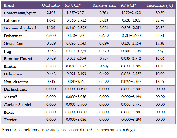
82 (21.92%) out of 374 suspected cases, were found positive for various types of CA. The most observable symptoms in presented dogs were exercise intolerance (39.02%), dyspnoea (12.19%), overweight 12.19%, and ascites 17.07%. The incidence of CA was highest in Pomeranian (30.70%) and lowest in Pug (9.67%). Whereas no CA was found in Dachschund, Mastiff, Cocker spaniel and Boxer breeds. On further analysis of risk factor and association at 95% confidence interval, the value of odd ratio and relative risk was highest in Pomeranian when compared with other breeds (Table 1).
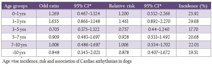
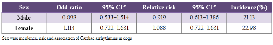
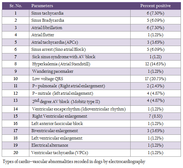
The highest incidence (29.68%) of CA was recorded in dogs of 1–3 year age group followed by 0–1years (25.92%), 7–10 years (22.03 %), 5–7 years (20.68 %), >10 years (19.51%) and 3–5 years (17.70 %) age groups. The sex wise analysis of data reveled higher incidence and risk with positive association of CA in female (22.98%) than males (21.13%). The detailed age wise and sex wise values of relative risk and odd ratio at 95% CI has been presented in Table 2 and 3. Cardiac Abnormalities Detected by ECG A total of 20 types of CA have been detected by ECG with most common finding of low voltage QRS complexes in 20.73% dogs (Table 4).
Biochemical Analysis The plasma concentration of chloride (Cl–) was found significantly (P < 0.05) elevated in animals with CA as compared to the healthy dogs. However, the plasma concentration of sodium (Na+), potassium (K+), calcium (Ca++), phosphorus (P), magnesium (Mg++), blood urea nitrogen (BUN) and creatinine in diseased animals were found statistically non–significant when compared with healthy dogs (Table–5).

DISCUSSION
The overall incidence of various types of CA in the present study was 21.92% (82/374) however much less incidence (7.67%) was reported in dogs (Sarita, 2008). Similarly, in another study 11% of dogs showed reliable signs of heart diseases and 9% had possible heart disease (Detweiler and Patterson, 1965). In the present study, the highest incidence of CA was found in Pomeranian when compared with other breeds. Our findings are in agreement with earlier studies in which the prevalence of cardiac arrhythmia and conduction disturbances in different breeds in India was found highest in Pomeranian (Changkija, 2007; Sarita, 2008). The possible reason for higher risk and incidence of CA in Pomeranian breed in present and other studies in India could also be due to more number of dogs of this particular breed presented due to preference of Indian dog lovers for Pomeranian than other breed of dogs.
On further analysis of breed wise risk factor, odd ratio values in case of Pomeranian indicate that they were 2.103 times more prone to CA when compared with other breeds of study. Similarly the value of relative risk in Pomeranian (1.764), German shepherd (1.081) and Labrador (1.033) indicated positive association of these breeds with CA. The risk of developing a particular type of heart diseases varies with breeds (Gülanber et al., 2005).
The age wise analysis of data revealed highest incidence of CA in 1–3 years of age group, followed by 0–1 years, 7–10 years, >10 years with lowest in 3–5 year age group for CA. Similarly the risk of development of cardiac diseases is more in 1–3 years of age group followed by 0–1 and 7–10 years of age group with relative risk value of more than 1 indicating positive association of these age groups with the CA. In another study, the highest age wise prevalence was in the 0–3 years (46.4%) followed by 31.1% in above 9 years of age (Sarita, 2008). However, the prevalence of arrhythmia as 80% in pups (up to 2month), 65.34% (2–6months), 58.75% (6 months to 1 year), 51.29% (1–3 years), 57.15% (3–5 years), 52.94% (5–10 years) and 57.69% in dogs of above 10 years of age (Changkija, 2007). In the present study the incidence as well as risk of development of CA was observed to be higher in females than the males. In contrast to our findings, the CA was higher in males (69.0%) than females (31.0%) in earlier study (Sarita, 2008).
The most common CA observed on ECG in present study was low voltage QRS complex followed by hyperkalemia, sinus arrhythmia and right ventricular enlargement. In another study of cardiac arrhythmia and conduction disturbances in normal canine population in India, 56.67% dogs showed various types of arrhythmias with sinus arrhythmia as most prevalent 45.97%, followed by supraventricular arrhythmia (6.10%), conduction disturbances (3.35%), multiple changes (0.83%) and ventricular arrhythmias in 0.42% dogs (Changkija, 2007).
The low voltage complex is suggestive of pericardial effusion and could also be found in cases of ascites (Tilley, 1985). The higher percentage of low voltage in present study correlate well with clinical signs as all dogs showing low voltage were found to be affected with ascites (Pandian, 2005). The hyperkalemia atrial standstill was recorded in 14.63% dogs in the present study. Contrary to our findings, atrial standstill is reported to be rare (Ettinger et al., 2000). In the present study, average potassium concentrations in dogs with CA were found to be higher than healthy dogs (4.12±0.23 and 3.10±0.31 mEq/L) which could be the possible reason for higher number of atrial standstill cases.
The sinus arrhythmia (6.09% sinus bradycardia and 7.30% tachycardia) recorded in this study has also been recorded by various other workers as one of the most common form of arrhythmia in young animals (Varshney and Tiwari, 2002; Changkija et al., 2006). The Deep Q–wave (greater than 0.5 mV in lead II) found in the present study could be result of right ventricular enlargement (Tilley, 1985).
The other types of CA with lower percentage has been reported previously and include atrial fibrillation (Meurs et al., 2001), atrial tachycardia (Varshney and Chaudhary, 2007), sinus arrest (Kumar et al., 2011), wandering pacemaker (Saelinger et al., 2008), p–pulmonale (Eckeenfels and Tribs, 1979), second degree AV block –Mobitz type II (Mertin, 2007), left anterior fascicular block (Gordon et al., 1995), and biventricular enlargement (Tilley, 1985).
In the present study, the biochemical parameters of dogs with various CA were compared with healthy dogs. The serum concentrations of diseased and healthy for Na+, K+, Ca++, Mg++, Phosphorus, BUN and creatinine did not show any significant difference than the apparently healthy dogs except serum Cl– concentration. The Previous studies on clinico– pathological changes in tachyarrhythmias showed increase in serum concentration of Na+, Ca++ and decrease in K+ and Mg++ while dogs with bradyarrthymias showed increased serum concentration of Na+, K+ and Mg++(Gupta et al., 2007). In another study, a non–significant decrease in serum K+ and Mg++ was found in dogs with ventricular fibrillation than the healthy control whereas the serum Ca++ was found significantly decreased than the healthy dogs (Salerno et al., 1987). The plasma K+ concentration in dogs with CA of present study was found higher than healthy dogs. In dogs, a mild increase in plasma K+ may lead to a variety of atrio–ventricular conduction abnormalities and changes in QRS which may simulate right bundle branch block or left bundle branch block in ECG recording if K+ is in range of 8.4mEq/L (Fisch et al., 1963). Only extreme increase in Ca++ concentration produces arrhythmia under natural conditions, however mild increase plays minor role in production of arrhythmia (Surawicz and Gettes, 1971). Similarly the arrhythmias due to hypo or hypernatremia are rare in clinical conditions (Garcia–Palmieri, 1962). The role of Mg++ in production of arrhythmia is not clear though; the studies suggest that hypomagnesaemia may enhance the susceptibility for arrhythmia (Tomiyasu et al., 1998). However, magnesium proved to play a vital role in the management of atrial, junctional and ventricular arrhythmia (Ho, 2008).
CONCLUSION
In nut shell it can be concluded that CA affects the significant proportion of dog population morbidity. The Pomeranian breed being the most favored pet among Indian animal lovers, the incidence and risk related to CA was also found higher than the other breeds of dog. The various types of arrhythmia and conduction disturbances were higher in young population than in the older ones which in future may lead to higher incidence of cardiomyopathy (dilated or hypertrophic) even in small breeds of dogs in India. Contrary to the earlier reports of male being at increased risk of CA, it was observed in the present study that females can also be at higher risk of developing CA. There was no significant difference in the biochemical parameters between dogs with CA and healthy control dogs however dogs were found to suffer with different type of CA therefore it is concluded that presence of normal level of biochemical parameters does not rule out CA .
ACKNOWLEDGEMENT
The financial assistance given to the first author in the form of INSPIRE FELLOWSHIP for Ph.D research work by DST, GOI to conduct the study is thankfully acknowledged. We also acknowledge the support of Joint Director (Academic) and Director, Indian Veterinary Research Institute, Izatnagar, Bareilly for providing necessary facilities to carry out research work.
REFERENCES
Bonnett BN, Egenvall A, Hedhammar A, Olson P (2005). Mortality in over 350,000 Insured Swedish dogs from 1995–2000: I. Breed–, Gender–, Age– and Cause–specific Rates. Acta. Vet. Scand. 46: 105–120.
http://dx.doi.org/10.1186/1751-0147-46-105
PMid:16261924 PMCid:PMC1624819
Changkija B (2007). Electrocardiographic studies in dogs with reference to management of cardiac tachyarrhythmia by alternate drugs. Ph.D Thesis, Indian Veterinary Research Institute, Izatnagar, Bareilly, India.
Chankija B, Varshney JP, Gopinathan A (2006). Myocardial infarction in a Pomeranian dog–A case report. Indian J. Vet. Med. 26:158–159.
Detweiler DK, Patterson DF (1965). Prevalence and types of cardiovascular disease in dogs. Ann. NY. Acad. Sci. 127: 481–516.
http://dx.doi.org/10.1111/j.1749-6632.1965.tb49421.x
PMid:5217276
Dove RS (2001). Nutritional therapy in the treatment of heart disease in dogs. Alt. Med. Rev. 6: S38–S45.
PMid:11591172
Eckeenfels A, Trib G (1979). The normal electrocardiogram of the conscious Beagle dog. Toxicol and Appl. Pharmacol. 47: 567–584.
http://dx.doi.org/10.1016/0041-008X(79)90527-1
Egenvall A, Bonnett BN, Olson P, Hedhammar A (2000). Gender, age and breed pattern of diagnoses for veterinary care in insured dogs in Sweden during 1996. Vet. Rec. 146 (19): 551–557.
http://dx.doi.org/10.1136/vr.146.19.551
PMid:10839449
Ettinger SJ, Le Bobinnec G, Cote E (2000). Electrocardiography. In: Textbook of Veterinary Internal Medicine: Diseases of Dog and Cat, Volume, I. 5th ed, Philadelphia, USA: W.B. Saunders, PP. 800–883.
Fisch C, Feigenbaum H, Bowers JA (1963). The effect of potassium on atrioventricular conduction of normal dogs. Am. J. Cardiol. 11: 487–492.
http://dx.doi.org/10.1016/0002-9149(63)90009-2
Garcia–Palmieri MR (1962). Reversal of hyperkalemic cardiotoxicity with hypertonic saline. Am. Heart. J. 64: 483–488.
http://dx.doi.org/10.1016/0002-8703(62)90033-9
Gaur T, Varshney JP (2002). Studies on hypertension in dogs. Indian J. Vet. Med. 22: 40–41.
Gordon BE, Tilley LP, Raymond RM (1995). Left ventricular function in a dog with left anterior fascicular block. Lab. Anim. Sci. 45: 686–689.
PMid:8746532
Gülanber EG, Gönenci R, Kaya U, Aksoy O, Biricik HS (2005). Vertebral Scale System to Measure Heart Size in Thoracic Radiographs of Turkish Shepherd (Kangal) Dogs. Turk. J. Vet. Anim. Sci. 29: 723–726.
Gupta DK, Singh JL, Kumar M (2007). Clinico–pathological changes in cardiac arrhythmia in dogs. Indian J. Vet. Med. 27: 91–94.
Ho KM (2008). Intravenous magnesium for cardiac arrhythmias: jack of all trades. Magnes. Res. 21:65–68.
PMid:18557136
Kumar A, Mahendran K, Dey S (2011). Sinoatrial block in a Dog and its Management. Intas Polivet. 12: 274–275.
Kumar YRS, Roa PM, Yathiraj S (2003). Electrocardiography studies in different age groups of dogs. Indian Vet. J. 80: 1125–1127.
Manczur F, Hetyey C, Reczigel J (2003). Occurrence of canine cardiological diseases in Hungary (1997–2000). Magy. Allatorvosok. 125: 669–682.
Mattera G, Vanoli E, Loi FM, Martinez V, Borsini F (2012). Borsini Detecting Drug–induced QT Interval. Prolongation in Healthy Dogs: A Practical Approach. Webmed. Central Pharmacol. 3: 3615.
Mertin M (2007). Small Animal ECGs: An Introductory Guide. 2nd ed. Oxford: Blackwell Publishing Ltd.
http://dx.doi.org/10.1002/9780470692080
Meurs KM, Miller MW, Wright NA (2001). Clinical features of dilated cardiomyopathy in Great Danes and results of pedigree analysis: 17 cases (1990–2000). J. Am. Vet. Med. Assoc. 218: 729–732.
http://dx.doi.org/10.2460/javma.2001.218.729
PMid:11280406
Pandian JS (2005). Clinical and ultrasonographic investigation of ascites in dogs. M.V.Sc. Thesis, Kerala Agriculture University, Thrissur, India.
Parker HG, Meurs KM, Ostrander EA (2006). Finding cardiovascular disease gene in the dog. J. Vet. Cardiol. 8:115–127.
http://dx.doi.org/10.1016/j.jvc.2006.04.002
PMid:19083345 PMCid:PMC3559124
Saelinger CA, Estrada AH, Maisenbacher HW (2008). ECG of the month. Exaggerated sinus arrhythmia and wandering pacemaker. J. Am. Vet. Med. Assoc. 233: 231–233.
http://dx.doi.org/10.2460/javma.233.2.231
PMid:18627224
Salerno DM, Elsperger KJ, Helseth P, Murakami M, Chepuri V (1987). Serum potassium, calcium and magnesium after resuscitation from ventricular fibrillation: a canine study. J. Am. Coll. Cardiol. 10: 178–85.
http://dx.doi.org/10.1016/S0735-1097(87)80177-8
Sarita D (2008). Epidemiological studies of canine cardiac diseases including clinicopathology, diagnosis and therapeutic. M.V.Sc Thesis, Anand Agriculture University, Gujarat, India.
Snedecor GW, Cochran WS (1994). Statistical Methods. 9th ed. IOWA State University Press Ames: IOWA.
Surawicz B, Gettes LS (1971). Effect of electrolyte abnormalities on the heart and circulation. In:Cardiac and Vascular Disease. Conn HL and Jr Horwitz O. Lea and Febiger, Philadelphia, USA, pp.539–576.
Tilley LP (1985). Analysis of P–QRS–T deflections. In: Essentials of canine and feline electrocardiogrphy. 2nd ed., Lea and Febiger, Philadelphia, USA, pp. 57–97.
Tomiyasu T, Chishaki A, Nakamura M (1998). Magnesium deficiency in adult rats promotes the induction of ventricular tachycardia by the administration of epinephrine. Heart and Vessels. 13:122–131.
http://dx.doi.org/10.1007/BF01747829
PMid:10328182
Varshney JP, Chaudhuri S (2007). Atrial Paroxysmal Tachycardia in Dogs and its Management with Homeopathic Digitalis—two case reports. Homeopathy. 96: 270–272.
http://dx.doi.org/10.1016/j.homp.2007.08.017
PMid:17954385
Varshney JP, Tiwari P (2002). Electrocardiographic and clinic–biochemical features in trypanosomiasis in dog with natural infection of Trypanosoma evansi. J. Canine Develop. Res. 2:51–54.

