
Advances in Animal and Veterinary Sciences


Research Article

Incidence of the Most Common Surgical Affections Among Sheep and Goats Admitted to a Referral Hospital in the State of Kuwait
Haithem Ali Mohamed Ahmed Farghali1*, Khaleifa Khalaf Ali2*, Ashraf Ali Eldesoky Shamaa1
1Department of Surgery, Anesthesiology and Radiology, Faculty of Veterinary Medicine, Cairo University, Egypt; 2Public Authority for Agriculture Affairs and Fish Resources, Kuwait City, Kuwait.
Abstract | Sheep and goats are a generational legacy in the State of Kuwait. So, this study was designed to record surgical affections in sheep and goats admitted to a referral hospital in Kuwait. From the obtained data, a total number of 658 small ruminants (385 sheep and 273 goats, representing 58.51% and 41.49% respectively) suffered from different surgical affections were admitted to the hospital from May/2017 to May/2020. The incidence of these affections varied in both species and different ages. The highest incidence was recorded in sheep aged 3 to 4 years old (27%) and in goats aged less than one-year-old (35.5%). Female affected animals (61.6%) were more recorded in both species than male ones (38.4%). Sixty surgical affections were recorded among sheep and goats (14 congenital and 46 acquired affections representing 17.9% and 82.1% respectively). Numerous surgical affections were recorded among sheep and goats in the state of Kuwait with the highest incidence of urogenital system affections in both species followed by udder and teat, digestive, integumentary, musculoskeletal, abdominal wall, eye, ear, and respiratory systems affections in sheep, while it followed by musculoskeletal, integumentary, udder and teat, digestive, eye, abdominal wall, ear, and respiratory systems affections in goats. Congenital anomalies are more prevalent in goats than sheep. Metabolic, nutritional, and infectious surgical conditions were more common in sheep, however traumatic affections more in goats. While sheep and goats share many surgical affections due to their anatomical and physiological similarities, there is a distinct nature of each species that makes some disorders more dominant in one species than the other.
Keywords | Surgical affections, Sheep, Goats, Abscess, Dystocia, Mastitis, Urethral obstruction, Kuwait
Received | July 13, 2020; Accepted | September 08, 2020; Published | September 20, 2020
*Correspondence | Haithem Ali Mohamed Ahmed Farghali and Khaleifa Khalaf Ali, Department of Surgery, Anesthesiology and Radiology, Faculty of Veterinary Medicine, Cairo University, Egypt; Public Authority for Agriculture Affairs and Fish Resources, Kuwait City, Kuwait; Email: dr_haithem0@yahoo.com, vet-1@live.com
Citation | Farghali HAMA, Ali KK, Shamaa AAE (2020). Incidence of the most common surgical affections among sheep and goats admitted to a referral hospital in the State of Kuwait. Adv. Anim. Vet. Sci. 8(s2): 41-57.
DOI | http://dx.doi.org/10.17582/journal.aavs/2020/8.s2.41.57
ISSN (Online) | 2307-8316; ISSN (Print) | 2309-3331
Copyright © 2020 Farghali et al. This is an open access article distributed under the Creative Commons Attribution License, which permits unrestricted use, distribution, and reproduction in any medium, provided the original work is properly cited.
INTRODUCTION
Worldwide, small ruminants are considered as a major source of the production of meat, milk, fur, and leather, in addition to the historical importance for the Arabs, especially the Arab Gulf countries, who consider sheep and goats as distinct animals and have special significance in these areas (Lambertz et al., 2018). Sheep and goats share many diseases and surgical injuries due to their closeness in anatomical and physiological features (Agrawal et al., 2014). However, there is a special nature of each species that makes some disorders more prevalent in one species than the other. The widespread of sheep and goats throughout the world with different breeds, rearing methods, and their purposes, as well as climatic conditions and the nature of nutrition, has led to a difference in the distribution and nature of surgical affections that afflict such animals, from one locality to another (Linklater and Smith 1993; Abdel-Hady et al., 2015). Sheep and goats are an important sector of livestock, food security, and a generational legacy in the state of Kuwait (homeland of the second author), where the number of these animals reached to 731.845 sheep and 182.039 goats in 2016 (Central Statistical Bureau, Kuwait, 2016; Ali et al., 2020a). The importance of these animals despite the absence of a faculty of veterinary medicine in the State of Kuwait and the poverty of published studies on the small ruminants’ field and surgical affections attracted the attention of the authors to the importance of studying, recording, and documenting of such affections in the State of Kuwait (Farghali et al., 2020).
Therefore, this study was designed to record the field surgical affections in sheep and goats admitted to referral hospital belonging to a public authority for agriculture affairs and fish resources in the State of Kuwait and to study which is more common in each species with their distributions among different body systems.
MATERIALS AND METHODS
Data collections
The present study was carried out on 658 small ruminants (385 sheep and 273 goats) who suffered from different surgical affections were admitted to a referral hospital belonging to a public authority for agriculture affairs and fish resources Kuwait from May/2017 to May/2020.
Establishment of the surgical affections and classification according to the affected system
Case history and clinical examination
The owner complaints and the full case history were taken from the animal’s owner according to previously designed sheet. The data regarding age, sex, species, breed, time of onset of the disease, previous interventions, and general health conditions were recorded (Pugh et al., 2020). Clinical signs including any changes in the animal behavior, appetite, nature of excretion and secretions, locomotion disorders, swellings, and expressions of pain and other alignments were recorded. General and local visual examinations of each case were performed for the detection of any structural and/or functional disorders of the affected region. Physical palpation of the affected parts and/or lesions was done to detect their nature, consistency, and tenderness (Abdel-Hady et al., 2015). The exploratory puncture was done whenever indicated to reveal the physical characters of the existence of fluids or contents in the examined lesions. General physical examination including pulse and respiratory rates, body temperature, and lymph nodes was performed to determine the health status of the animal. Digital photographs were obtained for each case. A complete description of the lesion was recorded to state the final diagnosis or directed to confirm the primary diagnosis using diagnostic tools (Matthews, 2016).
Diagnostic tools
The diagnostic imaging was selected according to the affections which needed to be confirmed. The diagnostic imaging tools used were radiography (Abu-Seida, 2014; Hashemiasl et al., 2016), ultrasonography (Gonzalez-Bulnes et al., 2010; Hakim et al., 2018), and endoscopy (Ali et al., 2019) which performed at the diagnostic imaging unit belonging to a public authority for agriculture affairs and fish resources in the State of Kuwait. Total blood picture and blood chemistry were conducted in cases when required to confirm the primary diagnosis (Pugh and Baird, 2012). Cytological examination of fluid obtained from exploratory puncture was done (Pugh et al., 2020). Microbiological examinations were performed in cases with septic conditions to confirm the diagnosis and state the pre and post-operative antibiotic of choice for treatment of infection. Diagnostic pathological examination was done when it is possible to confirm the primary diagnosis of some surgical conditions (Benavides et al., 2015).
The surgical affections of admitted sheep and goats were categorized according to the affected system (Linklater and Smith, 1993; Matthews, 2016).
RESULTS AND DISCUSSION
In the present study, a total number of 658 small ruminants (385 sheep and 273 goats, representing 58.51% and 41.49% respectively) (Table 1) suffered from different surgical affections were admitted to referral hospital belonging to a public authority for agriculture affairs and fish resources Kuwait in three years study period from May/2017 to May/2020.
Table 1: The number and the percentage of the affected sheep and goats.
| Total | Sheep | Goats |
| 658 | 385 | 273 |
|
100% |
58.51% | 41.49% |
The cases were distributed on the age of the affected animals as shown in (Table 2). The highest incidence was recorded in sheep aged 3 to 4 years old (27%) and in goats aged less than one-year-old (35.5%).
Table 2: The number and the percentage of the affected sheep and goats distributed among the age (per/years).
| Age | 0-1 | 1-2 | 2-3 | 3-4 | 4-5 | 5-6 | Total |
| Sheep | 61 | 34 | 82 | 104 | 68 | 36 | 385 |
| % | 15.8% | 8.8% | 21.3% | 27.0% | 17.7% | 9.4% | 100% |
| Goats | 97 | 18 | 43 | 63 | 29 | 23 | 273 |
| % | 35.5% | 6.6% | 15.8% | 23.1% | 10.6% | 8.4% | 100% |
| Total | 158 | 52 | 125 | 167 | 97 | 59 | 658 |
| % | 24.0% | 7.9% | 19.0% | 25.4% | 14.7% | 9.0% | 100% |
From the obtained data, male affected animals were 253 ones representing 38.4% (167 rams and 86 bucks representing 43.4% and 31.5% respectively). On the other hand, female affected animals were 405 ones representing 61.6% (218 ewes and 187 goats representing 56.6% and 68.5% respectively) (Table 3).
Table 3: The number and the percentage of the affected sheep and goats distributed among the gender.
| Gender | Male | Female | Total |
| Sheep | 167 | 218 | 385 |
| % | 43.4% | 56.6% | 100% |
| Goats | 86 | 187 | 273 |
| % | 31.5% | 68.5% | 100% |
| Total | 253 | 405 | 658 |
| % | 38.4% | 61.6% | 100% |
Regarding affected system, urogenital system (18.54%) (sheep, 20.5% and goats, 15.8%) was the highest affected one followed by udder and teat (14.29%) (sheep, 15.6% and goats, 12.5%), digestive system (13.37%) (sheep, 14.5% and goats, 11.7%), integumentary system (13.07%) (sheep, 12.2% and goats, 14.3%), musculoskeletal system (12.31%) (sheep, 10.1% and goats, 15.4%), abdominal wall (10.49%) (sheep, 10.6% and goats, 10.3%), eye (9.42%) (sheep, 8.3% and goats, 11.0%), ear (5.47%) (sheep, 4.2% and goats, 7.3%) and respiratory system (3.04%) (sheep, 3.9% and goats 1.8%) (Table 4).
Table 4: The number and the percentage of the affected sheep and goats distributed among the affected systems.
| Affected system | Sheep | % | Goats | % | Total | % |
| Urogenital system | 79 | 20.5% | 43 | 15.8% | 122 | 18.5% |
| Udder and teat | 60 | 15.6% | 34 | 12.5% | 94 | 14.3% |
| Digestive system | 56 | 14.5% | 32 | 11.7% | 88 | 13.4% |
| Integumentary system | 47 | 12.2% | 39 | 14.3% | 86 | 13.1% |
| Musculoskeletal system | 39 | 10.1% | 42 | 15.4% | 81 | 12.3% |
| Abdominal wall | 41 | 10.6% | 28 | 10.3% | 69 | 10.5% |
| Eye affections | 32 | 8.3% | 30 | 11.0% | 62 | 9.42% |
| Ear affections | 16 | 4.2% | 20 | 7.3% | 36 | 5.47% |
| Respiratory system | 15 | 3.9% | 5 | 1.8% | 20 | 3.04% |
| Total | 385 | 100% | 273 | 100% | 658 | 100% |
The urogenital affected cases in sheep (79 cases) were dystocia (38, 48.1%), vagina prolapse (19, 24.1%), scrotal hernia (12, 15.2%), chronic septic orchitis (4, 5.1%), penile affections (3, 3.8%), and scrotal hematocele (3, 3.8%), while the urogenital affected cases in goats (43 cases) were dystocia (21, 48.8%), hypospadias (5, 11.6%), vagina prolapse (4, 9.3%), penile affections (4, 9.3%), chronic septic orchitis (3, 7.0%), scrotal hematocele (3, 7.0%), and hermaphrodite (3, 7.0%) (Table 5, Figure 1).
Table 5: The numbers and the percentages of the urogenital system surgical affected cases among sheep and goats in the state of Kuwait.
| Urogenital system | Sheep | % | Goats | % | Total | % |
| Dystocia | 38 | 48.1% | 21 | 48.8% | 59 | 48.4% |
| Vagina prolapse | 19 | 24.1% | 4 | 9.3% | 23 | 18.9% |
| Scrotal hernia | 12 | 15.2% | - | - | 12 | 9.8% |
| Chronic septic orchitis | 4 | 5.1% | 3 | 7.0% | 7 | 5.7% |
|
Penile affections |
3 | 3.8% | 4 | 9.3% | 7 | 5.7% |
| Scrotal hematocele | 3 | 3.8% | 3 | 7.0% | 6 | 4.9% |
| Hypospadias | - | - | 5 | 11.6% | 5 | 4.1% |
| Hermaphrodite | - | - | 3 | 7.0% | 3 | 2.5% |
| Total | 79 | 100% | 43 | 100% | 122 | 100% |
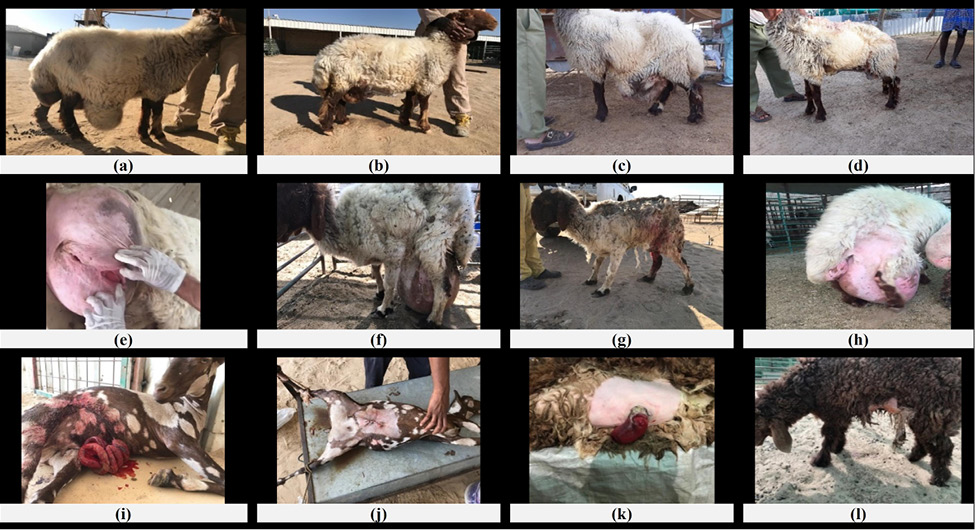
The udder and teat affected cases in sheep (60 cases) were chronic septic mastitis (27, 45.0%), gangrenous mastitis (19, 31.7%), supernumerary teats, (5, 8.3%), teat fistula (3, 5.0%), teat obstruction (5, 8.3%), and teat warts (1, 1.7%), while the udder and teat affected cases in goats (34 cases) were chronic septic mastitis (14, 41.2%), supernumerary teats (9, 26.5%), teat fistula (5, 14.7%), gangrenous mastitis (3, 8.8%), teat obstruction (2, 5.9%) and teat warts (1, 2.9%) (Table 6, Figure 2).
Table 6: The number and the percentage of the udder and teat surgical affected cases among sheep and goats in the state of Kuwait.
| Udder and teat | Sheep | % | Goats | % | Total | % |
| Chronic septic mastitis | 27 | 45.0% | 14 | 41.2% | 41 | 43.6% |
| Gangrenous mastitis | 19 | 31.7% | 3 | 8.8% | 22 | 23.4% |
| Supernumerary teats | 5 | 8.3% | 9 | 26.5% | 14 | 14.9% |
| Teat fistula | 3 | 5.0% | 5 | 14.7% | 8 | 8.5% |
| Teat obstruction | 5 | 8.3% | 2 | 5.9% | 7 | 7.4% |
| Teat warts | 1 | 1.7% | 1 | 2.9% | 2 | 2.1% |
| Total | 60 | 100% | 34 | 100% | 94 | 100% |
The digestive affected cases in sheep (56 cases) were ruminal foreign body (28, 50%), rectal prolapse (17, 30.4%), atresia ani (2, 3.6%), rectal carcinoma (3, 3.8%), abomasum displacement (3, 5.4%), ruminal fistula (2, 3.6%), and dental malocclusion (1, 1.8%), while the digestive affected cases in goats (32 cases) were ruminal foreign body (13, 40.6%), atresia ani (5, 15.6%), rectal prolapse (4, 12.5%), dental malocclusion (3, 9.4%), rectal carcinoma (2, 6.3%), ruminal fistula (2, 6.3%), tongue protrusion (2, 6.3%), and tongue tumor (1, 3.1%) (Table 7, Figure 3).
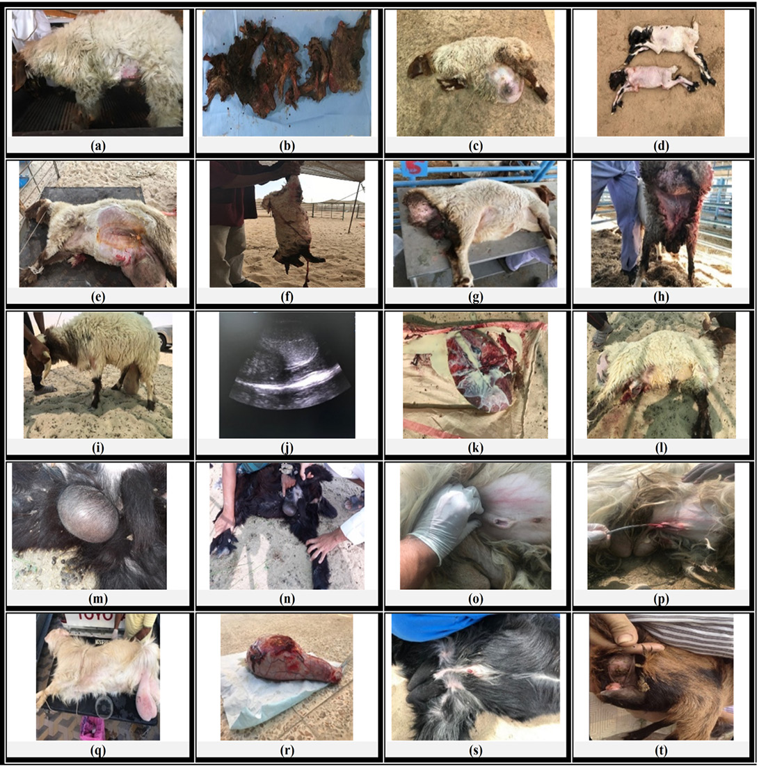
(a) A three years old ewe suffering from dystocia with anorexia and dullness. (b) After caesarean section of the same case, macerated foetus was recorded (which is a rare case in sheep) and the foetal bony parts were removed. (c) A four years old ewe suffering from dystocia and ventral abdominal hernia. (d) After caesarean section of the same case and hernia repair, the whole uterus was found in the hernia sac and contained two full term lamb. (e) A three and half years old ewe suffering from dystocia with anorexia and dullness. (f) After caesarean section of the same case, emphysematous foetus was recorded. (g) A two years old ewe suffering from vaginal prolapse before and, (h) after operation. (i) A three years old ram suffering from testicular swelling. (j) Ultrasongraphic examination of the same case tests showed hyperechogenic testicular parenchyma containing multiple anechoic patches. (k) The tests after open castration showed macroscopic features of chronic septic orchitis with accumulation of pus in multiple abscessations. (l) The ram after the castration. (m) and (n) A two years old buck suffering from urethral diverticulum. (o) A six months old buck suffering from congenital short penis. (p) The same case after excision of preputial excess skin. (q) A two years old buck suffering from left unilateral scrotal swelling. (r) The testis after castration showed unilateral scrotal haematocele. (s) A two months old buck suffering from hypospadias. (t) A three weeks old goat born as hermaphrodite.
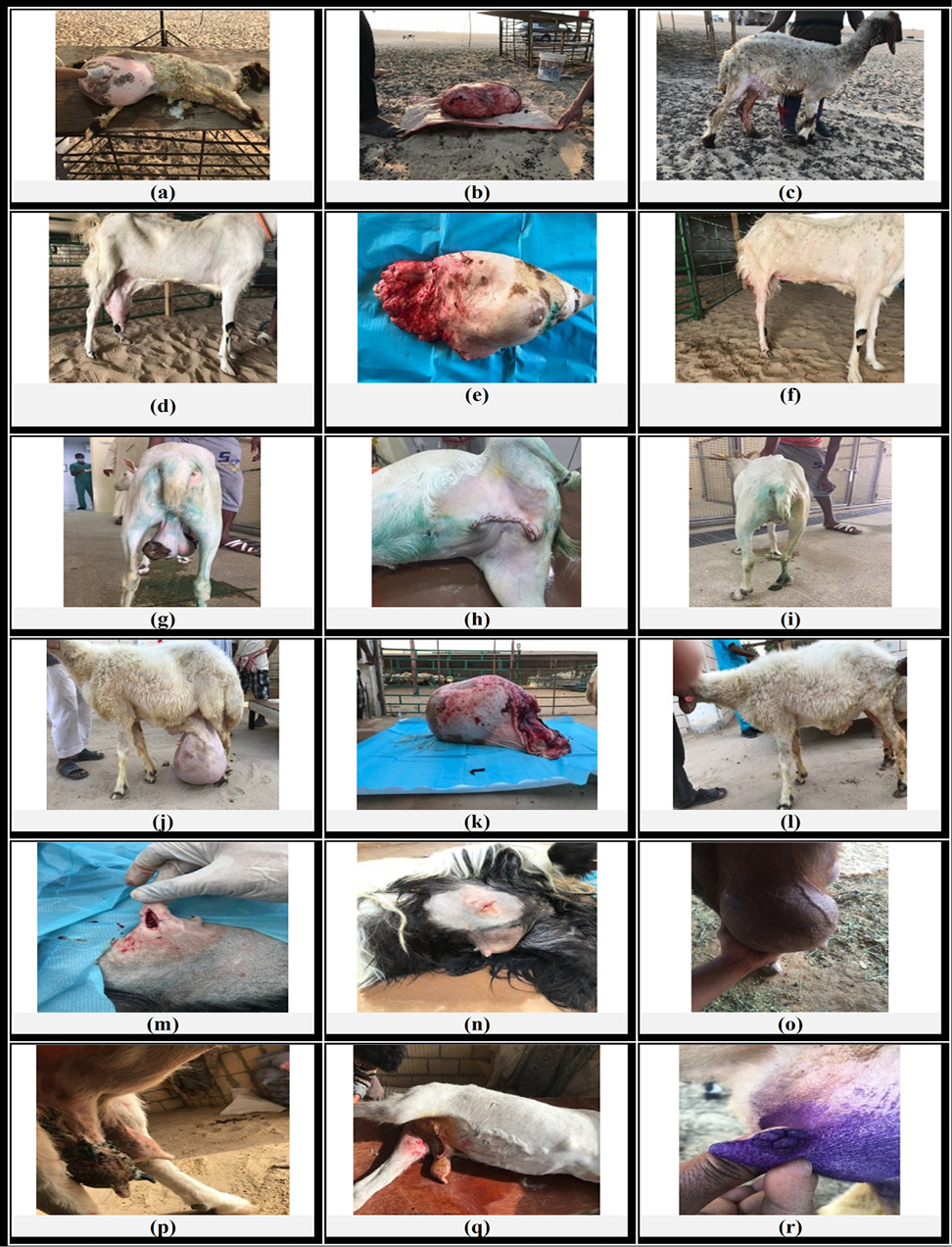
(a) A four years old ewe suffering from chronic septic mastitis. (b) The huge udder and (c) The ewe after total mastectomy. (d) A two years old goat suffering from chronic septic mastitis. (e) The udder and, (f) the goat after total mastectomy. (g) A three years old goat suffering from gangrenous mastitis. (h) and (i) after total mastectomy. (j) A three years old ewe suffering from gangrenous mastitis. (k) The udder weighting about 18 kg and (l) The same animal after total mastectomy. (m) A five years old goat suffering from teat fistula. (n) the same case refreshment and suturing of the fistula lips. (o) A one-year old goat suffering from teat obstruction (Clogged teat). (p) A three years old goat suffering from udder and teat warts. (q) The same case after unilateral mastectomy. (r) A five years old goat suffering from teat papilloma.
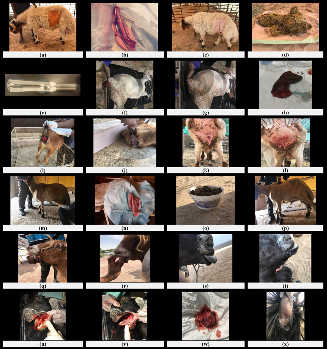
Figure 3: The digestive system surgical affected cases among sheep and goats in the state of Kuwait.
(a) A two years old ewe suffering from ruminal foreign body. (b) Rumenotomy of the same case. (c) The ewe postoperatively. (d) Two kg foreign body obtained from rumen. (e) The 75 mm selectable liner cutter with 3D staples (Ethison©, USA) used in the rumenotomy operation. (f) A three years old goat suffering from rectal prolapse. (g) The same case after resection of the (h) gangrenous prolapsed rectal part. (i) A nine months old goat suffering from partial rectal prolapse before and, (j) after reduction and application of purse string suture. (k) A three years old ewe suffering from partial rectal carcinoma pre and, (l) Post-operative. (m) A four years old ram suffering from left abomasopexy after incision and removing of (o) about 9 kg abomasal contains. (P) The same case after closure of the skin. (q) A three years old goat suffering from malocclusion of the incisor teeth. (r) The same case after teeth extraction. (s) A two years old Ardhi breed of the goat showing tongue portion (Sticking out of tongue). (t) The same case after surgical removal of hanging tongue portion. (u) A five years old goat suffering from tumour at the base of the tongue (Squamous cell carcinomn) before and (v) after surgical excision of (w) the tumour mass. (x) A three months old kid suffering from atresia ani.
Table 7: The number and the percentage of the digestive system surgical affected cases among sheep and goats in the state of Kuwait.
| Digestive system |
Sheep |
Goats |
Total |
|||
| Ruminal foreign body | 28 | 50.0% | 13 | 40.6% | 41 | 46.6% |
| Rectal prolapse | 17 | 30.4% | 4 | 12.5% | 21 | 23.9% |
| Atresia ani | 2 | 3.6% | 5 | 15.6% | 7 | 8.0% |
| Dental malocclusion | 1 | 1.8% | 3 | 9.4% | 4 | 4.5% |
| Rectal carcinoma | 3 | 5.4% | 2 | 6.3% | 5 | 5.7% |
| Ruminal fistula | 2 | 3.6% | 2 | 6.3% | 4 | 4.5% |
| Abomasum displacement | 3 | 5.4% | - | - | 3 | 3.4% |
| Tongue protrusion | - | - | 2 | 6.3% | 2 | 2.3% |
| Tongue tumor | - | - | 1 | 3.1% | 1 | 1.1% |
| Total | 56 | 100% | 32 | 100% | 88 | 100% |
The integumentary affected cases in sheep (47 cases) were skin neoplasms (20, 42.6%), abscess (15, 31.9%), wounds (6, 12.8%), cyst (3, 6.4%), abnormal horn growth (2, 4.3%), and horn fracture (1, 2.1%), while the integumentary affected cases in goats (39 cases) were abscess (9, 23.1%), cyst (9, 23.1%), skin neoplasms (8, 20.5%), wounds (8, 20.5%), and abnormal horn growth (5, 12.8%) (Table 8, Figure 4).
Table 8: The number and the percentage of the integumentary system surgical affected cases among sheep and goats in the state of Kuwait.
|
Integumentary system |
Sheep | % | Goats | % | Total | % |
| Skin neoplasms | 20 | 42.6% | 8 | 20.5% | 28 | 32.6% |
| Abscess | 15 | 31.9% | 9 | 23.1% | 24 | 27.9% |
| Wounds | 6 | 12.8% | 8 | 20.5% | 14 | 16.3% |
| Cyst | 3 | 6.4% | 9 | 23.1% | 12 | 14.0% |
|
Abnormal horn growth |
2 | 4.3% | 5 | 12.8% | 7 | 8.1% |
| Horn fracture | 1 | 2.1% | - | - | 1 | 1.2% |
| Total | 47 | 100% | 39 | 100% | 86 | 100% |
The musculoskeletal affected cases in sheep (39 cases) were fractures (22, 51.3%), hygroma (8, 2.6%), limb gangrene (3, 38.5%), arthritis (3, 7.7%) flexor angular deformities (2, 15.4%), and dislocation (1, 5.1%), while the musculoskeletal affected cases in goats (42 cases) were fracture (27, 64.3%), hygroma (5, 11.9%), limb gangrene (5, 11.9%), flexor angular deformities (3, 7.1%), arthritis (1, 2.4%), and dislocation (1, 2.4%) (Table 9, Figure 5).
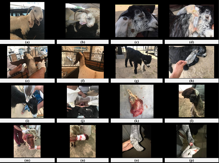
The abdominal wall affected cases in sheep (41 cases) were umbilical hernia (17, 41.5%), inguinal hernia (udder hernia) (11, 26.8%), ventral abdominal hernia (8, 19.5%), perineal hernia (4, 9.8%), and abdominal wall wound (intestinal prolapse) (1, 2.4%), while the abdominal wall affected cases in goats (28 cases) were ventral abdominal hernia (10, 35.7%), umbilical hernia (9, 32.1%), perineal hernia (8, 28.6%), and abdominal wall wound (intestinal prolapse) (1, 3.6%) (Table 10, Figure 6).
Table 9: The numbers and the percentages of the musculoskeletal system surgical affected cases among sheep and goats in the state of Kuwait.
| Musculoskeletal system |
Sheep |
Goats |
Total |
|||
| Fractures | 22 | 51.3% | 27 | 64.3% | 49 | 60.5% |
| Hygroma | 8 | 2.6% | 5 | 11.9% | 13 | 16.0% |
| Limb gangrene | 3 | 38.5% | 5 | 11.9% | 8 | 9.9% |
| Flexor angular deformities | 2 | 15.4% | 3 | 7.1% | 5 | 6.2% |
| Arthritis | 3 | 7.7% | 1 | 2.4% | 4 | 4.9% |
| Dislocation | 1 | 5.1% | 1 | 2.4% | 2 | 2.5% |
| Total | 39 | 100% | 42 | 100% | 81 | 100% |
Table 10: The numbers and the percentages of the abdominal wall surgical affected cases among sheep and goats in the state of Kuwait.
| Abdominal wall | Sheep | % | Goats | % | Total | % |
| Umbilical hernia | 17 | 41.5% | 9 | 32.1% | 26 | 37.7% |
| Ventral abdominal hernia | 8 | 19.5% | 10 | 35.7% | 18 | 26.1% |
| Perineal hernia | 4 | 9.8% | 8 | 28.6% | 12 | 17.4% |
| Inguinal hernia (udder hernia) | 11 | 26.8% | - | - | 11 | 15.9% |
| Abdominal wall wound (intestinal prolapse) | 1 | 2.4% | 1 | 3.6% | 2 | 2.9% |
| Total | 41 | 100% | 28 | 100% | 69 | 100% |
The sense organs affections among sheep and goats were recorded in eye and ear. The eye affected cases in sheep (32 cases) were pink eye (5, 15.6%), keratoconjunctivitis (4, 12.5%), hyphema (4, 12.5%), corneal dermoid (3, 9.4%), nictitating gland hypertrophy or prolapse (cherry eye) (3, 9.4%), entropion (3, 9.4%), eye tumor (2, 6.3%), cataract (2, 6.3%), lower eyelid wound (2, 6.3%), trichiasis (1, 3.1%), circumocular hypopigmentation (1, 3.1%), exophthalmos (1, 3.1%), and lower eyelid swelling (1, 3.1%), while the eye affected cases in goats (30 cases) were keratoconjunctivitis (6, 20%), eye tumor (4, 13.3%), pink eye (3, 10%), hyphema (3, 10%), corneal dermoid (3, 10%), nictitating gland hypertrophy or prolapse (cherry eye) (3, 10%), entropion (3, 10%), cataract (2, 6.7%), trichiasis (2, 6.7%), lower eyelid wound (1, 3.3%) (Table 11, Figure 7).
The ear affected cases in sheep (16 cases) were ear cartilage deformity (8, 50%), ear papilloma (warts) (2, 12.5%), ear hematoma (2, 12.5%), ear tumors (2, 12.5%), and ear abscess (2, 12.5%), , while the ear affected cases in goats (20 cases) were ear papilloma (warts) (12, 60%), ear hematoma (4, 20%), ear tumors (2,10%), and ear abscess (2, 10%) (Table 11, Figure 8).
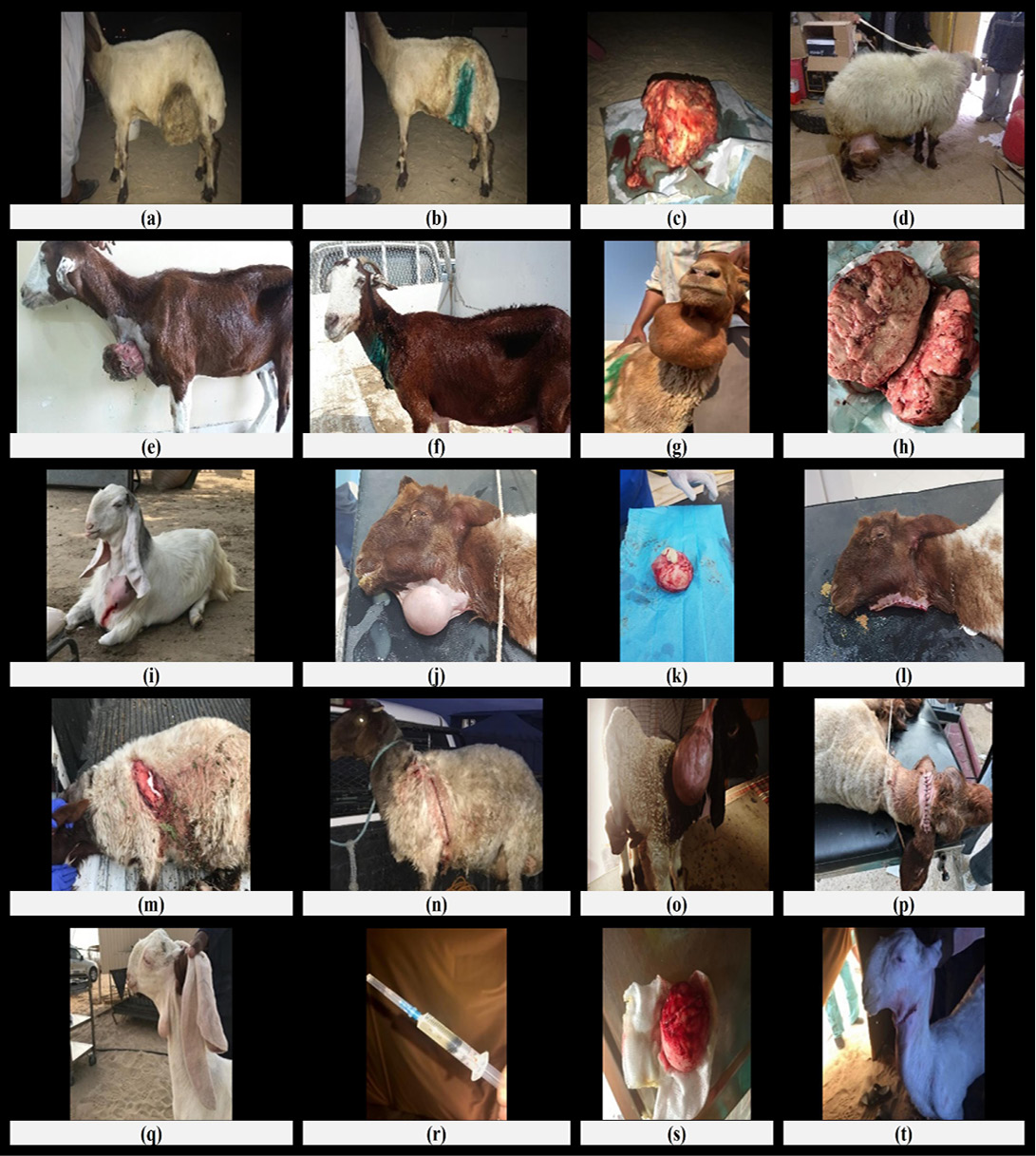
Figure 4: The integumentary system surgical affected cases among sheep and goats in the state of Kuwait.
(a) A five years old ewe suffering from cutaneous tumour at the level lift stifle fold. (b) The same case after surgical excision of (c) the huge tumour mass. (d) A five and half years old ewe suffering from cutaneous tumour at the level of right knee joint. (e) A four years old goat suffering from cutaneous tumour at the brisket region before. (f) after surgical excision. (g) A six years old goat suffering from submandibular suffering from perscapular abscess (Caseous lymphadenitis). (j) A three years old ewe suffering from submandibular abscess (Caseous lymphadenitis). (k) The circumscribed abscess after surgical excision and (l) The ewe postoperatively. (m) A four years old ewe suffering from left shoulder region recent incised skin would before and, (n) after suturing of the wound. (o) A two weeks old lamb suffering from subcutaneous cyst before and, (p) after surgical excision. (q) A three months old kid suffering from wattle cyst with (r) clear aspirated fluid. (s) the excised cyst and (t) the kid postoperatively.
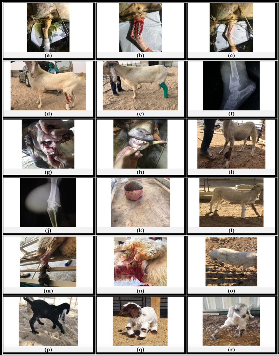
Figure 5: The musculoskeletal system surgical affected cases among sheep and goats in the state of Kuwait.
(a) A three years old ewe suffering from complete midshaft left tibial fracture. (b) Open reduction and internal fixation using extramedullary plate fixation. (c) The ewe postoperatively. (d) A two years old goat suffering from metatarsal fracture treated with (e) external fixation using poly acrylic cast (f) Radiographic image of a three years old goat suffering from complete distal shaft fracture of right tibia. (g) A four years old ram suffering from complete symphyseal fracture of the mandible treated with (h) cross pin technique. (i) A three years old ram suffering from carpal hygroma. (k) The circumscribed bursa and (l) The goat after surgical excision. (m) A four years old ewe suffering from right hind limb gangrene before and (n) after amputation (o) The same case two weeks postoperative. (p), (q) and (r) Three kids one month, seven and two days ages respectively suffering from Flexor Angular Deformities of the fore limbs.
(a) A three years old ram suffering from umbilical hernia (omphalocele) before and, (b) after surgical herniorrhaphy. (c) A four years old ewe suffering from ventral abdominal hernia before and, (d) after surgical herniorrhaphy. (e) A four years old ewe suffering from perineal hernia. (f) A six years old ewe suffering from inguinal hernia at the base of the udder before and, (g) after surgical repair of the hernia and mastectomy. (h) A three years old ewe suffering from inguinal hernia at the base of the udder. (i) A one-year old goat suffering from ventral abdominal wall wound (perforating wound) with intestinal prolapse without injury before and, (j) after closure of the abdominal wound. (k) A three months old lamb suffering from failure of umbilical closure with skin wound and abomasal prolapse before and, (l) after umbilical wound surgical repair.
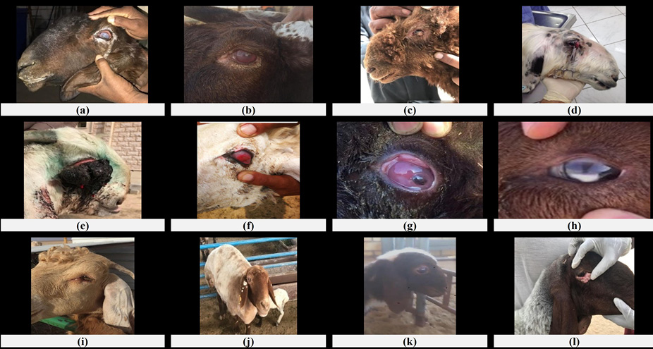
(a) A three years old buck suffering from superficial keratitis. (b) A two years old buck suffering from anterior uveitis and hyphaemia. (c) A four months old lamb suffering from corneal dermoid cyst. (d) A four years old buck suffering from 3rd eye lid hypertrophy and prolapse. (e) A six years old buck suffering from ocular neoplasm. (f) A six years old goat suffering from granuloma of the 3rd eye lid. (g) A two years old goat suffering from corneal ulcer complicated with corneal sequestration and granulation tissue. (h) A six years old goat suffering from mature cataract. (i) A four months old kid suffering from entropion. (j) A five years old ewe suffering from circumocular hypopigmentation. (k) A three months old lamb suffering from exophthalmia. (l) A one-year old ewe suffering from lower eye lid wound.
(a) A four years old goat suffering from warts on the outer surface of ear pinna before and, (b) after surgical excision of the warts. (c) A one-year old goat suffering from ear warts before and, (d) after cauterization. (e) A nine months old ewe suffering from ear pinna deformity before and, (f) after surgical correction. (g) A two weeks old kid suffering from ear hematoma before and, (h) after surgical intervention using incisional technique. (i) A six years old goat suffering from aural neoplasm before and, (j) after surgical excision. (k) The macroscopic features of the excised tumour similar to those of chondrosarcoma. (l) A four years old ewe suffering from tumour (squamous cell carcinoma) at the tip of the ear before and, (m) (n) after surgical ear cropping. (o) A two years old goat suffering from auricular abscess at the outer aspect of ear pinna before and, (p) after surgical opening and evacuation.
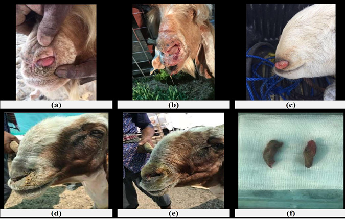
Figure 9: The respiratory system surgical affected cases among sheep and goats in the state of Kuwait.
(a) A three years old Pyrenean buck affected with enzootic nasal adenocarcinoma showing unilateral protrusion of fleshy mass. (b) The same case after radical surgical removing of the intranasal tumour mass. (c) A two years old Anglo-Nubian female goat suffering from unilateral fleshy mass protruded from the left nostril, persistent seromucous nasal discharge (d) A four years old ewe suffering from bilateral narrow nostril before and, (e) after surgical widening of the nostril. (f) The excised part of the nostril.
Table 11: The numbers and the percentages of the sense organs surgical (Eye and Ear) affected cases among sheep and goats in the state of Kuwait.
| Eye affections | Sheep | % | Goats | % | Total | % | |
| Keratoconjunctivitis | 4 | 12.5% | 6 | 20.0% | 10 | 16.1% | |
| Hyphema | 4 | 12.5% | 3 | 10.0% | 7 | 11.3% | |
| Pink eye | 5 | 15.6% | 3 | 10.0% | 8 | 12.9% | |
| Corneal dermoid | 3 | 9.4% | 3 | 10.0% | 6 | 9.7% | |
| Nictitating gland hypertrophy (cherry eye) | 3 | 9.4% | 3 | 10.0% | 6 | 9.7% | |
| Eye tumor | 2 | 6.3% | 4 | 13.3% | 6 | 9.7% | |
| Entropion | 3 | 9.4% | 3 | 10.0% | 6 | 9.7% | |
| Cataract | 2 | 6.3% | 2 | 6.7% | 4 | 6.5% | |
| Lower eyelid wound | 2 | 6.3% | 1 | 3.3% | 3 | 4.8% | |
| Trichiasis | 1 | 3.1% | 2 | 6.7% | 3 | 4.8% | |
| Circumocular hypopigmentation | 1 | 3.1% | - | - | 1 | 1.6% | |
| Exophthalmos | 1 | 3.1% | - | - | 1 | 1.6% | |
| Swelling lower eyelid | 1 | 3.1% | - | - | 1 | 1.6% | |
| Total eye affections | 32 | 100% | 30 | 100% | 62 | 100% | |
| Ear affections | Sheep | % | Goat | % | Total | % | |
|
Ear papilloma (warts) |
2 | 12.5% | 12 | 60% | 14 | 38.9% | |
| Ear cartilage deformity | 8 | 50% | - | - | 8 | 22.2% | |
| Ear hematoma | 2 | 12.5% | 4 | 20% | 6 | 16.7% | |
| Ear tumors | 2 | 12.5% | 2 | 10% | 4 | 11.1% | |
| Ear abscess | 2 | 12.5% | 2 | 10% | 4 | 11.1% | |
| Total ear affections | 16 | 100% | 20 | 100% | 36 | 100% | |
The respiratory affected cases in sheep (15 cases) were narrow nostril (12, 80%), and nasal adenocarcinoma (3, 20%), while the respiratory affected cases in goats (5 cases) were nasal adenocarcinoma (5, 100%) (Table 12, Figure 9).
Table 12: The numbers and the percentages of the respiratory system surgical affected cases among sheep and goats in the state of Kuwait.
| Respiratory system | Sheep | % | Goats | % | Total | % |
| Narrow nostril | 12 | 80% | - | - | 12 | 60% |
| Nasal adenocarcinoma | 3 | 20% | 5 | 100% | 8 | 40% |
| Total | 15 | 100% | 5 | 100% | 20 | 100% |
Regarding the overall surgical affected cases, the highest recorded affection among sheep was dystocia (9.9%) followed by the ruminal foreign bodies (7.3%), chronic septic mastitis (7.0%), fractures (5.7%), skin neoplasms (5.2%), vagina prolapse (4.9%), gangrenous mastitis (4.9%), umbilical hernia (4.4%), rectal prolapse (4.4%), abscess (3.9%), scrotal hernia (3.1%) and narrow nostril (3.1%). meanwhile, the highest recorded surgical affection among goats was fracture (9.9%) followed by dystocia (7.7%), chronic septic mastitis (5.1%), ruminal foreign body (4.8%), ear papilloma (warts) (4.4%), ventral abdominal hernia (3.7%), umbilical hernia (3.3%), abscess (3.3%), supernumerary teats (3.3%) and cyst (3.3%) (Table 13).
Table 13: The numbers and the percentages of the overall surgical affected cases among sheep and goats in the state of Kuwait.
| Affections | Sheep | % | Goats | % | Total | % |
| Dystocia | 38 | 9.9% | 21 | 7.7% | 59 | 9.0% |
| Fractures | 22 | 5.7% | 27 | 9.9% | 49 | 7.4% |
| Chronic septic mastitis | 27 | 7.0% | 14 | 5.1% | 41 | 6.2% |
| Ruminal foreign body | 28 | 7.3% | 13 | 4.8% | 41 | 6.2% |
| Skin neoplasms | 20 | 5.2% | 8 | 2.9% | 28 | 4.3% |
| Umbilical hernia | 17 | 4.4% | 9 | 3.3% | 26 | 4.0% |
| Abscess | 15 | 3.9% | 9 | 3.3% | 24 | 3.6% |
| Vagina prolapse | 19 | 4.9% | 4 | 1.5% | 23 | 3.5% |
| Gangrenous mastitis | 19 | 4.9% | 3 | 1.1% | 22 | 3.3% |
| Rectal prolapse | 17 | 4.4% | 4 | 1.5% | 21 | 3.2% |
| Ventral abdominal hernia | 8 | 2.1% | 10 | 3.7% | 18 | 2.7% |
| Supernumerary teats | 5 | 1.3% | 9 | 3.3% | 14 | 2.1% |
| Wounds | 6 | 1.6% | 8 | 2.9% | 14 | 2.1% |
| Ear papilloma (Warts) | 2 | 0.5% | 12 | 4.4% | 14 | 2.1% |
| Hygroma | 8 | 2.1% | 5 | 1.8% | 13 | 2.0% |
| Scrotal hernia | 12 | 3.1% | - | - | 12 | 1.8% |
| Cyst | 3 | 0.8% | 9 | 3.3% | 12 | 1.8% |
| Perineal hernia | 4 | 1.0% | 8 | 2.9% | 12 | 1.8% |
| Narrow nostril | 12 | 3.1% | - | - | 12 | 1.8% |
| Inguinal hernia (Udder hernia) | 11 | 2.9% | - | - | 11 | 1.7% |
| Keratoconjunctivitis | 4 | 1.0% | 6 | 2.2% | 10 | 1.5% |
| Teat fistula | 3 | 0.8% | 5 | 1.8% | 8 | 1.2% |
| Limb gangrene | 3 | 0.8% | 5 | 1.8% | 8 | 1.2% |
| Pink eye | 5 | 1.3% | 3 | 1.1% | 8 | 1.2% |
| Ear cartilage deformity | 8 | 2.1% | - | - | 8 | 1.2% |
| Nasal adenocarcinoma | 3 | 0.8% | 5 | 1.8% | 8 | 1.2% |
| Chronic septic orchitis | 4 | 1.0% | 3 | 1.1% | 7 | 1.1% |
| Penile affections | 3 | 0.8% | 4 | 1.5% | 7 | 1.1% |
| Teat obstruction | 5 | 1.3% | 2 | 0.7% | 7 | 1.1% |
| Atresia ani | 2 | 0.5% | 5 | 1.8% | 7 | 1.1% |
| Abnormal growth horn | 2 | 0.5% | 5 | 1.8% | 7 | 1.1% |
| Hyphema | 4 | 1.0% | 3 | 1.1% | 7 | 1.1% |
| Scrotal hematocele | 3 | 0.8% | 3 | 1.1% | 6 | 0.9% |
| Corneal dermoid | 3 | 0.8% | 3 | 1.1% | 6 | 0.9% |
| Nictitating gland hypertrophy (Cherry eye) | 3 | 0.8% | 3 | 1.1% | 6 | 0.9% |
| Eye tumor | 2 | 0.5% | 4 | 1.5% | 6 | 0.9% |
| Entropion | 3 | 0.8% | 3 | 1.1% | 6 | 0.9% |
| Affections | Sheep | % | Goats | % | Total | % |
| Ear hematoma | 2 | 0.5% | 4 | 1.5% | 6 | 0.9% |
| Hypospadias | - | - | 5 | 1.8% | 5 | 0.8% |
| Rectal carcinoma | 3 | 0.8% | 2 | 0.7% | 5 | 0.8% |
| Flexor angular deformities | 2 | 0.5% | 3 | 1.1% | 5 | 0.8% |
| Dental malocclusion | 1 | 0.3% | 3 | 1.1% | 4 | 0.6% |
| Ruminal fistula | 2 | 0.5% | 2 | 0.7% | 4 | 0.6% |
| Arthritis | 3 | 0.8% | 1 | 0.4% | 4 | 0.6% |
| Cataract | 2 | 0.5% | 2 | 0.7% | 4 | 0.6% |
| Ear tumors | 2 | 0.5% | 2 | 0.7% | 4 | 0.6% |
| Ear abscess | 2 | 0.5% | 2 | 0.7% | 4 | 0.6% |
| Hermaphrodite | - | - | 3 | 1.1% | 3 | 0.5% |
| Abomasum displacement | 3 | 0.8% | - | - | 3 | 0.5% |
| Lower eyelid wound | 2 | 0.5% | 1 | 0.4% | 3 | 0.5% |
| Trichiasis | 1 | 0.3% | 2 | 0.7% | 3 | 0.5% |
| Tongue protrusion | 0 | 0.0% | 2 | 0.7% | 2 | 0.3% |
| Dislocation | 1 | 0.3% | 1 | 0.4% | 2 | 0.3% |
| Abdominal wall wound (Intestinal prolapse) | 1 | 0.3% | 1 | 0.4% | 2 | 0.3% |
| Teat warts | 1 | 0.3% | 1 | 04% | 2 | 0.3% |
| Tongue tumor | - | - | 1 | 0.4% | 1 | 0.2% |
| Horn fracture | 1 | 0.3% | - | - | 1 | 0.2% |
| Circumocular hypopigmentation | 1 | 0.3% | - | - | 1 | 0.2% |
| Exophthalmos | 1 | 0.3% | - | - | 1 | 0.2% |
| Swelling lower eyelid | 1 | 0.3% | - | - | 1 | 0.2% |
| Total | 385 | 100% | 273 | 100% | 658 | 100% |
The congenital surgical affections among sheep (62 cases representing 16.1%) were Umbilical Hernia (17, 27.4%), Narrow Nostril (12, 19.4%), Ear Cartilage Deformity (8, 12.9%), Supernumerary Teats (5, 8.1%), Congenital Teat Obstruction (5, 8.1%), Congenital Cysts (3, 4.8%) Entropion (3, 4.8%), Corneal Dermoid (3, 4.8%), Atresia Ani (2, 3.2%), Flexor Angular Deformities (2, 3.2%), Dental Malocclusion (1, 1.6%), and Trichiasis (1, 1.6%), while the congenital surgical affections among goats (56 cases representing 20.5%) were Umbilical Hernia (9, 16.1%), Supernumerary Teats (9, 16.1%), Congenital Cysts (9, 16.1%), Atresia Ani (5, 8.9%), Hypospadias (5, 8.9%), Entropion (3, 5.4%), Corneal Dermoid (3, 5.4%), Flexor Angular Deformities (3, 5.4%), Dental Malocclusion (3, 5.4%), Hermaphrodite (3, 5.4%), Congenital Teat Obstruction (2, 3.6%), and Trichiasis (2, 3.6%) (Table 14).
Regarding the distribution of the affected cases among the causative group, metabolic and nutritional affections (36.2%) (sheep, 41.6% and goats, 28.6%) was the highest one followed by infectious group (19.8%) (sheep, 20.3% and goats, 19.0%), traumatic group (18.2%) (sheep, 14.3% and goats, 23.8%), congenital affections (17.9%) (sheep, 16.1% and goats, 20.5%) and neoplastic conditions (7.9%) (sheep, 7.8% and goats, 8.1%) (Table 15).
Table 14: The numbers and the percentages of the congenital surgical affected cases among sheep and goats in the state of Kuwait.
| Affections | Sheep | % | Goats | % | Total | % |
| Umbilical hernia | 17 | 27.4% | 9 | 16.1% | 26 | 22.0% |
| Supernumerary teats | 5 | 8.1% | 9 | 16.1% | 14 | 11.9% |
| Congenital cysts | 3 | 4.8% | 9 | 16.1% | 12 | 10.2% |
| Narrow nostril | 12 | 19.4% | - | - | 12 | 10.2% |
| Ear cartilage deformity | 8 | 12.9% | - | - | 8 | 6.8% |
| Congenital teat obstruction | 5 | 8.1% | 2 | 3.6% | 7 | 5.9% |
| Atresia ani | 2 | 3.2% | 5 | 8.9% | 7 | 5.9% |
| Entropion | 3 | 4.8% | 3 | 5.4% | 6 | 5.1% |
| Corneal dermoid | 3 | 4.8% | 3 | 5.4% | 6 | 5.1% |
| Hypospadias | - | - | 5 | 8.9% | 5 | 4.2% |
| Flexor angular deformities | 2 | 3.2% | 3 | 5.4% | 5 | 4.2% |
| Dental malocclusion | 1 | 1.6% | 3 | 5.4% | 4 | 3.4% |
| Hermaphrodite | - | - | 3 | 5.4% | 3 | 2.5% |
| Trichiasis | 1 | 1.6% | 2 | 3.6% | 3 | 2.5% |
| Total | 62 | 100% | 56 | 100% | 118 | 100% |
| Percentage out of total cases | 16.1% | 20.5% | 17.9% |
Table 15: The numbers and the percentages of the surgical affected cases among sheep and goats in the state of Kuwait distributed on the causative group.
| Affections | Sheep | % | Goats | % | Total | % |
| Metabolic and nutritional | 160 | 41.6% | 78 | 28.6% | 238 | 36.2% |
| Infectious | 78 | 20.3% | 52 | 19.0% | 130 | 19.8% |
| Traumatic | 55 | 14.3% | 65 | 23.8% | 120 | 18.2% |
| Congenital | 62 | 16.1% | 56 | 20.5% | 118 | 17.9% |
| Neoplastic | 30 | 7.8% | 22 | 8.1% | 52 | 7.9% |
| Total | 385 | 100% | 273 | 100% | 658 | 100% |
Effective project management in small businesses with a limited budget can obtain profit that could be considered high concerning the project unit expenses, especially when this unit has the multi-productivity power. According to this fact, the economic importance of small ruminant is established as a unit for setting up a small project with modest cost and great profits compared to large ruminants’ production as sources of meat, milk, fur, and leather which then directed to industrial productions (Kosgey, 2004). In the same direction, the goat is universally called “Poor man’s cow” (Iqbal et al., 2008). On the other hand, some societies consider sheep and goats a national heritage that reminds them of their ancient origins and civilizations of their country. This is clearly shown in the State of Kuwait, where the Arab Heritage Festival is held, and Kuwaitis celebrate their heritage and hold beauty competitions for many breeds of sheep and goats which indicate the importance of these animals as a generational legacy in the State of Kuwait (Farghali et al., 2020). According to the authors’ knowledge, there is no available literature that has studied the surgical affections among sheep and goats in the state of Kuwait.
The current study was conducted to illustrate and record the common surgical disorders among the sheep and goats aiming to set a map of such affections in the State of Kuwait under its environmental and managemental circumstances to be useful for global recording and studying of such conditions. To ensure the accuracy of the recorded incidence, the authors keened to do the study in a period of three-years in a referral hospital belonging to the public authority for agriculture affairs and fish resources, Kuwait.
From the obtained data, sheep (58.51%) were more frequently admitted with surgical affections than goats (41.49%). It may be contributed to the higher population of sheep (731.845) than goats (182.039) in the state of Kuwait (Central Statistical Bureau, Kuwait, 2016). Furthermore, these findings are agreed with many articles which stated that cattle and sheep had the tendancy to be affected more frequently than goats (Radostits et al., 2007).
Regarding the age of the affected animals, the incidence of the diseases varied with the ages. This is in agreement with the findings reported for surgical conditions affecting sheep and goats in several studies (Samad, 2000; Abdel-Hady et al., 2015). In the present study, the highest incidence was recorded in sheep aged 3 to 4 years old (27%) and in goats aged less than one-year-old (35.5%). It may have resulted from the frequent admission of congenitally affected cases in goats (20.5%) than in sheep (16.1%).
From the obtained data, female affected animals (61.6%) were more recorded in both species than male ones (38.4%). The findings parallel to that the highest affected system was urogenital (18.5%) (with the more prevalent female (67.3%) than male (32.7%) genital affections) followed by udder and teat affections (14.3%).
The urogenital system was the highest affected one in both species (sheep, 20.5%, and goats, 15.8%) because dystocia was the highest surgical indication of the admitted animals with nearly the same incidence in sheep (48.1%) and goats (48.8%). This similarity is in agreement with previous studies mentioned that generally, sheep and goats, as small ruminants, are very similar in several directions regarding reproduction performance even if there are little variations. The gestation period as will as number and delaviry procedure of offsprings seem to be parallel in both species as the initial phase of labor extending from 6 to 12 hrs., and the next phase from half to one hour with an incidence of dystocia may reach up to 50 % in these animals with more frequent incidence in ewes and does parteurate single and male offspring (Purohit, 2006; Ismail, 2017). The unforeseen low incidence of urine retention among sheep and goats in the present study in Kuwait was conflicting with several studies all over the world countries which stated that urine retention, resulted mainly from urolithiasis, is a common metabolic disease in all small ruminants (Pugh et al., 2020). On the other hand, these results parallel to those recorded in humans which mentioned that the incidence rate of a new stone former in Kuwait was significantly lower than that reported in Europe and the USA (El-Reshaid et al., 1997). This low incidence may be returned to the low rate of male sheep and goats’ castration and depending mainly on grazing feeding pattern in Kuwait which lowering urolithiasis formation. Another reason that may lead to the lacking of an accurate incidence rate is that the study was conducted in a referral hospital and that urine retention cases need interventions as soon as possible that may be carried out in small ruminant farms or in the nearest clinic.
In the present study, the affections of udder and teats ranked as the second affected system in sheep (15.6%) while in goats (12.5%) they were the fourth affected system. The findings which in the same direction of previous studies (Abdel-Hady et al., 2015). On the contrary, other research recorded that goats are more susceptible to udder and teat affections than sheep (Farghali, 2001). The most recorded cases belonging to udder and teat affections were chronic septic mastitis in both species. That is parallel to findings recorded in several articles which mensioned this affection as the most common udder affection in small ruminants which commonly caused by Staphylococcus spp. Other pathogens such as Streptococcus spp., Enterobacteriaceae, Pseudomonas aeruginosa, Mannheimia haemolytica, Corynebacteria, and fungi may contribute to the occurrence of the disease. Also, severe cases of mastitis related to incorrect preventative strategies have been attributed to the pathogens of Aspergillus fumigatus, Serratia marcescens, P. aeruginosa, or Burkholderia cepacian (Gonzalo et al., 2004; Abdel-Hady et al., 2015).
Regarding digestive system affections, it was the third affected system among sheep (14.5%), while in goats (11.7%), it was the fifth affected system. The superiority of sheep may be contributed to the higher incidence of ruminal foreign bodies in sheep (50%) than in goats (40.6%). The results which in agreement with previous researches which stated that among domestic ruminants, cattle are more susceptible to the development of ruminal impaction due to foreign materials followed by buffalo, sheep, and goat. (Priyanka and Dey, 2018). Goat is less predisposed to the ingestion of foreign bodies due to their well-developed sensitive prehensile organs and browsing and selective feeding behavior (Fromsa and Mohammed, 2011).
Another cause of increased incidence of digestive affections among sheep rather than goats was the higher repeating of rectal prolapse cases in sheep (30.4%) than in goats (12.5%). The findings that previously mentioned by other researches as rectal prolapse is more common in sheep than in goats (Pugh et al., 2020).
The integumentary system was the fourth affected system in sheep (12.2%), while in goats (14.3%), it was the third affected one. Meanwhile, sheep were more frequently affected with skin neoplasms (42.6%) and abscesses (31.9%) than goats (20.5% and 23.1%), the other recorded skin affections like wounds, cyst and horn malformations were higher in goats (20.5%, 23.1%, and 12.8%) than in sheep (12.8%, 6.4%, and 4.3%). Immune suppression, nutritional deficiencies, breed genetics, and arid conditions were predisposing factors of frequently recorded skin affections (Salib and Farghali, 2011).
The musculoskeletal system was placed as the fifth affected system in sheep (10.1%), while in goats (15.4%), it was the second affected one. In the current study, fracture cases put the goats (64.3%) in front of sheep (51.3%) as more musculoskeletal affected species. Fracture is the most common musculoskeletal surgical disorder in goats. These animals are frequently susceptible to fractures due to their abundant gathering in urban territories which predisposes them to trauma, canine bites leading to several musculoskeletal affections (Doijode, 2018).
Regarding abdominal wall affections, it was the 6th affected group in sheep (10.6%) and the 7th in goats (10.3%). In the present work, these affections could be divided into hernias (97.1%) and abdominal wall perforating wounds (2.9%). The recorded types of hernia were umbilical, ventral abdominal hernia, perineal hernia, and inguinal hernia (udder hernia) in addition to scrotal hernia (estimated with genital affections). These results parallel to previous studies stated that the types of hernia frequently recorded in ruminants are umbilical, abdominal, inguinal, scrotal, and diaphragmatic hernia (Abdel-Hady et al., 2015).
Sense organs (eye and ear) were more commonly affected in goats (11.0% and 7.3%) than in sheep (8.3% and 4.2%). These findings may be contributed to that most of the eye injuries are caused by foreign objects like thorns, bushes, stings, and another pointed object. Sharp traumas are caused by grass tips, bamboos, etc. Goats have more prominent eyes than sheep and do encounter such injuries while browsing or grazing (Khan et al., 2014) as well as the long pendulous goats’ ear.
The respiratory system seemed to be less commonly surgically affected among sheep and goats and the recorded affections were narrow nostrils (in sheep only) and nasal adenocarcinoma (3 sheep and 5 goats). Enzootic nasal adenocarcinoma in sheep and goats may be an economically critical contagious tumor of the nasal mucosal glands with a prevalence that may reach 10% in some localities (Ali et al., 2019). On the other hand, the narrow nostril of sheep is rare but Kuwait has sheep of Awassi breed which has a characteristic long head and frequently recorded narrow nostrils complain (Ali et al., 2020b).
From the overall obtained data, the highest recorded surgical affection among sheep was dystocia (9.9%) followed by a ruminal foreign body (7.3%), chronic septic mastitis (7.0%), fractures (5.7%), skin neoplasms (5.2%), vagina prolapse (4.9%), gangrenous mastitis (4.9%), umbilical hernia (4.4%), rectal prolapse (4.4%), abscess (3.9%), scrotal hernia (3.1%) and narrow nostril (3.1%). Meanwhile, the highest recorded surgical affection among goats was fracture (9.9%) followed by dystocia (7.7%), chronic septic mastitis (5.1%), ruminal foreign body (4.8%), ear papilloma (warts) (4.4%), ventral abdominal hernia (3.7%), umbilical hernia (3.3%), abscess (3.3%), supernumerary teats (3.3%) and cyst (3.3%).
Regarding the recorded congenital affections, fourteen affections were recorded and goats (20.5%) seemed to be more commonly affected with anomalies than sheep (16.1%). The findings in agreement with previous research (Pugh et al., 2020).
When the authors classified the cases according to causes, we found that the most common causes were belonging to metabolic and nutritional causes (41.6% in sheep and 28.6% in goats) followed by infectious causes in sheep (20.3%) and traumatic ones in goats (23.8%). The results clarified that sheep had a higher tendency to be affected by metabolic, nutritional, and infectious diseases than goats (Radostits et al., 2007). On the other hand, goats had a higher tendency to be affected with traumatic affections than sheep due to their svelte conformations, and the goat’s natural curiosity and independence, make them tend to get into more trouble than sheep (Pugh et al., 2020).
CONCLUSION AND RECOMMENDATION
Numerous surgical affections were recorded among sheep and goats in the state of Kuwait with the highest incidence of urogenital system affections in both species followed by udder and teat, digestive, integumentary, musculoskeletal, abdominal wall, eye, ear, and respiratory systems affections in sheep, while it followed by musculoskeletal, integumentary, udder and teat, digestive, eye, abdominal wall, ear, and respiratory systems affections in goats. Congenital anomalies were more prevalent in goats than sheep. Metabolic, nutritional, and infectious surgical conditions were more common in sheep, however traumatic affections more in goats. While sheep and goats share many surgical affections due to their anatomical and physiological similarities, there is a distinct nature of each species that makes some disorders more dominant in one species than the other.
Author’s Contributions
Haithem Ali Mohamed Ahmed Farghali contributed to the conception and design of the work in addition to writing of the manuscript. Khalifah Khalaf Khatam Ali contributed to the collection of the data, clinical examination, and surgery. Ashraf Ali Eldesoky Shamaa contributed to the conception and design of the work in addition to the revision of the manuscript.
Conflict of interest
The authors have declared no conflict of interest.
REFERENCES

