
Advances in Animal and Veterinary Sciences


Research Article
Advances in Animal and Veterinary Sciences. 1 (4S): 45 – 50Special Issue–4 (Progress in Research on Viruses and Viral Diseases)
Prevalence of Midges: Potent Vectors for Bluetongue Virus Infection in West Bengal, India
Arkendu Halder1, Siddhartha Narayan Joardar1,*, Panchanan Parui2, Dhriti Banerjee2, Vikas Kumar2, Indranil Samanta1, Chandan Lodh3
- Department of Veterinary Microbiology; West Bengal University of Animal and Fishery Sciences; Post Office –Belgachia, Kolkata–700037
- Diptera Section, Zoological Survey of India, Post Office –New Alipore, Kolkata–700059, West Bengal, India
- Department of Veterinary Medicine, Ethics and Jurisprudence, West Bengal University of Animal and Fishery Sciences; Post Office –Belgachia, Kolkata–700037
*Corresponding author:joardar69@gmail.com
ARTICLE CITATION:
Halder A, Joardar SN, Parui P, Banerjee D, Kumar V, Samanta I and Lodh C (2013). Prevalence of midges; potent vectors for bluetongue virus infection in West Bengal, India. Adv. Anim. Vet. Sci. 1 (4S): 45 – 50.
Received: 2013–11–17, Revised: 2013–12–12, Accepted: 2013–12–12
The electronic version of this article is the complete one and can be found online at
(
http://nexusacademicpublishers.com/table_contents_detail/4/156/html
)
which permits unrestricted use, distribution, and reproduction in any medium, provided the original work is properly cited
ABSTRACT
With the aim of identifying the potent vector(s) of bluetongue virus (BTV) that is circulating in eastern Indian state, West Bengal, attempts were made to detect and isolate BTV from Culicoides midges, collected from localities having good number of sero–positive animals. Prevalence of various species of Culicoides in the state along with the dynamics of Culicoides population, their habitat and weather parameters were also studied to reveal their possible relation. Culicoides midges were collected from three different agro–climatic zones of West Bengal in different post monsoon months. Meteorological data of collection–months of that area were obtained and incorporated to investigate the environmental conditions at the collection time. Habitat and probable breeding areas of Culicoides sp. were also noted. For isolation of BTV, non–engorged midges were pooled to prepare sample and virus was isolated using embryonated chicken eggs, followed by isolation on cell culture (BHK–21). Confirmation of BTV was made by RNA–PAGE and RT–PCR. The present study reveals the vector competence of Culicoides schultzei that harbored BTV–16. Typical association between the dynamics of Culicoides population, their habitat and weather parameters were observed in the present study. It is concluded that West Bengal state possesses potent vector of bluetongue virus that warrants serious attention in near future.
INTRODUCTION
The family Ceratopogonidae (Order Diptera) currently includes 6056 species; of which 1322 species belong to the genus Culicoides (Borkent, 2012). Most of the Culicoides midges are hematophagous, and are particularly important as a biting nuisance to humans and to livestock as vectors of several arboviruses of domestic and wild animals. Bluetongue Virus (BTV) and African Horse Sickness Virus (AHSV) are both Culicoides–transmitted and are formally classified by OIE (Office International des Epizootics). Bluetongue is an infectious, inoculable, viral disease of sheep. Cattle and goats may harbor the virus; however, they generally fail to show pronounced clinical symptoms. The BTV belongs to the genus Orbivirus, family Reoviridae (Borden et al., 1971). BTV is transmitted by biting of blood–feeding insect vectors of the genus Culicoides spp. (Diptera: Ceratopogonidae) (Mellor, 2000). Moreover, a new animal virus, not previously detected in Europe and initially named "Schmallenberg Virus (SVB)”, is a member of the Simbu serogroup within the genus Orthobunyavirus (family Bunyaviridae) that are predominantly transmitted by biting midges, mainly Culicoides spp., and mosquitoes (Hoffmann et al., 2011; Rasmussen et al., 2012). Out of > 1300 species present worldwide; at least 39 species have been reported in India. Very few species of Culicoides have been demonstrated as vectors for the BTV, and the principal vectors varying geographically. Midges collected from Harayana, Punjab, Rajasthan and Himachal Pradesh was identified as C. oxystoma (Jain et al., 1988) and C. imicola and C. oxystoma were found to be prevalent in Tamil Nadu. A detailed description of vector–species responsible for transmission of BTV in India is not comprehensively studied and therefore requires future studies to investigate virus–vector relationships in India.
Unlike southern and western India, the BTV is not detected in the eastern part of the country. In fact, West Bengal state has not experienced any outbreak or active disease incidences so far. However, anti–bluetongue antibodies could be detected in sheep, goat and cattle population of eastern and north–eastern states of India, viz. West Bengal (Chakraborti et al., 2007, Biswas et al., 2009, Panda et al., 2011, Mandal et al., 2012), Assam (Joardar et al., 2013), Manipur (Joardar et al., 2012), thus might be considered as sub–clinically infected. Moreover, BTV was isolated from sheep of Kolkata Maidan pasture (in West Bengal) and serotyped as 15 and 21 (Joardar et al., 2009).
In general, developmental sites of Culicoides species are poorly known. They can breed in a wide range of soils, if they provide enough moisture and organic matter to allow the development of the larvae (Kettle, 1962). The large range of breeding sites can be divided into three principal categories (Meiswinkel et al., 2004): (i) the water–saturated soil ecotone between aquatic and terrestrial habitats, (ii) dung pats (fresh dung), and (iii) moist, decaying organic matter (including manure). The immature stages of Culicoides spp. usually live in surface soil layers (0–5 cm depth), and rarely are found deeper than 8 cm (Uslu and Dik, 2006).
There are reports about significant correlations between rainfall and Culicoides activities in Ukraine (Zhadnova, 1975), Nigeria (Dipeolu, 1976), China (Chen and Lin, 1977), South Africa (Jupp et al., 1980) and Taiwan (Lien and Chen, 1981). Impact of thermo–aero–helio–hygrological weather parameters on population dynamics was also reported by Campbell and Kettle (1979), and Baylis et al. (1998).
In this context, it is quite expedient to explore the complex interaction of virus, vector (Culicoides midges) and environmental factors of this state (West Bengal) in a holistic manner for better understanding of the disease spectrum of bluetongue. Knowledge of suitable breeding sites of each species, particularly those implicated in the transmission of parasites or pathogens, is essential to evaluate the risk in an area and therefore to contribute in the development of integrated control strategies.
The state of West Bengal (latitude 21038′ N to 27010′ N, longitude 85050′ E to 89050′ E) has been divided into six agro–climatic zones, viz. Hill, Tarai, Old alluvial, New Alluvial, Red latterite and Coastal saline. The environmental parameters, viz. rainfall, temperature and relative humidity being quite conducive for propagation of Culicoides sp. in West Bengal (Biswas et al., 2011), these midges breed profusely during the monsoons (May to September).
In order to identify the potent vector of BTV, circulating in West Bengal state and causing subclinical bluetongue, attempts were made to detect and isolate BTV from Culicoides midges that were collected from localities harboring good number of sero–positive animals.
MATERIALS AND METHODS
Sampling and Identification of Culicoides Midges
Culicoides midges were collected from three different agro–climatic zones of West Bengal (New Alluvial, Red Laterite and Coastal Saline) in different post monsoon months. The collection was performed by sweep netting in early morning, before sunrise or afternoon and just before the sunset. Meteriological data (temperature, relative humidity and rainfall) were obtained and incorporated to know the environmental condition at the collection time. The collected Culicoides midges were sent to Zoological Survey of India, Kolkata for species identification. The trapped insects were sorted under a light microscope and the identification was made based on the morphological characteristics of the midges (Sen and Dasgupta, 1959, Dasgupta, 1995).
Table 1: Details of Culicoides collection vis–a–vis meteorological data in respect of collection months
Habitat of Culicoides Midges
Detailed observations were made of the collection area of midges, as described below. Probable breeding area was also sought and noted.
Isolation and Identification of Bluetongue Virus from Culicoides Midges
Collection of Midges
Midges were collected in batches (n=6) from Agarpara region (22067'N, 88037'E) of 24–parganas district of West Bengal state. The area was chosen on the basis of presence of high percentage of sero–positive animals Midges were collected from large ruminant (buffalo) shed in the evening hours (4.30–6.30 pm) in the month of October using traditional sweep net.
Isolation of BTV from Midges
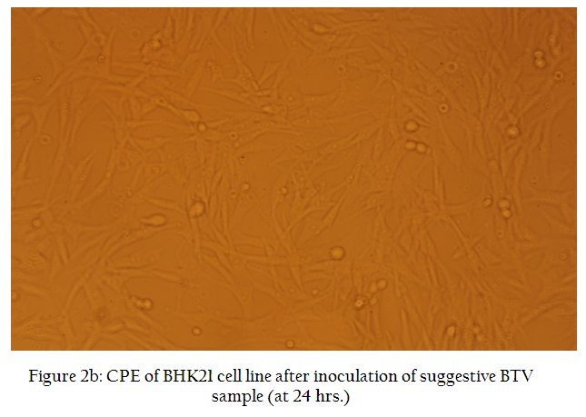
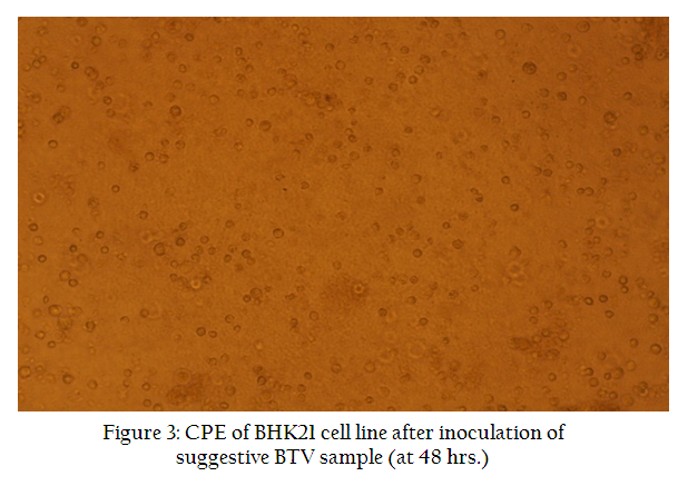
Each batch of non–engorged midges was pooled to prepare individual sample. The samples (n=3) each numbering up to 200 midges were ground in pestle and mortar with sterilized sand. After grinding, 10 ml of sterilized phosphate buffered saline (PBS, pH 7.2) was added into it and the cells were disrupted (at 0ºC) using ultrasonicator (Hielscher Ultrasonics GmBH, Germany) titanium probe, with the peak–to–peak wavelength of 8µ to 10µ for 40 cycles of one minute’s duration each, to release the virus. Intravenous inoculation of filtered aliquot (100µl) of the disrupted midge cell suspension into 10– to 12– day–old embryonated chicken eggs (ECE) was used to detect the presence of BTV (Foster and Luedke, 1968). For each sample, four ECE were used as test whereas two as control (where PBS, pH 7.2 was used instead of samples). The ECE were incubated at 33.5ºC for seven days. Embryos that died within seven days of inoculation showing subcutaneous haemorrhages were considered suspected. Embryos that died within 24 hours of inoculation were not considered for subsequent processing. Three blind passages in embryonated eggs were carried out for all the samples. Selected embryonic tissues, viz. spleen, kidney, heart, liver and muscle were collected and emulsified, using a sterilized pestle and mortar, with PBS having antibiotics (100IU/ml benzylpenicillin and 100µg/ml streptomycin sulphate) to make a 10% (w/v) suspension. This suspension was clarified by centrifugation and inoculated on to baby hamster kidney (BHK–21) confluent cell monolayer (Figure 2a) in 25cm2 tissue flask (Nunk) using Dulbecco’s modified Eagle’s medium (DMEM, Sigma, USA) supplemented with 10% fetal calf serum (Sigma, USA). Infected cells were observed under microscope for cytopathic effect (CPE) produced up to 72 hours post–infection (PI) and harvested for next cycle of infection. The harvested cells were filtered and 200 μl of inoculums were used to infect fresh monolayer. Thus three passages in BHK–21 cells were carried out for each case and observed for up to 72 hours PI for CPE.
Table 2: List of Culicoides spp. identified from different Districts and Agro–climatic zones of West Bengal
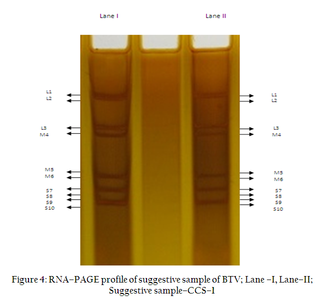
RNA–PAGE
Viral RNA was extracted from the BHK–21 cells using TRIzol® (Sigma, USA) (Chomczynski and Sacchi, 1987) and RNA–PAGE was performed as described by Squire et al. (1983).
RT–PCR
To further confirm the positivity of cell culture sample for BTV, the RT–PCR was performed using primers specific to the BTV NS1 gene. These primers (primer 1: 5’– GTT CTC TAG TTG GCA ACC ACC– 3’, primer 2: 5’– AAG CCA GAC TGT TTC CCG AT– 3’) (OIE, 2004), were used to amplify the partial sequence of NS1 gene.
RESULTS
Identification of Culicoides Midges
The midges collected in this study were identified as members of the Culicoides schultzei complex. The midges were medium sized flies with moderately hairy wings with numerous distinct pale spots including a pale spot over r–m cross–vein almost on the center of the vein. Paramere was broad at the base, curved and tapered towards the tip with apical hairs. These features are characteristics for Culicoides schultzei.
Habitat of Culicoides Midges
The principal habitat of different Culicoides spp. was mainly cattle or buffalo shed and decomposed manure. Besides, area–specific observation of the surroundings was noted during the study.
From Khatra (District– Bankura), midges were collected from cattle shed. The shed was situated just by the side of an open sewage disposal drain. In Belgachia (District Kolkata), Culicoides were found abundant in the cattle shed. Not only from cattle shed but also from nearby tree and bushes a huge number of midges was collected. It may be mentioned here that besides Culicoides schultzei,C. palpifer and C. definitus were also collected from Belgachia area. From Agarpara and Kamarhati (Dist –North 24 Parganas), Culicoides were collected from cattle and buffalo shed. The shed was surrounded by decomposed manure and straw. Open drain was present by the side of the shed and midges were present in both shed and decomposed manure. Sheep and goat sheds were the principal habitat of Culicoides in Sandeskhali (District North 24– Parganas). Maximum animal sheds were situated by the side of a river. Saline soil was present in the area. From Patelnagar (District– Birbhum), Culicoides were collected from cattle shed. The shed was situated by the side of a biogas plant. The whole area was covered with trees and bushes. In Hariharpara (District– Murshidabad), large number of sheep and goat sheds was presented in densely populated and congested area. The sheds were surrounded by bushes. Culicoides were found present in both the sheds and nearby area. From Malda Town (District– Malda), Culicoides were collected from organised cattle farm. Though it was a clean area, but surrounding sewage canals were open. Cow dung was also present in the farm premises. From all the above places Culicoides schultzei was collected.
From Nalpur (District–Howrah), Culicoides midges were collected from buffalo sheds and nearby bushes. Surrounding area was muddy and with water logged condition. From Panskura (District– Purba Mednipur), Culicoides was colllected from Buffalo shed. This was situated by the side of train tract. Surrounding muddy area is covered with bushes. More than one species of Culicoides were observed in these areas.
Isolation of BTV
Chicken embryos that died (in between 2 and 7 days of inoculation) in ECE showing subcutaneous haemorrhages (Figure 1) were suspected of having BTV. After processing, suspected samples (n=4) were inoculated in BHK–21 cell lines, one of the samples showed extensive CPE. Cellular destruction was evident as early as 24 hours, and the cells became enlarged, refractile and rounded (Figure 2b). After 48 hours inoculation, there was foamy degeneration, cell aggregation and vacuolation inside the cells (Figure 3). CPEs were seen throughout the monolayer by 72 hours post infection (PI), clumps of cells were detached from the flask and floated in the medium. This BHK–21 cell lysate sample (CCS–1) was chosen for further processing.
Figure 1: Pathognomonic lesion in chicken embryo after inoculating (I/V) suggestive sample of BTV from Culicoides
RNA–PAGE
Polyacrylamide gel electrophoresis of extracted RNA of the suggestive sample (CCS–1) showed 10–segmented profile characteristic of BTV (Figure 4).
RT–PCR
A band corresponding to 274bp was obtained in agarose gel when RT–PCR was performed using NS1 specific primers (Figure 5).
Figure 5: RT–PCR profile of suggestive sample in agarose gel; M– 100 bp DNA ladder; S– Sample (CCS–1)
DISCUSSION
With the aim of exploring the bluetongue epidemiology, attempts were made to study the role of Culicoides midges in BT infection in ruminants of West Bengal. In this direction, Culicoides biology along with virus isolation from haematophagous midges was sought. Culicoides midges were collected from different agro–climatic zones of West Bengal and identification up to species level was carried out with the help of Zoological Survey of India (ZSI). Culicoides species identified from three agro–climatic zones (new alluvial, red latterite and coastal saline) were Culicoides schultzei,Culicoides palpifer and Culicoides definitus. In India, BTV was isolated first time from Culicoides midges about 25 years ago. However, species was not identified (Prasad et al., 1988). Recently, from western state of India (Gujarat) BTV–1 was isolated from Culicoides oxystoma (Dadawala et al., 2011). In the present study, the BTV was isolated from C. schultzei collected from Agarpara area (North 24 District of West Bengal). The isolate was sent to AINP–BT (ICAR) typing centre (at Hissar) and found to be BTV–16. Previously, BTV–16 was isolated from sheep of Tamil Nadu, Maharashtra and Himachal Pradesh (Sreenivasulu et al., 2004). Recently, increasing trend for BTV–16 infection in different species of animals (sheep, goat and cattle) was observed from different states in India that suggest more studies on vector competence for transmission of infection (Anon, 2012). The present study confirms the vector competence of C. schultzei that harboured BTV–16. Recently, complete genome sequence of BTV–16 of goat origin from India was reported (Minakshi, 2012). Earlier, BTV–21 was isolated from Culicoides palpifer from Indonesia (Sendow et al., 1993).It may be mentioned here that Culicoides brevitarsis Keiffer and C. imicola Keiffer being proven vectors of BT virus, occur widely in India (Ilango, 2006). As virus transmitting species of Culicoides were observed in West Bengal, and high sero–positivity in ruminants were observed, the possibility of BTV circulation in West Bengal through vectors cannot be ruled out.
Prevalence of Culicoides midges and role in disease transmission were studied in Chittoor and Prakasam district of Andhra Pradesh during 2007 where disease was recorded previously during 2000 to 2003. A total of 1297 midges were collected using light trap method, out of which 982 (75.70%) were identified as female Culicoides and 315 (24.30%) as male species. Culicoides actoni, C. anophelis, C. inoxius, C. oxystoma and C. perigrinus were present in the catches (Reddy and Hafeez, 2008).
In the present study, habitat of collected midges was studied. The principal habitats were animal shed, surrounding manure and nearby bushes. Habitat, maximum and minimum temperature, rainfall and humidity of collection month were also observed in these districts from where midges were collected. It was noticed that collection of midges was huge just after winter and just after a rainfall. Maximum collection was done just before evening. Collection was low during too hot or cold climatic condition.
The prevalence of midges was attributed to the availability of breeding sites such as decaying vegetable matter, swamps, edges of ponds, boggy and semi–water logged areas, tree holes, rotten cacti etc. that is quite similar to the present findings. Well lighted cattle shed is one of the attractive sources of the flies which then adapted to feed on the same animal host. The water level of ponds, lakes, swamps roses high after the monsoon which helps the larva to migrate from slow making water to tamp soil, mud and other water logged heads and wooden particles etc. (Reddy and Hafeez, 2008).
It was established earlier that the environmental condition around the animal house with the heaps of manure, slow moving water, dazzling of rain fall in low lying areas are ideal breeding sites for Culicoides midges (Sen and Dasgupta 1959, Dasgupta 1995) which corroborates with the present findings. Besides these, wallow tanks, muddy sites of ditch, drainage, pond margin with moderate level of slow moving water incorporated with decaying vegetables or organic manure are more suitable for breeding of majority of Culicoides spp. (Howarth, 1985).
Typical association between the dynamics of Culicoides population, their habitat and weather parameters were observed in the present study. The seasonality in Culicoides population clearly indicated that monsoon season was most favorable for building up of population followed by winter. The sizeable population of Culicoides during winter season may act as a vector of BT disease which is of major concern in this area. These findings further reveal that the association of Culicoides with thermal and hygrological parameters of atmosphere and suggest that there are favourably good chances to control these parasites through improving the housing conditions of animals and regulation of their surrounding environment.
In short, it may be concluded that the eastern Indian state, West Bengal possesses potent vector of BTV that pose a menace and warrants control–based strategies.
ACKNOWLEDGEMENTS
Financial support from All India Network Programme on Bluetongue (Indian Council of Agricultural Research, New Delhi) is duly acknowledged. We are thankful to Dr. Minakshi, Principal Investigator, AINP–BT, Hissar centre for serotyping BTV isolate. Authors wish to thank the Vice Chancellor, West Bengal University of Animal and Fishery Sciences, Kolkata and the Director, Zoological Survey of India, Kolkata for providing necessary infrastructure facilities.
REFERENCES
Anon (2012). Annual Report of All India Network Programme on Bluetongue. 9th Annual Review Meeting. Indian Council of Agricultural Research, New Delhi. pp. 26.
Baylis M, Bouayoune H, Touti J and El–Hasnaoui H (1998). Use of climate data and satellite imagery to model the abundance of Culicoides imicola, the vector of African horse sickness virus in Morocco. Med. Vet. Entomol. 12: 255–266.
http://dx.doi.org/10.1046/j.1365-2915.1998.00109.x
PMid:9737597
Biswas M, Joardar SN, Samanta I, Isore DP and Aich R (2009). Seroprevalence of Bluetongue in West Bengal: Current Scenario. Indian J. Ani. Hlth. 48(1): 43–46.
Biswas M, Joardar SN, Samanta I, Isore DP, Aich R and Parui P (2011). Conducive environment for propagation of potent bluetongue vector exists in West Bengal. Indian J. Ani. Hlth. 50: 46–48.
Borden EC, Shope RE, Murphy FA (1971). Physical and morphological relationship of some arthropod borne viruses to bluetongue virus. J. Gen. Virol. 13: 262–271.
http://dx.doi.org/10.1099/0022-1317-13-2-261
Borkent, A., 2012. World Species of Biting Midges (Diptera: Ceratopogonidae). http://www.inhs.uiuc.edu/research/FLYTREE/CeratopogonidaeCatalog.pdf
Campbell MM and Kettle DS (1979). Abundance, temporal and spatial distribution of Culicoides brevitarsis Kieff (Diptera: Ceratopogonidae) on cattle in South East Queensland. Aust. J. Zool. 27: 60.
http://dx.doi.org/10.1071/ZO9790251
Chakraborti A, Lodh C, Joardar SN and Aich R (2007). Seroprevalence of bluetongue in West Bengal-current status. Indian J. Comp. Microbiol. Infect. Dis. 27: 63–64.
Chen CS and Lin YN (1977). A study on the seasonal succession of blood sucking midges (Culicoides spp.) at Nan Kang Taipei. J. Formosan Med. Assoc. 76: 631– 638
Chomczynski P and Sacchi N (1987). Single–step method of RNA isolation by acid guanidium thiocyanate–phenol–chloroform extraction. Anal Biochem. 162: 156–159.
http://dx.doi.org/10.1006/abio.1987.9999
http://dx.doi.org/10.1016/0003-2697(87)90021-2
Dadawala AI, Biswas SK, Rehman W, Chand K, De A, Mathapati BS, Kumar P,Cauhan HC,Chandel BS and Mondal B (2012). Isolation of bluetongue virus serotype 1 from Culicoides vector captured in livestock farms and sequence analysis of the viral genome sgment-2. Trans. Emeg. Dis. 59: 361–368.
http://dx.doi.org/10.1111/j.1865-1682.2011.01279.x
PMid:22151923
Dasgupta SK (1995). Morphotaxonomic features and species of Indian Culicoides. (Diptera: Ceratopogonidae). In: Prasad G. & Srivastava RN (eds), Bluetongue: Indian Perspective, HAU Press, Hisar, India. pp. 115–188
Dipeolu OO (1976). Species distribution and abundance of Culicoides Latr. (Diptera : Ceratopogonidae) in Nigeria. Bull. Entomol. Res. 66: 685–693.
http://dx.doi.org/10.1017/S0007485300010750
Foster N M. and Luedke AJ (1968). Direct assay for bluetongue virus by intravascular inoculation of embryonating chicken eggs. Am J Vet Res 29: 749–753.
PMid:4966159
Hoffmann A, Mettenleiter TC, Beer M (2011). Novel orthobunyavirus in cattle, Europe. Emerg. Infect. Dis. 18: 3.
Howarth FG (1985). Biosystemics of the Culicoides of Laos. Int. J. Entomol. 27: 1–96.
Ilango K (2006). Bluetongue outbreak in Tamil Nadu, Southern India: needs to study the Indian biting midge, vectors, Culicoides Lattreille (Diptera: Ceratopogoniae). Curr. Sci. 90: 163-167.
Jain NC, Prasad G, Gupta Y and Mahajan BK (1988). Isolation of bluetongue virus from Culicoides sp. In India. Rev. Sci. Tech. 7: 375–378.
Joardar SN, Lodh C, Chakraborti A, Baksi, S and Aich R (2009). Isolations of bluetongue serotypes 15 and 21 in West Bengal, India. Vet. Rec. 165: 751–752.
PMid:20023280
Joardar SN, Singh Ng I, Barkataki B, Lodh C, Chakraborti A and Pradhan NR (2012). Seroprevalence of bluetongue in ruminants of Manipur. Indian J. Comp. Microbiol. Immunol. Infect. Dis. 33: 54–56.
Joardar SN, Barkataki B, Halder A, Lodh C and Sarma D (2013). Seroprevalence of bluetongue in north eastern Indian state – Assam. Vet. World doi:10.5455/vetworld.2013:196–199.
Jupp PG, McIntosh BM and Nevill EM (1980). A survey of the mosquito and Culicoides faunas at two localities in the Karoo region of South Africa with some observations on the bionomics. Onderstepoort J. Vet. Res. 47: 1–6.
PMid:7454229
Kettle DS (1962). The bionomics and control of Culicoides and Leptoconops (Diptera, ceratopogonidae = Heleidae). Ann. Rev. Entomol. 7:401 –411.
http://dx.doi.org/10.1146/annurev.en.07.010162.002153
Lien JC and Chen CS (1981). Seasonal succession of some common species of genus Culicoides Latr. 1809 (Diptera: Ceratopogonidae) in Central Taiwan. J. Formosan Medical Assoc. 80: 673–682.
Mandal N, Joardar SN, Samanta I and Isore D (2012). Development of user–friendly diagnostic tool to detect anti–bluetongue antibodies. Indian J. Ani. Hlth. 51 (2): 63–68.
Meiswinkel R, Venter GJ and Nevill EM (2004). Vectors: Culicoides spp. In: Coetzer, J.A.W., Tustin, R. (Eds.), Infectious Diseases of Livestock., 2nd ed. Oxford University Press, Cape Town, pp. 93–136.
Mellor PS (2000). Replication of arboviruses in insect vectors. J. Comp. Pathol. 123: 231–247.
http://dx.doi.org/10.1053/jcpa.2000.0434
PMid:11041993
OIE 2004. Bluetongue. In Manual of Diagnostic Tests and Vaccines for Terrestrial Animals.www.oie.int/eng.normes/mmanual/A_0032.htm.Accessed August 10, 2003.
Panda M, Mondal A and Joardar SN (2011). Seroprevalence of bluetongue virus in sheep, goat and cattle in West Bengal, India. Ani. Sci. Rep. 5(3): 105–110.
Prasad M, Singh R, Ranjan K, Kumar P, Joshi CG, Reddy, YKM and Prasad G (2012). Complete genome sequence of bluetongue virus serotype 16 of goat origin from India. J. Virol. 86: 8337– 8338.
http://dx.doi.org/10.1128/JVI.01128-12
PMid:22787269 PMCid:PMC3421637
Reddy CVS and Hafeez Md (2008). Studies on certain aspects of prevalence of Culicoides species. Indian J. Anim. Sci. 78 92): 138– 142.
Sen P and Dasgupta SK (1959). Studies on Indian Culicoides. Ann Entomol Soc Am 52: 617–643.
Sendow I, Daniels PW, Soleha E, Erasmus BJ, Sukarishi, and Ronohardso P (1993). Isolation of bluetongue virus serotypes new to Indonesia from sentinel cattle from West Java. Vet. Rec. 133: 166 – 168.
http://dx.doi.org/10.1136/vr.133.7.166
PMid:8236706
Sreenivasulu V, Subba Rao MV, Reddy YN and Gard GP (2004). Overview of bluetongue disease, viruses, vectors, surveillance and unique features: the Indian subcontinent and adjacent regions. Vet. Ital. 40(3): 73–77.
PMid:20419638
Squire KRE, Chuang RY, Osburn BI, Knudson DL and Doi RH (1983). Rapid methods for comparing the double stranded RNA genomeprofiles of bluetongue virus. Vet Microbiol. 8:543–553.
http://dx.doi.org/10.1016/0378-1135(83)90003-2
Uslu U and Dik B (2006). Vertical distribution of Culicoides larvae and pupae. Med. Vet. Entomol. 20: 350–352.
http://dx.doi.org/10.1111/j.1365-2915.2006.00626.x
PMid:17044889
Zhadnova TG (1975). The activity of attack of blood sucking biting midges (Diptera: Ceratopogonidae) in relation to meteorological conditions in the left blank polesic of the Ukrane. Ve Strikzoologii 6: 58–64

