
Advances in Animal and Veterinary Sciences


Case Report

Pathological Femoral Fracture in a Doe Secondary to Osteomalacia: A Veterinary Clinical Case Report
Eric Lim Teik Chung1,2, Faez Firdaus Abdullah Jesse2,3*,Nurul Nadiah Mohamad1, Wan Mohd Sukri Wan Ishak3, Yusuf Abba4, Asinamai Athliamai Bitrus5, Innocent Damudu Peter3, 4, Idris Umar Hambali4, Mohd Azmi Mohd Lila2,3
1Department of Animal Science, Faculty of Agriculture, Universiti Putra Malaysia, 43400 Serdang, Selangor, Malaysia, 2Institute of Tropical Agriculture and Food Security, Universiti Putra Malaysia, 43400 Serdang, Selangor, Malaysia, 3Department of Veterinary Clinical Studies, Faculty of Veterinary Medicine, Universiti Putra Malaysia, 43400 Serdang, Selangor, Malaysia, 4Faculty of Veterinary Medicine, University of Maiduguri. P.M.B 1069 Maiduguri, Borno Nigeria, 5Research Unit, Microbial Food Safety and Antimicrobial resistance, Department of Veterinary Public Health, Faculty of Veterinary Science, Chulalongkorn University, 10330 Pathumwan Bangkok, Thailand.
Abstract | In this veterinary clinical case report highlights a clinical case of pathological femoral fracture in a doe secondary due to osteomalacia. An adult female Katjang goat aged 2 years old was presented to University Veterinary Hospital UPM with the complaint of recumbent and musculoskeletal problem. The doe weight was 18.3 kg with a body condition score of 2 out of 5. The history of the case indicates that the doe was found to fell through the floorboards and the left hindlimb was stucked and later the doe was on sternal recumbency and the most obvious sign was the swollen of the left hindlimb. Physical examination upon palpation revealed that crepitus and bone discontinuity can be felt at the left mid femur with warm and pain upon palpation. Diagnostic workouts of radiography and blood biochemistry were performed in this case. The result of blood biochemistry showed that the doe had hypocalcemia and hypophosphatemia which may indicate the doe suffering from a nutritional deficiency of vitamin D. The radiographic findings revealed bone discontinuity at the left mid femur and overriding of the proximal and distal femoral bone fractures with radiological diagnosis of oblique fracture of the left mid femur. Thinning of bone cortex and the presence of increased radiolucency was observed in bone medulla with radiological diagnosis of osteomalacia. Therefore, this clinical case was diagnosed as the doe suffered from pathological femoral fracture secondary to osteomalacia. The farmer was advised to cull the doe due to the welfare, poor prognosis of the case due to the severity and location of the fracture.
Keywords | Doe, Pathological fracture, Femur, Osteomalacia, Veterinary clinical case.
Received | January 29, 2019; Accepted | February 27, 2019; Published | April 23, 2019
*Correspondence | Faez Firdaus Abdullah Jesse, Department of Veterinary Clinical Studies, Faculty of Veterinary Medicine, Universiti Putra Malaysia, 43400 Serdang, Selangor, Malaysia; Email: jesse@upm.edu.my
Citation | Chung ELT, Jesse FFA, Mohammad NN, Ishak WMSW, Abba Y, Bitrus AA, Peter ID, Hambali IU, Lila MAM (2019). Pathological femoral fracture ina doe secondary to osteomalacia: a veterinary clinical case report. Adv. Anim. Vet. Sci. 7(6): 498-502.
DOI | http://dx.doi.org/10.17582/journal.aavs/2019/7.6.498.502
ISSN (Online) | 2307-8316; ISSN (Print) | 2309-3331
Copyright © 2019 Chung et al. This is an open access article distributed under the Creative Commons Attribution License, which permits unrestricted use, distribution, and reproduction in any medium, provided the original work is properly cited.
Introduction
Fracture is a break in the continuity of bone that may be caused by trauma, twisting due to muscle spasm, indirect loss of leverage or by a disease that result in decalcification of the bone (Blood et al., 2007). Pathological fracture occurs due to underlying bone or systemic disease such as nutritional deficiency, neoplasia or osteomyelitis that causes bones to be abnormal and thus more susceptible to fracture. On the other hand, a traumatic fracture is caused by some type of accident, fall or any kind of force (Trostle and Markel, 1996). Fracture of long bones is one of the major common orthopaedic condition encountered in small ruminants. In sheep and goats, most fractures occur in the femur, followed by the radius and ulna, the metatarsus, and the metacarpus (Kofler et al., 2017). According to Kushwaha et al. (2011), the location of fracture in long bones are proximal (28.6%), midshaft (42.8%) and distal (28.6%). There are more oblique or spiral fractures compared to any other type of fractures in the long bones. These fractures occur mostly in the hindlimb region which supports most of the body weight (Singh et al., 2017).
Femur bone is a very hard bone to break due to its’ structure of the cortical bone layer where the outer shell is covered by a tough fibrous sheath while under it is a hard and defence layer of compact bone which gives strength to the bone (Fubini and Ducharme, 2017). Femur fractures generally are identified with some form of trauma suggestive of predominant action of bending or compressive force on the bones. In young ruminants, femur fractures are associated with forced extraction during calving or being trampled by the dam; in older ruminants, fractures of the femur occur secondary to trauma in handling, transportation, or pathologic bone diseases (Trostle and Markel, 1996). The onset of lameness is rapid and the pain is usually severe with non-weight bearing lameness. Fractures of major long bones in adult ruminants usually are not treated due to the prolonged recovery and possible complications that may affect the prognosis of the condition (Kahn and Line, 2005).
In general, a goat requires around 0.3-0.8% of calcium in their diet (Hart, 2008). Calcium requirements are generally met in feeding small ruminants, however, supplementing adequate levels of calcium in lactating goats is necessary to prevent parturient paresis and other metabolic diseases related to bones. Johnson et al. (2000), stated that a higher incidence of fractures was observed in female animals. This condition is caused by a metabolic disorder that leads to a shortage of calcium in the blood due to calcium being used for milk production. As a result, bones will become soft and weak, resulting in fractures. Osteomalacia is a condition associated with bone pain, increased bone fragility and fractures (Walker et al., 2014). The softening of bones occurs as a consequence of impaired bone mineralisation, commonly due to vitamin D deficiency causing calcium resorption from the bone. Vitamin D is a fat-soluble vitamin available as a dietary supplement. Most animals especially ruminants require sunlight exposure to the skin to trigger vitamin D synthesis. Vitamin D promotes calcium absorption in the intestine and maintains adequate calcium and phosphate concentrations in the blood and bone (Hymoller and Jensen, 2010). The present veterinary clinical case report highlights a clinical case of pathological femoral fracture in a doe secondary due to osteomalacia.
Veterinary Case Report: Case Profiles, Clinical Examinations and Diagnosis.
An adult female Katjang goat aged 2 years old was presented to University Veterinary Hospital Universiti Putra Malaysia (UPM) with the complaint of recumbent and musculoskeletal problem. The doe weight was 18.3 kg with a body condition score of 2 out of 5and was managed intensively in a raised house system with slatted flooring. The vaccination and deworming status of the affected doe were up-to-date. The doe was fed with Mallotus paniculatus or known as “balikangin” leaves on a daily basis.
The history of the case indicates that the doe was found to fell through the floorboards and the left hindlimb was stucked and later the doe was on sternal recumbency and the most obvious sign was the swollen of the left hindlimb. Physical examination upon palpation revealed that crepitus and bone discontinuity can be felt at the left mid femur with warm and pain upon palpation. Diagnostic workouts of radiography and blood biochemistry were performed in this case. The result of blood biochemistry showed that the doe had hypocalcemia and hypophosphatemia which may indicate the doe suffering from a nutritional deficiency of vitamin D.
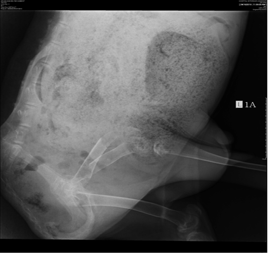
The left hindlimb was radiographed on both left-lateral view and ventral-dorsal view. The radiographic findings revealed bone discontinuity at the left mid femur and overriding of the proximal and distal femoral bone fractures with radiological diagnosis of oblique fracture of the left mid femur. The discontinued diaphysis exhibited at a different degree of radiolucency of the compact bone and endosteum suggesting an oblique fracture (Figure 1). Overriding of the proximal and distal femoral bone fracture was observed on the ventral-dorsal view. A sharp point of the proximal fracture protruded laterally was also observed (Figure 2). Thinning of bone cortex and the presence of increased radiolucency was observed in bone medulla with radiological diagnosis of osteomalacia. Therefore, the radiological diagnosis revealed that the doe was suffering from left hindlimb oblique fracture at the mid-femoral region due to osteomalacia.
Therefore, this veterinary clinical case was diagnosed as pathological femoral fracture secondary to osteomalacia. The farmer was advised to cull the doe due to the welfare, poor prognosis of the case due to the severity and location of the fracture.
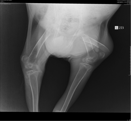
Figure 2: Overriding of the proximal and distal femoral bone fractures on the ventral-dorsal view. Thinning of bone cortex and the presence of increased radiolucency was observed in the bone medulla.
Discussion
Fractures of long bones are common in small ruminants and require either conservative management or surgical interference. The aim of fracture treatment is anatomic reduction and immobilization of fracture bones which is accomplished by either internal or external fixation (Sama et al., 2018). Treatment choices may vary between stall rest, external coaptation, external fixation and open reduction with internal fixation, depending on the type of fracture and the bone involved. (Mulon, 2013). Fractures of the axial skeleton are often treated by stall rest alone because external or internal fixation is not required (Bendrey, 2014). In cases of distal fractures, the conservative treatment with immobilization is the method of choice. Immobilization with plaster associated with Thomas splint, or just with wooden splints in young animals, was effective in reducing fractures. In cases of proximal fractures which was demonstrated in this clinical case report, the affected animals need to be treated surgically using either external or internal fixation (Camara et al., 2014) as casts cannot adequately immobilize fractures proximal to the distal radial physis or the distal tibial physis. In ruminants when there is reduced and restricted in movements, the affected animal more prone to bloat condition.
The main limitations in treating bone discontinuity in sheep and goats are the cost involved in surgical procedures, anaesthetics, implants, and post-operative care. Nevertheless, the choice between treatment and euthanasia also depends on the economic or genetic value of the animal and the prognosis associated with the particular fracture (Mulon, 2013). The treatment for severe fractures is not economical to be carried out on a production animal as it includes long term physiotherapy, splints, or surgeries such as external stabilization using dental acrylics or joint reduction surgery (Abdullah et al., 2015). According to Anderson and Jean (2008), the following questions must be answered in all fracture cases: (1) Is treatment required? (2) Can the fracture be acceptably reduced closed or is internal reduction required? (3) Can the fracture be adequately immobilized using external coaptation alone, or is an internal fixation, with or without external coaptation, required? and (4) What is the cost-benefit analysis? In the present case in the veterinary case report, the required treatment is to have reduction using internal fixation method. By considering all the criteria stated above, the decision to cull the doe was made in this case due to the welfare, poor prognosis of the case due to the severity and location of the fracture.
For future alternative options for fractures management in small ruminants, the external skeletal fixation (ESF) can opt as a successful, economic, and alternative to internal fixation. It is an external coaptation technique used to stabilize bone fragments or joints with percutaneous wires or pins held together by an external frame. It will help to stabilize bone fragments or joints with percutaneous pins or wires held together by an external frame which can be modified according to the economic feasibility (Anderson and Jean, 2008). ESF is gaining popularity since it is less invasive, relatively easy to perform and requires minimal equipment (Harari et al., 1998). The most common clinical complications associated with ESF are instability at the fracture site, pin loosening, pin tract osteolysis, pin tract infections, implant failure, osteomyelitis, and delayed union or nonunion of the fracture. However, with the proper pre and post-operative care of ESF, these complications can be prevented or minimized. This method of management was not opted in this case due to severity and poor prognosis of the case as the affected doe was suffering from osteomalacia as well.
One of the possible causes of osteomalacia is diet. According to Uhl (2018), the most common causes of osteomalacia in animals are dietary deficiencies of vitamin D or phosphorus. Calcium dietary deficiency is really unlikely to cause the condition because any decrease in extracellular concentration of calcium will be rapidly corrected by the action of PTH and vitamin D (Dittmer and Thompson, 2011). In addition, green grasses are poor sources of vitamin D. In relation to the case, the patient was only fed with leaves with a low nutritional value which is believed to be the cause of vitamin D deficiency in this case. Next, the possible cause of osteomalacia may due to the design of the goats’ house. According to a study by Hymoller and Jensen (2010), revealed that cows which were exposed to sunlight had a higher concentration of vitamin D3 in the plasma compared with unexposed cows. Ruminants are very dependent on sunlight exposure for vitamin D synthesis and therefore the design of house or animal shed are very important. For this case, the affected farm had a bad design of goats’ house where the roof does not allow any sunlight to enter and very shady. Therefore, the design of the house on this farm could also contribute to the occurrence of osteomalacia in this doe. For prevention and control, the exposure of sunlight could be considered by either increasing the height of the goat house or by providing transparent roof and supplement of feed with vitamin D.
Conclusions
Good farm design and nutrition intake are required to maintain the welfare and health status of the animal and avoid the osteomalacia condition that was observed in this case leading to pathological fracture.
Acknowledgements
The authors wish to acknowledge the staff from University Veterinary Hospital, Faculty of Veterinary medicine, Universiti Putra Malaysia for their technical assistance.
Conflict of Interest
There exists no conflict of interest.
Authors’ Contribution
All authors contributed equally and approved the final manuscript.
References

