The Journal of Advances in Parasitology
Case Report
Chronic Dermatitis Complicated with Otitis Due to Notoedres cati in a Persian Cat
Sirigireddy Sivajothi1*, Bhavanam Sudhakara Reddy2, Ramanujula Venkatasivakumar2
1Department of Veterinary Parasitology; 2Department of Veterinary Medicine, College of Veterinary Science, Sri Venkateswara Veterinary University, Proddatur - 516360, Andhra Pradesh, India.
Abstract | Notoedres cati is one of the most common mites causing dermatitis in cats. Otitis along with generalised dermatitis was observed in an eight months Persian cat. Cat had history of head shaking, intense pruritus, alopecic and erythematous areas on the ears. The skin at the lesion area was scrapped and examined under a light microscope. Notoedres mites were observed in otic discharges and skin scrapings. It was treated with doramectin at weekly intervals, ciprofloxacin otic drops daily twice for five days. After therapy, marked improvement was noticed and by re-growth of hair over the affected region within 14 days.
Keywords | Cat, Dermatitis, Doramectin, Notoedres cati, Otitis
Editor | Muhammad Imran Rashid, Department of Parasitology, University of Veterinary and Animal Sciences, Lahore, Pakistan.
Received | March 30, 2015; Revised | April 30, 2015; Accepted | May 08, 2015; Published | May 16, 2015
*Correspondence | Sirigireddy Sivajothi, Sri Venkateswara Veterinary University, Proddatur, Andhra Pradesh, India: Email: sivajothi579@gmail.com
Citation | Sivajothi S, Reddy BS, Venkatasivakumar R (2015). Chronic dermatitis complicated with otitis due to Notoedres cati in a Persian cat . J. Adv. Parasitology. 2(1): 19-22.
DOI | http://dx.doi.org/10.14737/journal.jap/2015/2.1.19.22
ISSN | 2311-4096
Copyright © 2015 Sivajothi et al. This is an open access article distributed under the Creative Commons Attribution License, which permits unrestricted use, distribution, and reproduction in any medium, provided the original work is properly cited.
INTRODUCTION
Dermatological problems are one of the most common clinical entities in domestic pets. Among them cats are affected with variety of parasitic infections and incidence of mange is high. After mite infestation, the skin becomes thickened, wrinkled, folded and finally covered with dense, tightly adhering, and yellow to grey crusts (Scott et al., 2001). Notoedric mange in cats is very common type of contagious parasite skin infestation. Occasionally humans in contact with affected animals may show a mild dermatitis due to a transient infestation. Previously notoedric mange was reported in cats and humans by Chakrabarti A, (1986) and Sivajothi et al., (2014). Foreign bodies or ecto parasites are the common causes for development of the primary otitis in cats. Feline otitis can be a challenging clinical problem to the clinicians. Cats affected with mange mites causes severe itching sensation and when they affect the ears causes otitis externa by exhibition of erythematous ear canal, otic exudates, scratching the ears (Ahaduzzaman et al., 2014). Present communication reports the notoedric mange causing otitis in a Persian cat in Andhra Pradesh of India.
CASE HISTORY AND OBSERVATIONS
An eight month old short haired Persian cat was referred to the Teaching Veterinary Clinical Complex, College of Veterinary Science, Proddatur with head shaking, chronic dermatitis and crusty lesions on the margins of both the ears. The skin at the neck, on the legs was corrugated, also observed the presence of inflammation over the margins of the ears resulting from self-inflicted trauma induced by pruritus (Figure 1) and otitis in the both ears (Figure 2). Cat was treated for one month at surrounding dispensaries around the Proddatur without any significant clinical improvement.
Multiple skin scrapings were collected from the skin lesions, cleared by 10% KOH solution and examined under low power of microscope. Cytological examination of the ear swabs were done to rule out the cause for otitis. Smears of ear discharge were made for parasitological, bacteriological and fungal examinations. Samples from the ears were obtained with the use of a small tipped cotton applicator. The swab was then rolled onto a new, clean microscope slide by rolling the collected material from both the ears separately. The slide was stained with Giemsa staining and Methylene blue staining. Some of the ear swabs were rolled in a drop of mineral oil on a microscope slide and examined for the mange mites (Sivajothi and Reddy, 2014; Reddy et al., 2014a).
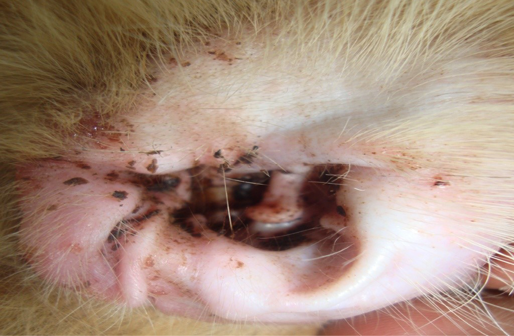
Figure 2: Cat showing the otitis
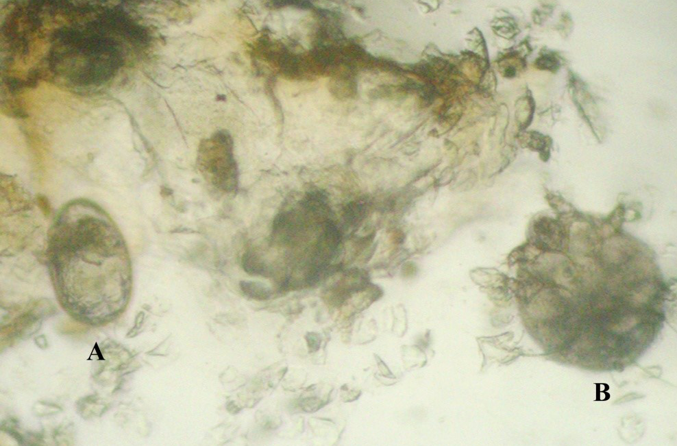
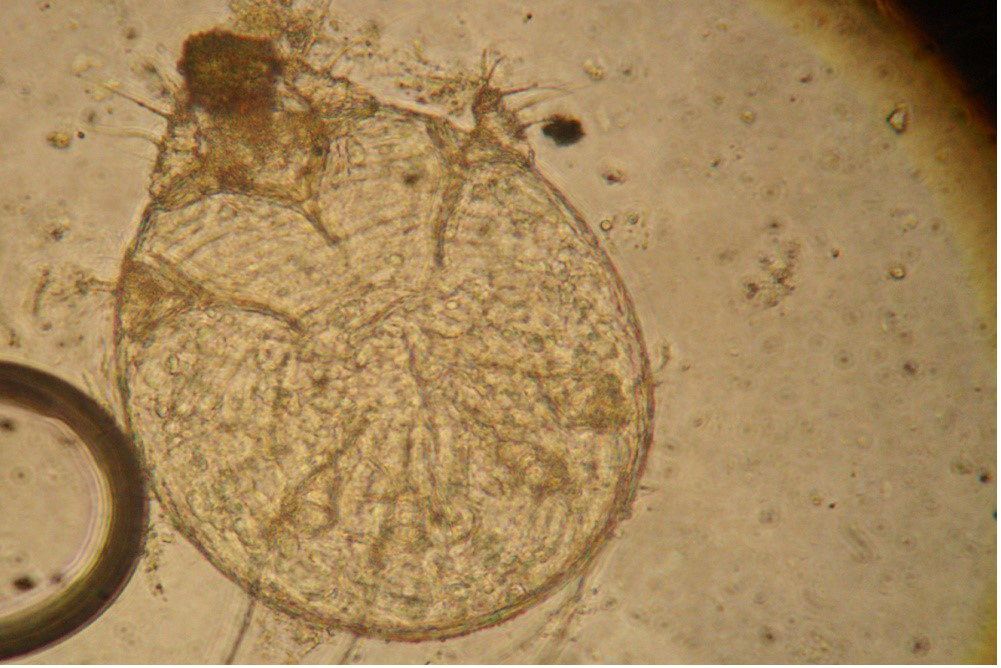
Under low power examination of the slides prepared from the ear cerumen revealed crawling mites across the field along with the typical dark coloured eggs (Figure 3). Examination of the cleared skin scrapings revealed different stages of Notoedric mites (Figure 4). Observation of the stained slides under higher power examination reveals bacteria and few neutrophils which indicate the presence of secondary invaders. Cat had haemoglobin of 9.8 g/dL, total erythrocyte count of 4.2x10⁶/cubic mm, total white blood cells of 18120/cubic mm with neutrophilia (72%) and eosinophilia (7%).
TREATMENT AND DISCUSSION
The cat was treated with doramectin (Dectomax® Injectable solution containing 1% w/v doramectin, 10 mg/mL, Pfizer Animal Health Ltd.) @ 0.2 mg / Kg body weight, subcutaneously at weekly interval for four weeks. Zincovit (Zincovit drops, Apex Ltd. It contains Each ml of Zincovit Drops contains Vitamin A (as palmitate) 2500 I.U., Vitamin E as (dl α-tocopheryl acetate) 2.5 I.U., Cholecalciferol (Vitamin D3) 200 I.U., Thiamine Hydrochloride 1 mg, Riboflavin Phosphate Sodium 1 mg, Pyridoxine Hydrochloride 0.5 mg, D- panthenol 1 mg, Nicotinamide 10 mg, Ascorbic Acid 40 mg, Lysine Hydrochloride 10 mg, Zinc sulphate 13.3 mg.) was administered orally 0.5 ml twice daily, Ciprofloxacin ear drops (Cyprin-D Intra Labs, ciprofloxacin 0.3% and dexamethasone) was used two times a day for five days after cleaning with warm sterile saline. Response to the treatment was assessed weekly once by clinical and laboratory assessment of skin scrapings. After five days of post therapy cat ear became normal without exudation. Skin scrapings collected from the same sites were negative for mites after the 2nd week of therapy. Cat was free from pruritus, skin lesions and improvement in the body condition was noticed (Figure 5). Present cat was suffering with chronic dermatitis so to avoid re infection, cat was treated with two more doses of doramectin after that complete resolution of the skin lesions was observed.
Notoedric mange is a contagious parasite disease of cats (Scott et al., 2001). The mite primarily attacks cats but may also infest foxes, dogs, rabbits and human which cause transient lesions (Foley, 1991). Management of otitis in cats mainly depends on the accurate diagnosis of cause for development of the otitis. Cytology is an important diagnostic tool to evaluate the perpetuating factors for development of the otitis. Observed clinical signs in this study were itching, hair loss and inflammation at the margins of the ears was in accordance with the previous studies (Itoh et al., 2004). But, in this study additionally otitis was also noticed. It might be due to the entry of the mites in to the ear canal due to chronicity of the infection. Foil (2003) also stated that characteristic itching and hair loss pattern was often all that was needed to diagnose notoedric mange in the cat.
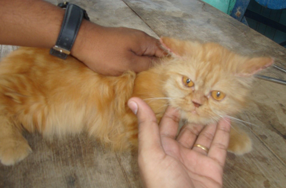
Figure 5: Cat after three weeks of therapy
Scraping collected from the skin lesions, revealed ova and adult stages of Notoedres cati mites in the present study. The mites were identified as per the reports of Walker (1994), based on their shape and the presence of dorsal anus, which distinctly differentiated the Notoedres cati from Sarcoptes spp. The mite was similar to Sarcoptes scabiei, and it was considerably smaller and more circular. The size of the mites was between 150 μm to 220 μm. There were no projecting scales but mid dorsally, the striae were broken into a scale like pattern; the stout setae were absent (Figure 4). Observed neutrophilia and eosinophilia in this study was according with previous studies (Reddy et al., 2014c).
Previously notoedric mange in cat was treated with doramectin, ivermectin and selamectin by the different workers (Delucchi et al., 2000; Itoh et al., 2004; Reddy and Sivajothi, 2014). In this study, we selected doramectin to treat mange mites. Doramectin is a novel avermectin that is produced by mutational biosynthesis and differs from ivermectin by having a cyclohexyl group in the C25 position of the avermectin ring. Doramectin has a broad range of activity against endoparasites and ectoparasites, and although only approved for use in cattle, sheep, goats and swine and off-label use has been widely used and reported in other species (Yas-Natan et al., 2003). Like other macro cyclic lactones, doramectin has two modes of action. The primary mode of action is binding of the doramectin molecule to postsynaptic glutamate-gated chloride ion channels in the synapses between inhibitory interneuron’s and excitatory motor neurons in nematodes, and in myoneural junctions in arthropods. Avermectins also enhance the release of gamma-aminobutyric-acid (GABA) in presynaptic neurons, which, in turn, open postsynaptic GABA-gated chloride channels. In either case, the influx of chloride ions reduces cell membrane resistance, which prevents the potential hyperpolarisation of neural stimuli to muscles, resulting in flaccid paralysis and death (Reinemeyer and Courtney, 2001).
Secondary bacterial and fungal infections are very common in cats affected by the ear mite. Topical use of antibiotics for treating otitis cases is a common practices followed by practioners. Mosges et al. (2011) stated that fluroquinolones had high success rate than non-quinolones in curing the otitis. Enrofloxacin is a drug of choice against Staphylococci in the recurrent and chronic skin problems in our local area (Reddy et al., 2014b).
CONCLUSION
Present communication reports the case of chronic dermatitis complicated with otitis due to Notoedres cati in a Persian cat in Andhra Pradesh of India.
ACKNOWLEDGEMENTS
Authors are thanking full to the authorities of the S.V.V.U for providing the facilities to carry out the present work. Corresponding author thank full to the animal owner for his co-operation.
REFERENCES


