Advances in Animal and Veterinary Sciences
Case Report
Efficacy of Auto-Hemotherapy in Bovine Teat Papillomatosis: A Case Report
P. Arun Nehru1*, S. Sunandhadevi2, T. Rama3, N. Muniyappan4
1Assistant Manager, KS Cattle Feeds, KSE Limited, Dindigul, Tamil Nadu, India - 642113; 2Teaching Faculty, TVCC, College of Veterinary Science, Prodattur, Andhra Pradesh, India; 3Teaching Faculty, Department of Veterinary Biochemistry,College of Veterinary Science, PVNRTVU, Telangana, India; 4Assistant Professor, Department of Veterinary Physiology, Madras Veterinary College, Vepery, Chennai.
Abstract | A seven year old female Holstein Friesian cross bred cow was presented with signs of various sizes of pedunculated cutaneous warts on teat, pain, bleeding and interference in milking. Based on the history and clinical picture, the case was diagnosed as bovine papillomatosis. The animal was treated with its own blood and repeated once in a week. The warts on udder and teats shrunk/regressed, dried and dropped away after a follow up period of four weeks.
Keywords | Papillomatosis, Teat, Auto-hemotherapy, Cross bred cow, Venous blood
Editor | Kuldeep Dhama, Indian Veterinary Research Institute, Uttar Pradesh, India.
Received | July 13, 2017; Accepted | August 11, 2017; Published | August 25, 2017
*Correspondence | P. Arun Nehru, KS Cattle Feeds, KSE Limited, Dindigul, Tamil Nadu, India - 642113; Email: vettyarun@gmail.com
Citation | Nehru PA, Sunandhadevi S, Rama T, Muniyappan N (2017). Efficacy of auto-hemotherapy in bovine teat papillomatosis: A case report. Adv. Anim. Vet. Sci. 5(8): 350-351.
DOI | http://dx.doi.org/10.17582/journal.aavs/2017/5.8.350.351
ISSN (Online) | 2307-8316; ISSN (Print) | 2309-3331
Copyright © 2017 Nehru et al. This is an open access article distributed under the Creative Commons Attribution License, which permits unrestricted use, distribution, and reproduction in any medium, provided the original work is properly cited.
INTRODUCTION
Cutaneous papillomatosis is a benign proliferative neoplasm caused by papilloma virus, which usually appear as multiple, sessile or pedunculated, circumscribed grey-white to dark brownish black outgrowth may appear on skin over different body parts. However, neck, eyelids, teats and lower line of abdomen are the most common sites. It is a contagious disease, usually transmitted via direct contact, contaminated food and equipment, flies, castration and injections. Papillomas on teats may cause difficulty in milking and suckling by calf and sometimes, pedunculated papillomas snap-off causing mastitis and teat infections (Singh and Somvanshi, 2010). The present study deals with therapeutic and management of teat and udder warts by auto-hemotherapy.
Case History and Observations
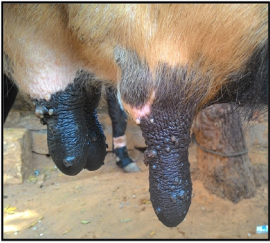
A seven year old female Holstein Friesian cross bred cow suffered in an organized cattle farm, Dindigul District, Tamil Nadu with the clinical signs of various sizes of pedunculated cutaneous warts on various parts of the body with teat involvement, causing pain, bleeding and interference in milking (Figure 1). The physiological parameters were within the normal range. The case was diagnosed as bovine papillomatosis.
Treatment and Discussion
The present case was decided to undertake auto-hemotherapy. Accordingly the animal was administered with its own blood. The venous blood was drawn @ 20 ml from the Jugular vein by using 18G hypodermic needle in a disposable syringe in that 10ml of venous blood was injected subcutaneously in the lateral neck region and 10ml was injected deep intramuscularly in the gluteal region by taking all sterile precautions. The treatment was repeated once in a week for four weeks continuously. After third injection, the papilloma growths showed signs of regression. The animal was under observation for six weeks. By the end of six weeks all the papilloma growths were completely reduced and only light black colored scars were seen at the sight of the growth.
Bovine papillomatosis is a contagious disease of cattle occurring as warts on skin and mucosa, caused by Bovine Papilloma Virus types 1 to 10 Vidhya et al. (2009).The virus infects the basal cells of the epithelium causing the excessive growth, which is characteristic of wart formation. Venugopalan (2000) and O’Conor (2001) have suggested remedial measures for removal of warts such as use of autogenous vaccine, wart enucleation, burning with hot iron or eraser, ligation and surgical removal of wart (excision) with surgical knife, application of salicylic acid ointment, di methtyl sulfoxide ointment and potential caustics. Surgical removal of one or two warts was proposed but surgical intervention and vaccination may increase the size of the residual warts and prolong the course of the disease Wadhwa et al. (1992). In the present case of teat papillomatosis, surgical intervention or vaccination would not have been the right choice of treatment, since it may have aggravated the condition. In auto-hemotherapy after fourth injection, the papilloma growths showed signs of regression. The findings were in accordance with those of Ganesh Hedge (2011) treated cutaneous papillomatosis in a non-descript cow by auto-hemotherapy and Chetan Kumar (2011) use auto-hemotherapy for the treatment of bovine papilloma.

Figure 1: Teat warts of varying size and shape
Hence the finding of this study concluded that without using any chemical agent, auto-hemotherapy can be effectively employed to treat bovine teat papillomatosis.
ACKNOWLEDGEMENTS
The authors are highly thankful to Deputy General Manager, KS Cattle Feeds, KSE Limited, Swaminathapuram, Dindigul District, Tamil Nadu, India for providing necessary facilities and financial support for carrying out this research.
CONFLICT OF INTEREST
There is no conflict of interest.
AUTHORS CONTRIBUTION
All the authors have contributed in terms of giving their technical knowledge to frame the article.
References


