The Journal of Advances in Parasitology
Research Article
Point Prevalence and Intensity of Gastrointestinal Parasite Ova/Oocyst and Its Association with Body Condition Score (BCS) of Sheep and Goats in Maiduguri, Nigeria
Paul Bura Thlama1,2*, Biu Abubakar Abdullahi2, Gadzama Mercy Ahmed2, Ali Mohammed2, Mana Hope Philip1, Jairus Yusuf1
1Veterinary Teaching Hospital, University of Maiduguri, Nigeria; 2Department of Veterinary Microbiology and Parasitology, University of Maiduguri, Nigeria.
Abstract | A survey of sheep and goats was conducted to investigate the prevalence and intensity of gastrointestinal parasites ova/oocyst and their effects on body condition scores. A total of 100 faecal samples were randomly collected from 72 sheep and 28 goats and subjected to standard saturated sodium chloride floatation technique to detect ova/oocyst while faecal egg counts were estimated using modified McMaster technique, and body condition score was estimated using standard methods. Seventy two percent (72.0%) of sheep and goats examined in this study were positive for various types of gastrointestinal parasite ova/oocyst. Prevalence rate was higher in sheep 53 (73.6%) than goats 19 (67.9%). Male (75%) and younger (70%) goats had higher prevalence rates compared to their female (65%) and adult (66.7%) counterparts while higher prevalence was recorded in adult (82.4%) and male (80%) sheep. Among different breed of sheep examined, the highest total prevalence was recorded in ouda (100%). Strongyles were generally the most prevalent in sheep (36.1%) and goats (35.7%) while Trematodes and Cestodes had the lowest frequencies (1.4%) in sheep but were not recorded in goats. Generally, a severe degree of EPG was observed in both sheep (1347.2 ±597.95) and goats (1257.9 ±542.18) examined in this study. There was no significant differences (P>0.05) in mean ±SD of EPG between different sexes and age groups of sheep and goats examined in this study. However, a significant difference (P<0.05) in mean ±SD of EPG was observed between sheep with emaciated, thin, average and fat body condition scores. A high prevalence and intensity of gastrointestinal parasite ova/oocyst was encountered in this study, and the most prevalent group was Strongyles. It was also established from this study that faecal egg counts significantly affected the body condition scores of sheep.
Keywords | Prevalence, Intensity, Gastrointestinal Parasite Ova/oocyst, Egg per Gram, Body Condition Score
Editor | Muhammad Imran Rashid, Department of Parasitology, University of Veterinary and Animal Sciences, Lahore, Pakistan.
Received | December 30, 2015; Revised | March 23, 2016; Accepted | March 28, 2016; Published | April 26, 2016
*Correspondence | Paul Bura Thlama, Veterinary Teaching Hospital, University of Maiduguri, P.M.B 1069, Bama Road, Maiduguri, Borno State, Nigeria; Email: bpaulgadzama@yahoo.com
Citation | Paul BT, Biu AA, Gadzama MA, Ali M, Mana HP, Jairus Y (2016). Point prevalence and intensity of gastrointestinal parasite ova/oocyst and its association with Body Condition Score (BCS) of sheep and goats in Maiduguri, Nigeria. J. Adv. Parasitol. 3(3): 81-88.
DOI | http://dx.doi.org/10.14737/journal.jap/2016/3.3.81.88
ISSN | 2311-4096
Copyright © 2016 Paul et al. This is an open access article distributed under the Creative Commons Attribution License, which permits unrestricted use, distribution, and reproduction in any medium, provided the original work is properly cited.
Introduction
Nigeria has an estimated 34.5 million goats and 22.1 million sheep of various breeds found predominantly in the northern region due to the favorable microclimatic conditions (Blench, 1999). Sheep and goats have been recognized as important livestock that contributes significantly to the food security and economy of developing countries (Lawal-Adebowale, 2012) and contribute about 5-6% of the gross domestic product of Nigeria (Opasina and David-West, 1989). Their socio-cultural values varies across the country, but they are generally used for cultural ceremonies such as weddings, burials, ritual and various types of religious sacrifices (Blench, 1999; Lawal-Adebowale, 2012). Sheep and goats are currently recognized as an important sources of animal protein in many countries of the world, contributing 30% of the total meat consumption in Nigeria (Opasina and David-West, 1999; Lawal-Adebowale, 2012; Ovutor et al., 2012) and are an important source of income earning for farmers, household keepers, animal traders, butchers and other stake holders involved in trading their products and by-products (Lawal-Adebowale, 2012).
Gastrointestinal parasites have been recognized as a serious threat to sheep and goat production worldwide (Regassa et al., 2006; Biu et al., 2009; Ovutor et al., 2012; Idika et al., 2012) and their impact on productivity is greater in sub-Saharan Africa due to the availability of suitable ecological factors that favors epidemiology (Shah-Fisher and Say, 1989; Ovutor et al., 2012). The economic impact of gastrointestinal parasites in sheep and goats may be direct through mortality or indirect through decreased production of milk, reduced weight gain, poor carcass quality, increased susceptibility to secondary infections and condemnation of affected organs at slaughter (Soulsby, 1982; Regassa et al., 2006; Biu et al., 2009; Ovutor et al., 2012; Idika et al., 2012). The prevalence and effects of gastrointestinal parasites of sheep and goats varies widely across different parts of the world due to local differences in ecological conditions such as temperature, humidity, rainfall, vegetation and management practices (Shah-Fischer and Say, 1989; Biu et al., 2009; Edosomwan and Shoyemi, 2012; Ovutor et al., 2012; Idika et al., 2012). Various reports have highlighted the prevalence and importance of gastrointestinal parasites in Nigeria (Fagbemi and Dipeolu, 1982; Biu et al., 2009; Edosomwan and Shoyemi, 2012; Idika et al., 2012). There is also paucity of information on the relationship between prevalence and intensity of gastrointestinal parasite ova/oocyst and body condition scores of sheep and goats in this area. This study was therefore conducted to determine the incidence, faecal egg/oocyst counts (FEC) of gastrointestinal parasite and their relationship with body condition scores in Maiduguri, Nigeria.
Materials and Methods
Study Area and Population
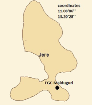
This study was carried out in Federal Government College Maiduguri staff quarters located along Bama Road in Jere Local Government Area of Borno State, Nigeria. Maiduguri is the capital and the largest city of Borno State and sits along the seasonal Ngadda River which disappears into the Firki swamps in the areas around Lake Chad. Borno State is located in the North East Geo-Political Zone of Nigeria and lies within latitude 10.200 – 13.400 N and longitude 9.800 – 14.140 E, sharing international boundaries with Republic of Niger and Chad in the north and Cameroon in the east. Borno State has a total land mass of 6,943,659 km2 and is characterized by hot-dry climate in the North and central parts of the State where rainfall lasts between July and September, while milder climatic conditions prevail in the southern parts where rainfall may last till October with humidity of about 49% (Udoh, 2003). The State derives great economic activity from its rich livestock and fishery products (NPC, 2006) (Figure 1).

Figure 1: Map of Jere showing study area
The sheep and goats used for this study were exclusively backyard flocks kept under the traditional husbandry system of semi-intensive management consisting of small flock sizes ranging between 2 and 10 in number. Animals were aged by dentition as described by Hassan and Hassan (2003). Animals aged1 1/2 years or younger were regarded as young while those older than 1 1/2 years were considered adults. Sex differentiation was based on the appearance of external genitalia and presence or absence of udder and or testes while breed differentiation was based on morphometric features such as the length of ears, shape of head, coat color and linear body measurements as described by Yunusa et al. (2013). Body condition score (BCS) was measured on a scale of 1 - 5 assessed by estimating the amount of muscling and fat cover on the lumber spinous processes and floating ribs as described by Jefferies (1961). Animals with body scores of 1 to 5 were categorized as emaciated, thin, average, fat and obese, respectively.
Sampling Procedure
A total of 100 animals comprising of 72 sheep and 28 goats were randomly selected for the study after estimating the sample size. The sample size was estimated using the formula given by Thrusfield (2005).
Faecal Collection and Examination
Approximately 5 grams of faeces was randomly collected from the rectum of sheep and goats using polythene gloves and placed into clean 30ml sterile bottles containing 2% formalin as preservative. Each sample was labeled appropriately with the approximate age, sex, breed and body condition score of sheep and goats examined. Standard saturated sodium chloride floatation technique was used for qualitative faecal examination as described by Brar et al. (1999). Parasite ova or oocyst were identified based on structural and morphometric criteria such as size, shape, color and presence or absence of polar cap or operculum as described by Soulsby (1982). Positive faecal samples were quantitatively analyzed to determine ova/oocyst per gram (EPG) using the modified McMaster technique as described by Kaufmann and Pfister (1990). The degree of infection was categorized as light with 50-799, moderate with 800-1200 or severe with >1200 eggs per gram of faeces as described by Urquhart et al. (1994).
Statistical Analysis
Data generated during the collection and laboratory examination of samples were summarized on Microsoft excel spread sheet 2007. Prevalence was calculated as P (%) = d/n where; P= prevalence, d= number infected and n= number examined (Thrusfield, 2005). The student t-test was used to compare the EPG between age and sex of animals. One way analysis of variance (ANOVA) was used to determine the relationship between EPG and body condition scores (BCS) of sheep and goats using GraphPad InStat statistical software version 3.0 and P<0.05 was considered significant.
Table 1: Overall prevalence of gastrointestinal parasite ova/oocyst in indigenous sheep and goats and their mean ±SD ova/oocyst output per gram of faeces
|
Animals |
No. Examined |
No. (%)Infected |
Mean (range) ±SD EPG |
|
Sheep |
72 |
53 (73.6) |
1347.2(600-3500)±597.95a |
|
Goats |
28 |
19(67.9) |
1257.9(550-2200)±542.18a |
|
Overall |
100 |
72(72.0) |
1323.6(550-3500)±581.34 |
*Column means represented by the same superscript were not significantly different (P>0.05)
Results
The overall prevalence of gastrointestinal parasite ova/oocyst of sheep and goats examined is presented in Table 1. Out of a total of 100 sheep and goats examined in this study, 72 (72.0%) were positive for various types of gastrointestinal parasite ova/oocyst with a mean ova/oocyst output per gram of 1323.6 ± 581.34. Out of 72 sheep examined, 53 (73.6%) were positive for ova/oocyst of various types of gastrointestinal parasites with a mean ova/oocyst output per gram of 1347.2 ± 597.95, while 19 (67.9%) out of the 28 goats examined in this study were positive for various types of gastrointestinal parasite ova/oocyst with mean ova/oocyst output per gram of 1257.9 ± 542.18. There was no significant differences (P<0.05) in the mean ±SD of ova/oocyst counts between sheep and goats examined in this study.
The prevalence of gastrointestinal parasite ova based on the sex, age, and body condition score of Sahel goats is presented in Table 2. Male and younger goats had higher prevalence rates of 75% and 70% compared with their female and adult counterparts who had prevalence rates of 65% and 66.7%, respectively (P<0.05). Goats with thin body condition scores had the highest prevalence rate (83%) for various types of gastrointestinal parasite ova while emaciated, fat and average goats had prevalence rates of 80%, 71.4% and 62.5% respectively (P<0.05). By contrast, obese goats were not infected with gastrointestinal parasite ova in this study.
Table 2: Prevalence of gastrointestinal parasite ova/oocyst in sahel goats examined based on sex, age and body condition scores
|
Variables |
No. Examined |
No. (%) Infected |
|
Sex |
||
|
Male |
8 |
6(75) |
|
Female |
20 |
13(65) |
|
Age |
||
|
Adult |
18 |
12(66.7) |
|
Young |
10 |
7(70) |
|
BCS |
||
|
Emaciated |
5 |
4(80) |
|
Thin |
6 |
5(83.3) |
|
Average |
8 |
5(62.5) |
|
Fat |
7 |
5(71.4) |
|
Obese |
2 |
0(0.0) |
|
Overall |
28 |
19(67.9) |
*BCS= Body condition score (Emaciated=1, Thin=2, Average=3, Fat=4 and Obese=5)
The spectrum of gastrointestinal parasites of Sahel goats identified in this study is presented in Table 3. Out of a total 19 (67.9%) positive cases recorded, 10 (35.7%) were positive for ova of various species of Strongyles. Mixed infections with Coccidia oocyst and Strongyle ova was recorded among 7 (25%), while 2 (7.1%) were positive for Coccidia oocyst alone. Mixed infections were more prevalent in male (10.5%) than female goats (5.6%) examined in this study. The prevalence of Strongyle ova was higher in young (14%) than adults (7.8%). Conversely, Coccidia oocyst were most prevalent in adult than young goats. The prevalence of Strongyle type ova was found to be higher in thin (14.0%), average (14.0%) and emaciated goats (11.2%) when compared with fat (3.9%) and obese goats (0.0) examined in this study. The prevalence of mixed infection with Strongyle ova and Coccidia oocyst was highest among fat goats (11.9%) when compared with thin (9.3%), emaciated (5.6%) and average goats (3.5%).
Table 3: Spectrum of various groups of gastrointestinal parasites ova/oocyst detected in sahel goats examined based on sex, age and body condition scores
|
Variables |
No. Examined |
No (%) Infected with |
||
|
Coccidia oocyst |
Strongyle ova |
Mixed infection |
||
|
Sex |
||||
|
Male |
8 |
0 (0.0) |
3 (10.5) |
3 (10.5) |
|
Female |
20 |
2 (2.8) |
7 (9.8) |
4 (5.6) |
|
Age |
||||
|
Adult |
18 |
2 (3.1) |
5 (7.8) |
5 (7.8) |
|
Young |
10 |
0 (0.0) |
5 (14.0) |
2 (5.6) |
|
BCS |
||||
|
Emaciated |
5 |
1 (5.6) |
2 (11.2) |
1 (5.6) |
|
Thin |
6 |
0 (0.0) |
3 (14.0) |
2 (9.3) |
|
Average |
8 |
0 (0.0) |
4 (14.0) |
1 (3.5) |
|
Fat |
7 |
1 (3.9) |
1 (3.9) |
3 (11.9) |
|
Obese |
2 |
0 (0.0) |
0 (0.0) |
0 (0.0) |
|
Total |
28 |
2 (7.1) |
10 (35.7) |
7 (25.0) |
*BCS= Body condition score (Emaciated=1, Thin=2, Average=3, Fat=4 and Obese=5)
Table 4: Mean ova/oocyst per gram output of sahel goats examined based on sex, age and body condition scores
|
Variables |
No. Infected |
Mean (Range) ±SD EPG |
|
Sex |
||
|
Male |
6 |
1008.3(550-1700)±440.93a |
|
Female |
13 |
1296.2(600-2200)±565.86a |
|
Age |
||
|
Adult |
12 |
1283.3(550-220)±589.43a |
|
Young |
7 |
1214.3(600-2000)±491.35a |
|
BCS |
||
|
Emaciated |
4 |
1312.5(1000-1850)±370.53a |
|
Thin |
5 |
1660.0(1000-2000)±384.71a |
|
Average |
5 |
1080.0(600-2200)±641.87a |
|
Fat |
5 |
990.00(550-1900)±570.53a |
|
Overall |
19 |
1257.9(550-2200) ±542.18 |
*BCS= Body condition score (Emaciated=1, Thin=2, Average=3, Fat=4 and Obese=5); Column means represented by the same superscript were not significantly different (P>0.05)
The mean ±SD ova/oocyst output per gram of faeces of Sahel goats based on their sex, age and body condition scores is presented in Table 4. There was no significant differences (P>0.05) in mean±SD of ova/oocyst counts between different sexes, age groups and body condition scores of goats examined in this study. The overall mean ±SD ova/oocyst output per gram of faeces of Sahel goats was 1257.9±542.18 >1200, thus indicating a severe infection.
The prevalence of gastrointestinal parasites ova/oocyst in sheep based on sex, age, breed body condition and scores is presented in Table 5. Between different sexes and age groups, the highest prevalence was recorded in adult (82.4%) and male (80%) sheep. Among different breeds of sheep examined, the highest total prevalence was recorded in Ouda (100%), followed by Yankasa (77.1%) and Balami (66.7%). Emaciated and thin sheep both had a prevalence of 80% for various species of gastrointestinal parasites while fat sheep had a prevalence of 89.5%.
Table 5: Prevalence of gastrointestinal parasites ova/oocyst in sheep examined based on their sex, age and body condition scores
|
Variables |
No. Examined |
No. (%) Infected |
|
Sex |
||
|
Male |
26 |
21(80.8) |
|
Female |
46 |
32(69.6) |
|
Age |
||
|
Adult |
34 |
28(82.4) |
|
Young |
38 |
25(65.8) |
|
Breed |
||
|
Yankasa |
35 |
27(77.1) |
|
Balami |
33 |
22(66.7) |
|
Ouda |
4 |
4(100) |
|
BCS |
||
|
Emaciated |
5 |
4(80.0) |
|
Thin |
10 |
8(80.0) |
|
Average |
38 |
24(63.2) |
|
Fat |
19 |
17(89.5) |
|
Overall |
72 |
53(73.6) |
*BCS= Body condition score (Emaciated=1, Thin=2, Average=3, Fat=4 and Obese=5)
The spectrum of gastrointestinal parasites ova/oocyst detected in sheep in this study based on sex, age, breed and body condition score is presented in Table 6. Out of a total of 53 (73.6%) positive cases, 26 (36.1%) were positive for ova of various species of Strongyle nematodes representing the most prevalent group of gastrointestinal parasites in sheep. Coccidia Oocyst were most commonly encountered among male (19.4%), young (15.2%). Balami breed (13.1%) and fat sheep (18.9%). Only 1 (1.4%) was positive for tapeworm and fluke ova respectively. Mixed infections with Coccidia oocyst and Strongyle ova was recorded among 13 (18.1%), while 12 (16.7%) were positive for Coccidia oocyst alone. Infection with Strongyle type nematode ova was
Table 6: Spectrum of gastrointestinal parasites ova/oocyst detected in sheep examined based on sex, age, breed and body condition scores
|
Variables |
No. Examined |
No (%) Infected with |
||||
|
Coccidia Oocyst |
Cestode Ova |
StrongyleOva |
TrematodeOva |
Mixed Infection |
||
|
Sex |
||||||
|
Male |
26 |
7 (19.4) |
0 (0.00) |
7 (19.4) |
1 (2.8) |
6 (16.6) |
|
Female |
46 |
5 (7.8) |
1 (15.7) |
19 (29.7) |
0 (0.00) |
7 (10.9) |
|
Age |
||||||
|
Adult |
34 |
4 (8.5) |
1 (2.1) |
17 (36.0) |
0 (0.00) |
6 (12.7) |
|
Young |
38 |
8 (15.2) |
0 (0.00) |
9 (17.1) |
1 (1.9) |
7 (13.3) |
|
Breed |
||||||
|
Balami |
33 |
6 (13.1) |
0 (0.00) |
10 (21.8) |
1 (2.2) |
5 (9.4) |
|
Ouda |
4 |
0 (0.00) |
0 (0.00) |
2 (36.0) |
0 (0.00) |
2 (36.0) |
|
Yankasa |
35 |
6 (12.3) |
1 (2.1) |
14 (28.8) |
0 (0.00) |
6 (12.3) |
|
BCS |
||||||
|
Emaciated |
5 |
0 (0.00) |
0 (0.00) |
3 (43.2) |
0 (0.00) |
1 (14.4) |
|
Thin |
10 |
1 (7.2) |
0 (0.00) |
4 (28.8) |
0 (0.00) |
3 (21.6) |
|
Average |
38 |
6 (11.4) |
1 (1.9) |
11 (20.8) |
1 (1.9) |
5 (9.5) |
|
Fat |
19 |
5 (18.9) |
0 (0.00) |
8 (30.3) |
0 (0.00) |
4 (15.2) |
|
Total |
72 |
12 (16.7) |
1 (1.4) |
26 (36.1) |
1 (1.4) |
13 (18.1) |
*BCS= Body condition score (Emaciated=1, Thin=2, Average=3, Fat=4 and Obese=5)
most frequently observed in female (29.7%), adult (36.0%), Ouda breed (36.0%) and emaciated sheep (43.2%) examined in this study while mixed infections were most prevalent in male (16.6%), young (13.3%), Ouda breed (36.0%) and thin bodied sheep (21.6) examined in this study.
Table 7: Mean ova/oocyst per gram output in sheep examined based on sex, age, breed and body condition scores
|
Variables |
No. Infected |
Mean (Range)±SD EPG |
|
Sex |
||
|
Male |
22 |
1227.3(700-2200)±426.70a |
|
Female |
31 |
1400.0 (600-3500)±674.80a |
|
Age |
||
|
Adult |
27 |
1403.6(600-3500)±680.13a |
|
Young |
26 |
1273.1(600-2200)±489.54a |
|
Breed |
||
|
Balami |
24 |
1212.5(600-2200)±447.60a |
|
Ouda |
04 |
1650.0(1200-2000)±341.57a |
|
Yankasa |
25 |
1408.0(600-3500)±712.93a |
|
BCS |
||
|
Emaciated |
04 |
2550.0(1800-3500)±793.73a |
|
Thin |
08 |
1187.5(800-2000)±387.07b |
|
Average |
24 |
1337.5(600-2000)±477.14c |
|
Fat |
17 |
1152.9(600-2000)±486.21d |
|
Overall |
53 |
1347.2(600-3500)±597.95 |
*BCS= Body condition score (Emaciated=1, Thin=2, Average=3, Fat=4 and Obese=5); Column means represented by different superscripts are significantly different (P<0.05).
The overall mean ±SD ova/oocyst output per gram of faeces in sheep examined in this study was 1347.2 ± 597.95 > 1200, thus indicating a severe infection. The mean ±SD ova/oocyst output per gram of faeces in sheep based on sex, age, breed and body condition score is presented in Table 7. There was no significant differences (P>0.05) in mean±SD of ova/oocyst counts between different sexes, ages and breeds of sheep examined in this study. Conversely, the mean ±SD ova/oocyst output per gram of faeces of indigenous sheep based on their body condition scores were significantly different (P<0.05).
Discussion
This study has revealed an overall prevalence of 72% for ova/oocyst of gastrointestinal parasites of sheep and goats. The highest prevalence and EPG was recorded in sheep (73.6%; 1347.2 ±597.95) compared with goats (67.9%; 1257.9 ±542.18). The high prevalence agrees with previous reports (Regassa et al., 2006; Biu et al., 2009; Maichomo et al., 2009; Idika et al., 2012; Ovutor et al., 2012). The higher prevalence in sheep compared to goats contradicts earlier reports by Regassa et al. (2006) and Biu et al. (2009) who reported higher prevalence in goats than sheep. The study also showed that both sheep and goats were severely (EPG>1200) infected with ova/oocyst of gastrointestinal parasites, and Strongyle ova and Coccidia oocyst were the most prevalent. These findings are consistent with Regassa et al. (2006) who reported an overall prevalence of 69.6% for gastrointestinal parasites of cattle, sheep and goats in Ethiopia and observed that Strongyle ova and Eimeria oocyst were most prevalent. Our finding also concurs with Ovutor et al. (2012) who reported an overall prevalence of 73.2% and 81.6% for helminths in exotic and indigenous goats examined in Southern part of Nigeria. Our result also agrees with Maichomo et al. (2004) who reported a prevalence of 80% and 82% in sheep and goats respectively in Kenya. However, the results disagree with Biu et al. (2009) who reported a lower prevalence of 54% and 58% in sheep and goats examined in Maiduguri. The discrepancies could be explained on the basis of differences in management of the animals (Regassa et al., 2006). While this study examined typical backyard flocks on semi intensive system t received little or no Veterinary care, the previous study examined animals on the University research farm on nearly zero grazing and received adequate Veterinary care. Our result is also at variance with Idika et al. (2012) who reported a higher overall prevalence of 99% for gastrointestinal nematodes in slaughtered goats. The observed differences could be explained on the basis of local differences in ecological conditions such as temperature, rainfall and humidity which influence the bionomics, distribution and intensity of infection with gastrointestinal parasites of ruminants in different parts of the country (Soulsby, 1982; Shah-Fischer and Say, 1989). Furthermore, seasonal influences associated with ecological conditions could account for the observed differences as the two studies were carried out in different seasons of the year.
This study also reports higher prevalence of gastrointestinal parasites ova/oocysts in male and young goats as well as adult and male sheep when compared with their counterparts. Variations were also observed in prevalence rates among different breed of sheep examined with Ouda having the highest prevalence (100%). Sex, age and breed difference in prevalence were previously observed and reported by several authors working in different parts of the world (Soulsby, 1982; Regassa et al., 2006; Mbaya et al., 2009; Shimelis et al., 2011; Idika et al., 2012). It is generally believed that younger animals show clinical symptoms of gastrointestinal parasitism during their first challenge, may accumulate heavy parasitic burdens and subsequently develop resistance under favorable conditions (Soulsby, 1982; Regassa et al., 2006; Mbaya et al., 2009). However, certain conditions such as pregnancy and parturition in female, nutritional stress and concurrent infection, may compromise immunity and exacerbate gastrointestinal parasitism in adult ruminants (Fakete and Kellems, 2007), hence the finding of higher prevalence in adult than young sheep. Male animals are also known to have high natural tendencies of acquiring diseases generally because they tend to move in search of mates for courtship and breeding purposes.
Generally, we observed a severe degree of EPG (>1200 ova/gram) in sheep and goats examined in this study with higher but not significant EPG (P>0.05) in adult than young sheep and higher but not significant EPG (P>0.05) in young than adult goats. This finding may be associated with poor development of immunity in adult sheep and goats due to stress such as poor nutrition and concurrent infections. The unavailability of grasses during the dry season may subject these animals to stress since there is virtually no supplement provided; this leads to poor immunity and rebound of infections. It has been established that poor nutrition may weaken or cause poor development of the immune system and exacerbate parasitic infections (Fakete and Kellems, 2007). Female sheep and goats had a severe (>1200 ova/oocyst/gram) but not significant EPG (P>0.05) compared with their male counterparts. This finding does not agree with Idika et al. (2012) but could be explained on the basis of differences in management systems under which the animals are raised. While the previous study examined trade animals pooled from various sources, this study examined backyard flock on semi-intensive management where female animals are usually kept for longer periods for breeding purposes and could accumulate worm burdens over time. Severe (>1200 ova/oocyst/gram) but not significant EPG (P>0.05) was observed in Ouda, Balami and Yankasa breeds, with Ouda having the highest EPG. This finding may be attributed to genetic differences in susceptibility and resistance to gastrointestinal parasites among various breeds of sheep. Stear and Welkin (1998) reported that the ability of animals to resist infections with parasites is genetically determined and therefore varies between individuals or breeds of a given host species.
This study reports an association between body condition scores and EPG in sheep and goats which does not agree with Regassa et al. (2006) who reported no association between BCS and EPG. However, it agrees with Idika et al. (2012) who reported an inverse high correlation between BCS and EPG. Severe (>1200 ova/oocyst/gram) but not significant EPG (P>0.05) was observed in both sheep and goats in this study. Significant EPG (P <0.05) was observed between fat, thin, emaciated and average sheep examined in this study. Sheep with emaciated and average body condition scores (BCS) had severe EPG while sheep with thin and moderate BCS had moderate EPG. On the other hand thin and emaciated goats had severe but not significant EPG (P >0.05) while average and fat goats had moderate but not significant EPG (P>0.05). This finding may be associated with pathogenic effects of gastrointestinal parasites in sheep and goats. It is widely reported that the main features of infection with gastrointestinal parasites in ruminants include anemia, diarrhea, and haemorrhagic gastroenteritis which lead to protein-losing enteropathy, hypoproteinaemia, and reduced growth rate in young animals, poor weight gain, and loss of body condition (Soulsby, 1982; Shah-Fischer and Say, 1989; Mbaya et al., 2009; Idika et al., 2012).
The spectrum of gastrointestinal parasites (Strongyles, Coccidia, Cestodes and Trematodes) in this study was previously reported (Biu et al., 2009; Mbaya et al., 2009; Edosomwan and Shoyemi, 2012). Strongyle, Coccidia and mixed infections with Strongyle and Coccidia were common in both sheep and goats while Trematodes and Cestodes ova was found only in sheep, however, Strongyle ova was the most prevalent. This finding is consonant with previous reports by Maichomo et al. (2004), Biu et al. (2009), Shimelis et al. (2011) and Kantzoura et al. (2012). Trematodes and Cestodes infections had the lowest frequencies in sheep and were not encountered in goats in this study. Generally speaking, infection with Trematodes is rarely diagnosed in sheep and goats (Khoramian et al., 2014) especially in the Sahel zone (Mbaya et al., 2010) while the detection rates of Cestodes ova is very low using faecal examination (Hansen and Perry, 1994).
Conclusion
A high prevalence and intensity of gastrointestinal parasite ova/oocyst was encountered in this study and the most prevalent were Strongyles and Coccidia. It was also established from this study that faecal egg counts significantly affected the body condition scores of sheep.
Recommendations
It is therefore recommended that all animals in the Federal Government College, Maiduguri should be treated against gastrointestinal parasites routinely.
Acknowledgement
The authors are very grateful to Mr. Yusuf Jairus of Veterinary Teaching Hospital, University of Maiduguri and Mallam Ali Mohammed of the Department of Veterinary Microbiology and Parasitology, University of Maiduguri for their technical support.
Conflict of Interest
The authors don’t have any conflict of interest to declare concerning this article.
Authors’ Contribution
Paul Bura Thlama conceived the idea, designed and carried out field and laboratory investigations as well as the preparation of the draft. Gadzama Mercy Ahmed, Mohammed Ali and Mana Hope Philip assisted in the field investigation, literature review and data analysis, while Jairus Yusuf helped with the laboratory analysis of samples. Professor Biu Abubakar Abdullahi supervised and scrutinized the final draft of this manuscript before submission.
References


