Advances in Animal and Veterinary Sciences
Research Article
Expression of Porcine Reproductive and Respiratory Syndrome Virus Nucleocapsid Protein in Nicotiana benthamiana for Diagnostic Applications
Boonyasit Porngarm1, Aktsar Roskiana Ahmad1, Kate Neelasawee3, Pichai Joiphaeng3, Tawatchai Hoonsuwan3, Kaewta Rattanapisit1,2, Balamurugan Shanmugaraj1,2*, Waranyoo Phoolcharoen1,2*
1Department of Pharmacognosy and Pharmaceutical Botany, Faculty of Pharmaceutical Sciences, Chulalongkorn University, Bangkok, Thailand; 2Research unit for Plant-produced Pharmaceuticals, Chulalongkorn University, Bangkok, Thailand; 3B.F. Feed Company Limited, Thung Song Hong, Lak Si, Bangkok, Thailand.
Abstract | Porcine reproductive and respiratory syndrome virus (PRRSV), the causative agent of Porcine reproductive and respiratory syndrome (PRRS) in pigs, is an enveloped positive strand RNA virus belongs to the family Arteriviridae. PRRSV poses severe threat to swine industry worldwide. Routine surveillance, rapid timely diagnosis of PRRSV infection is crucial for effective monitoring and disease control. Serological tests based on highly conserved viral nucleocapsid (N) protein are widely used for the detection of antibodies to PRRSV. Here, we investigated the potential of plant system to produce PRRSV N-protein in order to use as a reagent for the diagnosis of PRRSV infection. In this regard, the codon-optimised PRRSV N-protein coding sequence was cloned in geminiviral vector and expressed in Nicotiana benthamiana. The transient expression conditions such as effective gene construct, leaf harvesting time and Agrobacterium cell density were optimized for maximal protein production. Further, by using western blot assay, with a plant-produced protein we sought to detect the antibodies against PRRSV in pig sera. Preliminary testing revealed that the recombinant protein was identified by antibodies present in pig sera. However sensitivity/specificity and correlation of results with other assays needs to be evaluated. This proof of concept study indicated that plant-produced N-protein can likely be used as diagnostic reagent and demonstrate the potential of plant expression system for the production of recombinant PRRSV antigens.
Keywords | Nicotiana benthamiana; Nucleocapsid protein; Plant-produced protein; Porcine reproductive and respiratory syndrome virus; Transient expression
Received | October 27, 2020; Accepted | November 10, 2020; Published | February 20, 2021
*Correspondence | Balamurugan Shanmugaraj, Waranyoo Phoolcharoen, Department of Pharmacognosy and Pharmaceutical Botany, Faculty of Pharmaceutical Sciences, Chulalongkorn University, Bangkok, Thailand; Email: Balamurugan.S@chula.ac.th; Waranyoo.P@chula.ac.th
Citation | Porngarm B, Ahmad AR, Neelasawee K, Joiphaeng P, Hoonsuwan T, Rattanapisit K, Shanmugaraj B, Phoolcharoen W (2021). Expression of porcine reproductive and respiratory syndrome virus nucleocapsid protein in nicotiana benthamiana for diagnostic applications. Adv. Anim. Vet. Sci. 9(4): 581-587.
DOI | http://dx.doi.org/10.17582/journal.aavs/2021/9.4.581.587
ISSN (Online) | 2307-8316; ISSN (Print) | 2309-3331
Copyright © 2021 Shanmugaraj et al., This is an open access article distributed under the Creative Commons Attribution License, which permits unrestricted use, distribution, and reproduction in any medium, provided the original work is properly cited.
Introduction
Porcine reproductive and respiratory syndrome virus (PRRSV) is an economically important infectious pathogen causing respiratory disease and reproductive failures in pigs leading to substantial economic losses and poses severe threat to swine industry. It is an enveloped virus belongs to the genus Arterivirus, family Arteriviridae, and order Nidovirales. It contains positive strand RNA genome of about 15 Kb encoding for 11 known open reading frames (ORFs) (Lunney et al., 2016). The virus was first reported in North America in 1987 and in Western Europe in 1990 (Collins et al., 1992; Wensvoort et al., 1992). Initially the syndrome was recognized as mystery swine disease (Lunney et al., 2010). The ORF1a and ORF1ab comprises ~60–80% of the genome which encodes for non-structural proteins with protease and replicase functions and ORFs 2–7 encodes for viral structural proteins ie., GP2, GP3, GP4, GP5, membrane, envelope, nucleocapsid. The nucleocapsid protein (N) is the most abundant, immunogenic part of the virus which forms nucleocapsid core of 20–30 nm diameter and is considered as best target for serodiagnosis. Pigs infected with PRRSV produce detectable antibody 5 days after infection and can be detected up to 300 days post infection (Nelson et al., 1994; Yoon et al., 1995). The commercial ELISAs were developed based on N-protein for serodiagnosis of PRRSV.
Several protein production platforms have been developed to produce recombinant viral antigens including E. coli, insect, yeast, and mammalian cell cultures. Although bacterial system is considered as efficient, inexpensive production system, the lack of post-translational machinery and high cost, safety issues related to protein production in mammalian system demands to explore for other efficient alternative system. Recently plants are considered as potential platform, as it offers many advantages including time efficiency, high yield, scalability, low or no risk of pathogen contamination and eukaryotic-specific post-translational modifications of recombinant proteins (Ma et al., 2003). In recent decade, enormous progress has been made in recombinant protein production in plants to overcome the limitations associated with traditional protein production platforms (Ma et al., 2015; Fox, 2012; Lomonossoff and D’Aoust 2016). Furthermore, the recombinant antigens and therapeutic antibodies can be rapidly produced in plant expression system by transient expression meditated by agroinfiltration and is always a method of choice over time-consuming stable expression (Shanmugaraj et al., 2020a).
The major goal of this study was to evaluate the feasibility of transient expression of N- protein in N. benthamiana that can be used as a diagnostic reagent for the detection of PRRSV infection in pigs. To investigate the influence of the presence of signal peptide and location of histidine tags either at N-terminal or C-terminal, we designed four different constructs and the construct that showed high expression was selected and used to optimize the transient expression conditions for high protein production. The PRRSV N-protein in the plant crude extract was detected by anti-histidine antibodies and serum antibodies collected from pigs.
Materials and Methods
Plant material
All the wild-type N. benthamiana plants used in the study were grown in a greenhouse at 25 °C under a 16 h light/8 h dark cycle. Six to eight weeks old N. benthamiana plants were used for the experiments.
Bacterial strains and expression vector
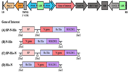
Agrobacterium tumefaciens strain GV3101 harboring plant expression vector was used for transient transformation. The geminiviral vector (Diamos and Mason 2019; Chen et al., 2011) was kindly provided by Professor Hugh S Mason at Arizona State University, USA. All plant expression constructs used in this study are listed in Figure 1.

Figure 1: Schematic diagram of the plant expression vector used in the present study. LB and RB, the left and right borders of the gene region transferred by Agrobacterium into plant cells; Pin2 3′: the terminator from potato proteinase inhibitor II gene; P19: the RNA silencing suppressor from tomato bushy stunt virus; TMVΩ 5′-UTR: 5′ untranslated region of tobacco mosaic virus Ω; P35S: Cauliflower Mosaic Virus (CaMV) 35S promoter; LIR: long intergenic region of BeYDV; Gene of Interest: (A) SP-N-His, (B) N-His, (C) SP-His-N, (D) His-N; Ext3′FL: 3′ region of tobacco extension gene; Rb7 5′ del: tobacco RB7 promoter; SIR: short intergenic region of BeYDV; C1/C2: Bean Yellow Dwarf Virus (BeYDV) ORFs C1 and C2 encoding for replication initiation protein (Rep) and RepA.
Cloning of PRRSV N sequence into plant expression vector
The sequence which encodes for PRRSV N-protein was retrieved from the Genbank (Accession No.: ABU87671.1) and codon-optimised for expression in N. benthamiana by invitrogen GeneArt® Gene Synthesis (Thermo Fischer Scientific, USA). The codon-optimised gene was used as a template for making different plant expression constructs which differs in the presence of signal peptide and location of 8x histidine tag (N or C-terminal). Primers based on the synthetic gene were designed that include cleavage sites for restriction enzymes. There was a restriction site for XhoI in the forward primer (5′ – CTCGAGCATCATCACCACCATCACCATCATATGGCCGGCAAGAACCAGTC–3′) and a restriction site for SacI in the reverse primer (5′ – GAGCTCTCAAAGCTCATCCTTTTCAGAACTAGCACCCTGTGAAGCAG–3′). The polymerase chain reaction (PCR) reaction was performed with Q5 high fidelity DNA polymerase (New England Biolabs, UK) under the following conditions: 94 °C for 10 min, then 30 cycles of 94 °C for 30 sec, 52 °C for 30 sec, and 72 °C for 30 sec, and a final extension of 72 °C for 10 min. The PCR product was separated on a 1% agarose gel and gel purified using fragment DNA purification kit (iNtRON Biotechnology, South Korea), according to the manufacturer’s instructions. The PCR product was cloned into expression vector pBYR2eK2Md (pBYR2e) by T4 DNA ligase (New England Biolabs, UK). The plant expression constructs were transformed into E. coli strain DH10B competent cells by heat-shock method and the recombinant clones were confirmed by using gene specific primers by following the standard PCR protocol (as mentioned above). The plasmid was isolated from the positive clones and used for Agrobacterium transformation.
Agrobacterium mediated transient expression of PRRSV N-protein
The plant expression constructs containing N-protein coding gene was transformed into Agrobacterium tumefaciens strain GV3101 by electroporation. Transformed cells were plated on luria bertani (LB) agar plates supplemented with kanamycin (50 µg/mL), rifampicin (50 µg/mL), and gentamicin (50 µg/mL) and incubated for 2 days at 28°C. Recombinant clones were analyzed by PCR and the positive colonies were cultured in LB broth containing the above mentioned antibiotics on a shaker (200 rpm) at 28°C overnight. Then the cells were harvested by centrifugation and resuspended in 1x infiltration buffer (10 mM 2-N-morpholino-ethane sulfonic acid (MES) and 10mM MgSO4, pH 5.5). The cell density was adjusted to 0.4 at OD600 and infiltrated into the leaves N. benthamiana wild type plants.
The infiltrated leaves were harvested on 4 day post-infiltration (dpi) and the plant crude extract was collected from the harvested leaves by grinding with extraction buffer phosphate buffered saline (PBS, pH 7.4). The crude extracts were centrifuged at 26,000 g at 4°C for 45 min and the supernatant was collected. The supernatants were analyzed by sodium dodecyl sulfate-polyacrylamide gel electrophoresis (SDS–PAGE) and western blot.
Optimization of recombinant protein expression in N. benthamiana
In order to obtain the maximal protein production, the leaf harvesting time post infiltration was optimized. Agroinfiltration was performed same like mentioned above and the infiltrated N. benthamiana samples were collected on 2, 3, 4 and 5 dpi. The plant crude extract was collected from the infiltrated leaves and analyzed by SDS–PAGE and western blot.
Similarly, the effect of Agrobacterium cell density on infiltration efficiency was also assessed. N. benthamiana plants were infiltrated with different cell suspension densities of Agrobacterium (OD600) of 0.2, 0.4, 0.6, 0.8, and 1.0 and then the infiltrated leaves were collected at 4 dpi. The plant crude extract was collected from the infiltrated leaves and analyzed by SDS–PAGE and western blot.
SDS-PAGE and Western Blot
The proteins were denatured under non-reducing condition by heating for 5 min with loading buffer (125mM Tris-HCl, 12% (w/v) SDS, 10% (v/v) glycerol, and 0.001% (w/v) bromophenol blue) and separated in 15% acrylamide gel. Proteins were electroblot transferred to nitrocellulose membrane (Thermo Fischer Scientific, USA). The membrane was blocked for 60 min in 5% skim milk dissolved in 1x PBS. The membrane was washed thrice with 1x PBST (PBS with 0.05% tween 20) and incubated with anti-histidine antibody conjugated with HRP (Abcam, UK) diluted 1:5000 in 3% skim milk in 1x PBS. After washing three times with 1x PBST, the membrane was developed by chemiluminescence using ECL plus detection reagent (GE Healthcare, UK).
Western Blot of recombinant N-protein using sera samples collected from pigs
The pig sera samples were kindly provided by B.F. Feed Company Limited, Thailand. The western blot was performed as described previously. After electroblot transfer, the membrane was blocked with 5% skim milk in 1x PBS for 1 h. After washing with 1x PBST, the blots were cut into 10 strips and the different pig sera diluted 1:500 in 1x PBS was added and incubated for 90 min. Commercial porcine anti-PRRS serum (IDEXX, USA) was used as primary antibody in the positive control. After washing step, anti-porcine IgG antibody conjugated with horseradish peroxidase (HRP) (IDEXX, USA) at a dilution of 1:5000 was added and incubated for 1 h. After incubation, the membrane was washed and the protein fractions recognized by the sera was revealed by using chemiluminescence using ECL plus detection reagent.
Results
Cloning of N-protein coding gene in plant expression vector
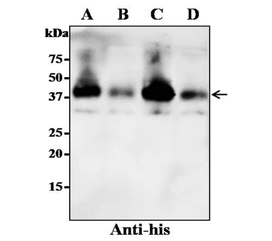
The PRRSV nucleocapsid gene encoding nucleocapsid protein was codon-optimised and cloned into plant expression geminiviral vector pBYR2e. Four different expression vector constructs SP-N-His, N-His, SP-His-N, His-N were constructed that differs in signal peptide and location of histidine tag. The 8x histidine tag either C-terminal or N-terminal was included for detection by western blot. The restriction sites used for cloning respective fragments into plant expression vector pBYR2e are mentioned in the Figure 1. The expression vector contains p19 PTGS suppressor, ER retention signal SEKDEL and the gene of interest was cloned under the transcriptional control of CaMV 35S promoter. The identity of recombinant PRRSV-N protein was confirmed by western blot probed with the anti-histidine antibodies conjugated with HRP. The band size of N-protein (∼37kDa) was observed on the western blot which could possibly be the dimeric form of the N-protein. The results showed that the construct SP-His-N showed high expression compared to other constructs. Hence this construct was used for further experiments.
Expression of recombinant N-protein in N. benthamiana
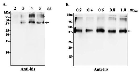
The levels of recombinant protein expression at different time points were evaluated. We evaluated the nucleocapsid expression in plants on 2, 3, 4 and 5 dpi to obtain a time course of expression. The strongest expression was detected on 4 dpi (Figure 2). Similarly, we investigated the effect of Agrobacterium cell density during infiltration; Agrobacterium cell suspension harboring the construct pBYR2e- SP-His-N, at a concentration of OD600 of 0.2, 0.4, 0.6, 0.8, and 1.0 was tested by infiltrating into the leaves of N. benthamiana independently. Following infiltration, the infiltrated plant leaves were collected at 4 dpi and the protein expression was assessed by western blot. As shown in Figure 3, western blot data on the infiltrated samples collected at 4 dpi, showed highest expression at OD600 of 0.6.

Figure 2: Expression of recombinant N-protein in N. benthamiana by using four different expression cassettes. The leaves were infiltrated with A. tumefaciens strain GV3101 containing either one of the four gene constructs. The crude proteins were extracted from the infiltrated leaves and the protein was separated on SDS-PAGE gel and transferred to nitrocellulose membrane. The proteins on the blot were probed with a rabbit polyclonal anti-his antibody conjugated to horseradish peroxidase. Lane A: SP-N-His; Lane B: N-His; Lane C: SP-His-N; Lane D: His-N. The arrowhead indicates the position of the recombinant protein.

Figure 3: Optimization of leaf harvesting time and A. tumefaciens cell density for optimum transgene expression. (A) N. benthamaina leaves were infiltrated with A. tumefaciens containing plasmid pBYR2e-SP-His-N and the leaves were harvested on day 2, 3, 4 and 5 post infiltration. The protein was extracted from the infiltrated leaves and western blot was performed with rabbit polyclonal anti-His antibody conjugated to horseradish peroxidase. (B) N. benthamaina leaves were infiltrated with A. tumefaciens containing plasmid pBYR2e-SP-His-N at OD600 of 0.2, 0.4, 0.6 and 0.8. The leaves were harvested 4 dpi and western blot was performed with rabbit polyclonal anti-his antibody conjugated to horseradish peroxidase. The arrowhead indicates the position of the recombinant protein.

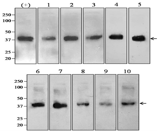
Figure 4: Western blot analysis of plant-produced recombinant N-protein detected with sera collected from pigs. The plant-produced N-protein was separated on SDS-PAGE gel and the proteins were transferred to nitrocellulose membrane. The blot (1-10) was incubated with panel of porcine sera at the dilution of 1:500 and then the bound antibody was detected with anti-porcine IgG antibody conjugated with HRP (1:5000). Commercial porcine anti-PRRSV serum was used as primary antibody in the positive control. The arrowhead indicates the position of the recombinant protein.
Detection of recombiant N-protein with serum specimens from pigs.
The ability of plant-produced recombinant N-protein to use as a diagnostic antigen was demonstrated by western blot. To determine whether the purified plant-produced PRRSV N- protein can detect antibodies in serum from the pig samples, plant-produced N-protein was separated on SDS-PAGE gels and detected with pig sera. All sera samples recognized a dimeric N-protein band at ∼37kDa (Figure 4).
Discussion
PRRSV affect swine health and productivity thereby have great economic and social impact. PRRSV surveillance is essential for early diagnosis, implementing control measures and effective disease management (Nathues et al., 2017). The potent immune response against PRRSV antigens have been documented (Molina et al., 2008) and enzyme-linked immunosorbent assay, immunofluorescence assay, immunoperoxidase monolayer assay based assays are available for detection of antibodies to PRRSV infection in laboratory setting (Ouyang et al., 2013; Seuberlich et al., 2002). N-protein elicits rapid humoral immune response after PRRSV infection. Hence, the available diagnostic tests employ N-protein to detect PRRSV-specific antibodies and regarded as potential biomarker for early diagnosis.
The recombinant antigens are mainly produced in mammalian expression system (Shanmugaraj and Ramalingam 2014). For virological studies, recombinant proteins are widely used as capturing antigens for the diagnosis of many diseases (Loa et al., 2004; Ndifuna et al., 1998; Yu et al., 2006; Marques et al., 2020). Although PRRSV N-protein has been produced in E. coli (Seuberlich et al., 2002), the improper folding and aggregation of recombinant proteins might have an impact on diagnostic utility and assay sensitivity (Peternel and Komel, 2011). Recombinant proteins produced in plants are shown to be properly folded and functional; with rapid scalability, safety, speed and most likely cost saving benefits, plants are considered as alternative protein production platform (Rattanapisit et al., 2020; Schillberg et al., 2019; Nandi et al., 2016; Shanmugaraj et al., 2020b).
Extensive evidences are available that showed the possibility of expressing viral antigens in plants in order to use as a vaccine candidate or diagnostic reagent (Rybicki, 2017; Marques et al., 2020; Shanmugaraj et al., 2020a). In this study, we have successfully cloned the PRRSV N- protein coding gene in plant expression geminiviral vector and expressed the N-protein in N. benthamiana. The expression conditions were optimized and the recombinant N-protein in the plant crude extract was detected with anti-histidine antibodies. The size of the plant-produced N-protein was as expected. The results indicated that the harvesting time of 4 dpi was suitable for maximum protein production and Agrobacterium cell density of 0.6 at OD600 was optimal for infiltration and maximal transgene delivery. Ten sera samples collected from pigs were used to evaluate the diagnostic capability of plant-produced protein. Western blotting confirmed that the recombinant N-protein reacted with antibodies present in pig serum. Specific ∼37kDa bands were obtained with all of the sera tested. Recent studies demonstrated the potential of plant-produced NS1 protein for dengue diagnosis (Xisto et al., 2020; Marques et al., 2020). Similarly, Hepatitis E Virus (HEV) capsid protein and Rift Valley fever virus (RVFV) nucleoprotein produced in N. benthamiana have also shown to detect the infection in the sera collected from respective infected organism (Mbewana et al., 2018; Mazalovska et al., 2017). Our results confirmed that the plant-produced N-protein was intact and truly the recombinant version of PRRSV N-protein having diagnostic potential. The future tasks should be the assessment of diagnostic applicability, specificity, sensitivity of the recombinant plant-produced N-protein in comparison with other methods for PRRSV diagnosis.
In conclusion, we have produced N-Protein of PRRSV in N. benthamiana by using geminiviral vector. Preliminary testing of plant-derived N-protein by western blot showed that the protein was detected by the pig sera suggesting the feasibility of the plant-produced antigen for the diagnosis of PRRSV. However, other parameters including the sensitivity, specificity in comparison with those of other serologic methods needs to be investigated. This proof of concept study helps to utilize plant expression platform for the production of diagnostic antigens against infectious diseases.
Acknowledgement
We would like to thank Professor Hugh Mason (Arizona State University) for providing geminiviral vector. The authors would like to acknowledge Research and Researchers for Industries (RRI)-Thai Science Research and Innovation (TSRI) (MSD62I0091), the 90th Anniversary of Chulalongkorn University Scholarship (B.P), Graduate School, Ratchadapisek Somphot Fund (K.R) and Second Century Fund (C2F) (B.S), Chulalongkorn University for financial support.
Author Contribution
Conceptualization, B.S. and W.P.; literature review, B.S.; methodology: B.P., A.R.A., K.N., P.J., T.H., K.R. and B.S.; writing-original draft preparation, B.S.; writing—review and editing, B.S. and W.P.; supervision, B.S. and W.P.; funding acquisition, W.P. All authors have read and agreed to the published version of the manuscript.
Conflicts of Interest
The authors declare that no conflict of interest.
References


