

Immunolocalization and Expression of Androgen Receptor in the Oviduct of Laying Hens


Immunolocalization and Expression of Androgen Receptor in the Oviduct of Laying Hens
Bian Xunguang1,2, Li Jiancheng1, Xu Chu1 and Yang Li2,3,4,*

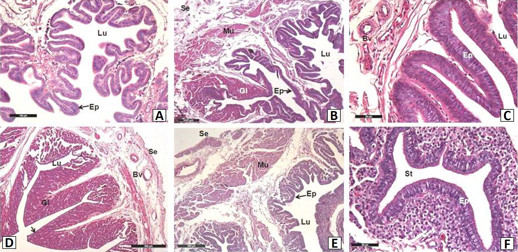
Light micrographs in different regions of the hens oviduct. A, infundibulum; B, magnum; C, isthmus; D, uterus; E, vagina; F, the SST in the anterior vagina. Ep, epithelium; Gl, gland; Lu, lumen; St, sperm-storage tubul; Mu, muscle; Bv, blood vessel; Se, serous. Bar=100 μm (A), bar=500 μm (D), bar=200 μm (B, E), bar=50 μm (C, F).
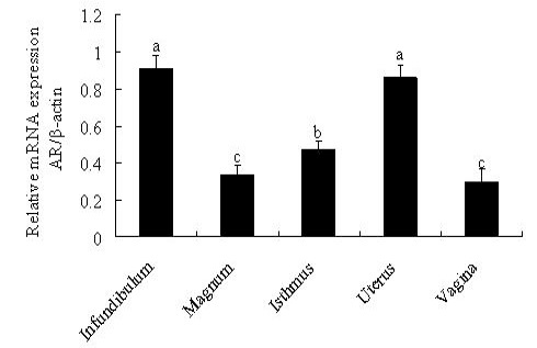
Relative mRNA expression levels of AR in different regions of oviduct, determined by qPCR. The β-actin gene was used as an internal standard. Different letters indicate significant differences between regions (p<0.05). Values represent means ± SE (n=5).

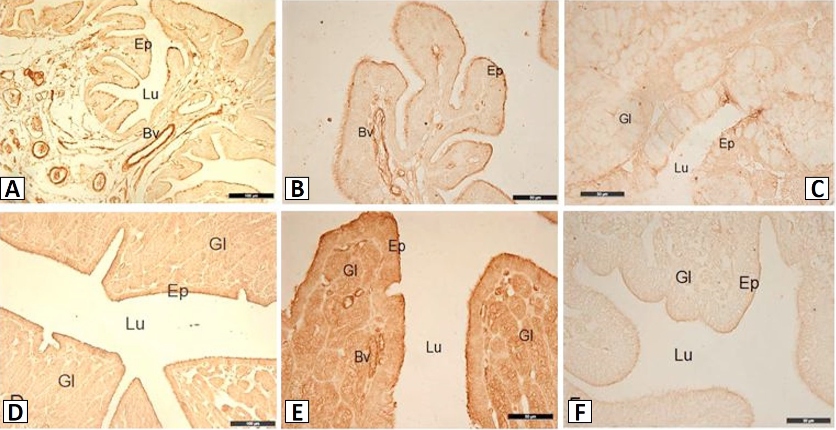
Immunostaining of AR in five regions of the hens oviduct. A and B, infundibulum; C, magnum; D, isthmus; E, uterus; F, vagina. Ep, epithelium; Gl, gland; Lu, lumen; Bv, blood vessel. Bar=100 μm (A, D); bar=50 μm (B, C, E, F).


