Research Article · Volume 14, Issue 3, 17 June 2026 · Open Access
Toxicological Effect of Lead Acetate on the Retina and Brain of Chick Embryo: A Comparative Histometric and Histopathological Study
· · · · · ·
Affiliations & Notes ⌄
1Department of Anatomy and Histology, Faculty of Veterinary and Animal Science, Hajee Mohammad Danesh Science and Technology University, Dinajpur-5200, Bangladesh
2Department of Medicine, Surgery and Obstetrics, Faculty of Veterinary and Animal Science, Hajee Mohammad Danesh Science and Technology University, Dinajpur-5200, Bangladesh. *Correspondence | Md. Abu Hassan, Department of Anatomy and Histology, Hajee Mohammad Danesh Science and Technology University, Dinajpur-5200, Bangladesh
However, there is insufficient data related to the effects of Pb-acetate on the retinal histology of chicken embryos.
Lead (Pb) act as a both retinotoxic and neurotoxic agent and present findings showed that Pb-acetate exposure can be altered neural dentate gyrus and cerebellar thickness with histopathological alterations like hypertrophy, necrotic foci and vacuolation.
On the contrary, all Pb induced group in retina showed altered retinal histology such as detachment of the inner plexiform layer with vacuolation, reduced ganglionic cells etc., that completely indicate the retinotoxic effect.
So, the results of the present experiment clearly indicate that exposure of Pb-acetate causes the significant histo-morphometrical alteration in the developing chick embryo.
Selected from the article's own text.
Abstract
This study was aimed to examine the toxic effects of in ovo lead (Pb)-acetate administration on developing chicken brain and retina advancement using histometric and histological approaches. A total of 150 fertilized eggs were assigned to 4 groups: I (untreated control), II (vehicle control), and III (7 µg/0.1 ml/ egg Pb-acetate); IV (14 µg/0.1 ml/egg Pb-acetate) with at least 3 replicates and incubated for up to 3 days to assess embryo viability. After 48 hours of incubation, the eggs were sterilised properly, and injecting via air sac route, and sealed the hole by using liquid paraffin, then again placed the injected eggs in an incubator. After 12 days of incubation, the eggs were carefully opened, and the pieces of brain and retina tissues were obtained. Moreover, pictures of the intact embryo for gross lesions were taken, and body weight was also recorded. All Pb-acetate treated groups showed the remarkable decrease in body weight (p < 0.05), along with pin point haemorrhages in the neck, body, and limbs. Histologically, the thickness of total retina, thickness of photoreceptor layer (PR), the outer nuclear layer (ONL), the outer plexiform layer (OPL) inner nuclear layer (INL), ganglion cell layer (GL), and nerve fibre layers (NFL), as well as a notable decline in the quantity of ganglion cells (p < 0.05), and degenerative changes such as vacuolar degeneration also noticed in GL, NFL and inner plexiform layer (IPL) respectively. Furthermore, IPL detachment attributed to vacuolation were also observed. In the brain, Pb-acetate-treated groups (III and IV) showed a significant increase in dentate gyrus thickness and granular layer thickness compared with the control (p < 0.05). In contrast, the thickness of white matter (WM) and the molecular layer (ML) was significantly decreased (p < 0.05) in these groups. Additionally, necrotic foci and vacuolations, indicative of degenerative changes, were observed in the ML. Overall, these results clearly indicate that in ovo administration of Pb-acetate adversely affects the development of the retina and brain, disrupts normal embryonic growth, and leads to abnormal embryo development.
Lead (Pb) is a heavy metal and a significant environmental toxicant sometimes found in contaminated water consumed by chicks and other animals. It is a potent neurotoxin that produces behavioral, neurological, and cognitive impairments even at low exposure levels ; ; ; . Pb is considered a global environmental concern that affects both humans and animals due to its persistence in the ecosystem ; ; . Birds may be exposed to Pb from polluted air, water, food, Pb shots, batteries, paint, pesticides, and petrol ; ; . Once absorbed, Pb enters the bloodstream and accumulates in soft tissues such as the liver, kidneys, bones, ovaries, and brain ; ; ; . Even at low concentrations, Pb can disrupt normal physiological functions and cause severe developmental abnormalities, particularly during the early stages of life . Pb passes through the blood-brain barrier (BBB) and blood-retinal barrier (BRB) to reach the brain and retina, often by mimicking calcium ions or interacting with transport proteins ; ; and interferes with cellular processes such as oxidative balance, neurotransmitter regulation, and gene expression. Because the embryonic BBB is still immature, developing embryos are extremely vulnerable to Pb-neurotoxicity. As a result, exposure during embryonic development may Pb to irreversible damage in neural and retinal tissues ; . In addition, Pb has been reported to exert harmful effects on ocular structures, particularly the retina, which is sensitive due to its complex neuronal organization and high metabolic activity. Retinal damage during development may impair visual function ; . Significantly, research on humans has shown that Pb easily moves through the placenta beginning the third month of pregnancy, and its distribution in embryonic organs is quite similar to that of adults . Pb alter can synaptic plasticity and induces neuronal degeneration in several kinds of species, including humans, rodents, and birds ; . Pb is also a genotoxic agent because it destabilizes chromosomes and promotes carcinogenic processes ; ; . For instance, Pb inhibits hippocampus neurogenesis, alters dendritic morphology, increases apoptosis, and decreases cerebellar Purkinje cell density ; . Additionally, studies on rodents show that adolescents exposed to Pb had thinner growth plates, altered trabecular histomorphometry, decreased mechanical endurance, and reduced body and bone mass ; . These results are aligned with research on chicks and birds, where Pb-exposure results in reduced embryo weight, microphthalmia, hydrocephalus, cranioschisis, and overall central nervous system damage ; ; ; ; but still lacking information on comparative histometric and histopathological changes due to the effect of Pb-acetate on the brain and retina of chicken embryo. In chickens, Pb-toxicants have been demonstrated to have adverse effects on early neurological development, especially in the brain and retina. Neurotoxicity during organogenesis is confirmed by histopathological investigations in chick embryos, which show neuronal disintegration, cytoplasmic vacuolation, mitochondrial swelling, and focal hemorrhage in the brain . But there is an inadequate relationship between early neuronal histopathological alteration and the retinal histoarchitecture. The avian central nervous system is highly vulnerable during embryogenesis, as indicated through the evidence that heavy metal exposure can also result in severe brain malformations, abnormal neural tube development, including disrupted neuronal and retinal histoarchitecture, increased cell death and impaired cerebellar formation ; ; ; . Although several studies have documented the neurotoxic effects of Pb-acetate, no detailed information on its dose-dependent histopathological impact and a comparative study on the hippocampus, cerebellum, and, more especially, histological zone-wise alteration in the retina of chick embryos due to Pb-toxicity is still unexplored. Most existing studies focus on general neurotoxicity and retino-toxicity on different animal models in several separate articles, while comparative histological zone/layer-wise histo-microstructural alteration in developing retinal and neural tissues during embryogenesis remains poorly understood. Therefore, further investigation is needed to clarify how different doses of Pb-acetate affect the embryonic retina and brain at the histopathological level. Understanding these histoarchitectural changes is crucial for assessing the developmental risks of Pb-exposure and for establishing safer environmental limits. Accordingly, the current work aims to investigate how different doses of Pb-acetate alter the histo-retinal layers, histopathology, and particular brain regions of chick embryos under in ovo conditions, using a comparative histopathological assessment.
MATERIALS AND METHODs
Experimental design and preparation of test solution
For the experiment, 150 Cobb 500 fertilized eggs (50–55 g), obtained from Cobb 500 laying hens (Gallus gallus domesticus), were sourced from Kazi Farms, Dinajpur, Bangladesh. Lead (Pb)-acetate (≥99% purity) was purchased from Sigma-Aldrich, USA. After that, the fertilized egg in the incubator (HT-352, HITOP®, China) was incubated at 37.5°C with 65% relative humidity for 48hrs for confirming viable embryo by using the candling method. Then Lead (Pb)-acetate doses were fixed for 55 g/egg weight. Pb-acetate was diluted with sterilized distilled water. Pb-acetate doses (7 µg/0.1 ml/ egg and 14 µg/0.1 ml/ egg) were prepared. The eggs were divided into four groups: Group-I (untreated control, 25 eggs), which includes non-treated eggs; Group II (vehicle control, which was injected only with normal saline, 25 eggs); and two Pb-acetate injected groups as Group III (7 µg/0.1 ml/ egg Pb-acetate, 50 eggs); Group IV T2 (14 µg/0.1 ml/egg Pb-acetate, 50 eggs). The number of eggs in each group was used to measure embryonic fatalities.
Treatment administration
After 48 hours of incubation, the eggshell surface was sterilized with 10% povidone iodine, followed by 70% ethyl alcohol solution. All injections were performed in the air sac area, confirmed by light candling. The test solution was injected through a sterile insulin injector after a hole was drilled on the expanded side of the egg (air chamber verified by candling) using a specialized egg driller. The hole was then sealed with liquid paraffin, and the eggs were put back in the incubator under ideal conditions (37.5 ◦C temperature and 65% relative humidity). Up to the 12th day of incubation, the eggs were held at 45-degree turning angles and rotated 12 times per day. The stages of embryos were identified using the Hamburger-Hamilton categorization scheme .
Tissue collection
When the eggs reached the 12th day of incubation, they were removed from the incubator and allowed to equilibrate to room temperature. Embryonic developmental stages were determined using the Hamburger et al. (1992) staging system (H–H scale). A large blood vessel on the chorioallantoic membrane (CAM) was identified by candling, and a small opening was made in the shell membrane to expose the vessel. Anesthetic (sodium pentobarbital) was then injected directly into the CAM vasculature, and the embryo was left for 2–5 minutes. Subsequently, the embryo was carefully removed and placed in clean Petri dishes containing 10% phosphate-buffered saline (PBS) solution. Embryos were decapitated using sterile scissors. Both eyes and the brain were carefully dissected. The anterior segment of the eye and a portion of the brain (hippocampus and cerebellum) were collected and placed into the 10% formalin solution for histological analysis. Photographs of embryos were taken to document gross morphological changes.
Histopathological examination
For an entire day, tissue samples have been preserved in 10% formalin solution. Then, dehydration was performed at an ascending grade of ethanol solution (70%, 80%, 90%, 95%, and 100%), each for one hour. After two changes in xylene for about 90 minutes, the collected specimens were embedded in paraffin. Using a microtome (MU 509, Euromex, Japan), sections with a thickness of 6 µm were created. They were then floated in a water bath at 45°C, mounted carefully on clean glass slides, and stained with Haematoxylin and Eosin (H and E) in compliance with standard methods. Prepared sections were viewed under a Leica DM-2500 type light microscope for histological analysis. An attached DFC-320 model was used to take digital pictures of the necessary regions, which were then examined for histometric measurements. The Leica IM50 measuring program (Leica, Leica Microsystems GmbH, Wetzlar, Germany) was used for all histometric measurements, and numerical data of the parameters under investigation were acquired. A 100 µm line length in one field per section was used to count the number of ganglion cells.
Statistical analysis
The normality of the data was examined by the application of the Shapiro-Wilk test. All the data had an equal variance and were distributed normally. For statistical Analysis, IBM SPSS (Version 27.0, SPSS, IBM Corp., Released 2020, Armonk, NY, USA) was used to measure all the data produced throughout the study. ANOVA and the Tukey test were used to assess the thickness of total retinal, various retinal layer thicknesses, and the quantity of ganglion cells. The results were expressed as Mean ± Standard Deviation (SD), and the level of significance was considered at p<0.05.
RESULTS
Body weight and macroscopic changes
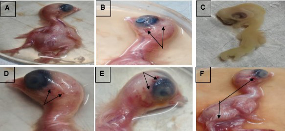
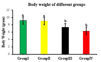
Following 12 days of incubation, the embryos of the Group I and Group II (Figure 1A), displayed normal body weight, growth patterns, and external morphology consistent with their developmental stage. There were no abnormalities or gross lesions found in this group. Reduced growth was accompanied by a number of gross abnormalities. In the Group III and IV, embryos showed almost similar macroscopic changes like as pinpoint haemorrhagic lesions on the eye and hindbrain region (Figure 1B, D, E, F), as well as scattered haemorrhages along the neck, body, and limbs. In case of, Pb treated group few early embryonic mortalities and mal development were also observed (Figure 1C). These results indicate that lead acetate produces reductions in embryo body weight and causes haemorrhagic and developmental abnormalities, with the both treated doses causing the almost severe and similar macroscopic changes. Embryos from Group I and II shows normal and almost similar body weight (Figure 2); but both Group III and IV show a significant (P < 0.05) declined body weight than the unexposed groups (Group I and II) (Figure 2).
Histopathological alterations in the retina
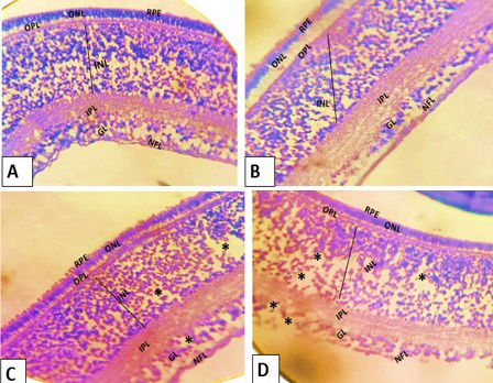
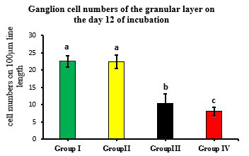
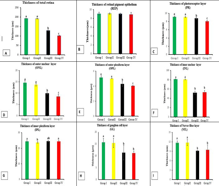
Histopathological examination of chick embryo retinas under 40x magnification and 50µm scale bar showed dose-dependent changes after lead acetate treatment, as evaluated by using haematoxylin and eosin (H and E) staining (Figure 3). The retina was found to have 8 layers on the twelfth day of incubation, arranged from the outer inward.: retinal pigment epithelium (RPE), photoreceptor layer (PR), outer nuclear layer (ONL), outer plexiform layer (OPL), inner nuclear layer (INL), inner plexiform layer (IPL), ganglion cell layer (GL), and nerve fibre layer (NFL) (Figure 3). In the Group I and Group II, both retinal histology showed a normal retinal layered structure with intact all cell layers (Figure 3A, B). The ganglion cell count was significantly (p < 0.05) reduced in the GL layer (Figure 4) and this layer was vacuolated and reduced (black star in Figure 3C, D) and for all Pb-acetate treated groups (Group III and Group IV) (Figure 4). In the Pb-acetate treated group (Group III and Group IV), the total retinal thickness decreased significantly (p < 0.05) compared with the Control (Group I and II) (Figure 5A), the thickness of the different retinal layers, especially the PR, ONL, OPL, INL, GL, and NFL layers also decreased significantly (p < 0.05, Figure 5). Furthermore, in these groups’ retinal degeneration such as IPL layer detachment (Figure 3C, D) and vacuolization in the IPL, INL, GL, and NFL layers were also observed (Figure 3C, D).
Histopathological alterations in the brain hippocampus
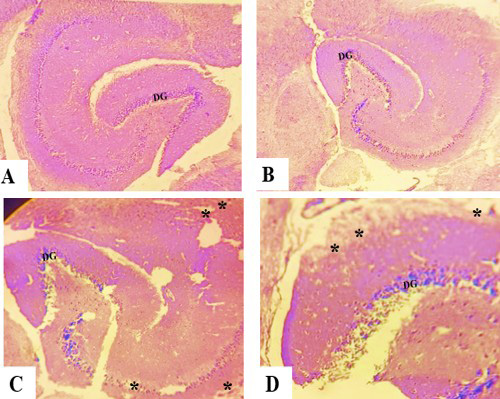
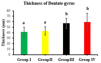
In Group I and Group II, the hippocampus showed normal histological features, including intact neuronal organization and absence of necrosis or vascular abnormalities. The thickness of the dentate gyrus (DG) was also normal; however, the cornu ammonis (CA) region was not clearly visible at day 12 of chick embryo development (Figure 6A–D). There were severe histological alterations in the both Group III and IV shown the cellular changes such as vacuolation, cell swelling, hypertrophy suggested that starting early reactive activity (star marks) and also increased the thickness (Figure 6C–D). The thickness of DG of Group III shown increased significantly (p < 0.05) and similar to the Group IV (Figure 7). These results suggested that lead acetate dosages cause neuroinflammatory reactions and progressive neuronal damage in the hippocampus.
Histopathological alterations in the cerebellum
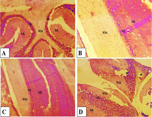
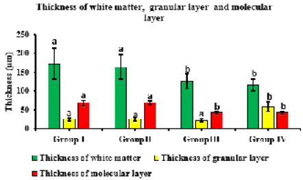
In the Group I and Group II, the cerebellum showed normal histoarchitecture, with intact White Matter (WM), Molecular Layer (ML), and Granular Layer (GL) (Figure 8A, B and 9). The Group III, showed the significantly decreased (p < 0.05) thickness of WM of both Group III and Group IV (Figure 9). Additionally, a nearly similar thickness of the granular layer (GL) was observed among Groups I, II, and III, whereas Group IV showed a significant increase (p < 0.05) in thickness compared with the other groups (I–III) (Figure 9). Furthermore, the decreased ML thickness of Group III and Group IV were shown statistically significant (p < 0.05) than the Group I and Group II (Figure 9). In Group IV, more severe histological alterations were observed, including vacuolation (black star) and necrotic foci (white star) (Figure 8D), which were absent in Groups I, II, and III. These findings suggest that higher levels of lead acetate induce neurotoxic effects, including neuroinflammation, and disrupt normal cerebellar development.
DISCUSSION
A frequent model used to study embryotoxicity is chicken embryos ; Bölükba et al., 2021), neurotoxicity ; ; ; , eye diseases , retinal development , and retinal pathologies . Therefore, chicken embryos are good models for the investigation of the effects of many environmental pollutants during embryogenesis because embryonic development is fully described and the individual developmental stages are clearly visible and easily accessible ; . Other than the exchange of gasses, the chicken egg is a closed system with no interaction with its surroundings. The embryonic nervous system and retina is more vulnerable to heavy metal than the mature brain and retina which can lead to persistent structural and functional damage ; ; ; ; Szabo et al., 2024; ; . Environmental exposure to heavy metals like Lead (Pb) is therefore of particular concern, as it has been strongly associated with neurodevelopmental abnormalities and cognitive impairments in children as well as in experimental animal models ; ; ; ; ; ; Bölükba et al., 2023). Early embryonic exposure to metals can have teratogenic, neurotoxic, and growth-retarding effects, according to recent reviews that emphasized on avian embryos ; ; ; ; Bölükba et al., 2023) that is strongly aligned with our investigation. Pb hinders embryonic growth by disrupting nutrient utilization, energy metabolism, and hepatic and gastrointestinal function. Lead also reduces skeletal growth and overall body mass by interfering with chondrocyte and osteoblast activity; these results are consistent with earlier studies demonstrating reduced growth performance and body weight in animals exposed to lead ; ; ; ; ; ; . Furthermore, lead inhibits cell proliferation and activates apoptosis in rapidly dividing embryonic tissues, which may cause developmental arrest or early embryonic mortality ; .
Heavy metal can alter the retinal histopathology by inducing retinal toxicity ; . Numerous investigations have looked at the typical retinal anatomy of chickens ; . However, there is insufficient data related to the effects of Pb-acetate on the retinal histology of chicken embryos. It was also noted that in the Pb-treated groups, total retinal thickness decreased significantly (p < 0.05, Figure 5A) on the incubation days 12 examined compared with the control groups, and thickness of the PR, ONL, OPL, INL, GL, and NFL layers on day 12, was significantly reduced (p < 0.05, Figure 5C-F, H-I) and aligned with group of researches finding . These Pb-acetate groups showed that degenerative changes, such as vacuolar degeneration and INL detachment, and markedly reduced ganglion cell numbers in comparison to the control groups (p < 0.05, Figure 4). These observations were consistent with rat, mouse, and chicken studies ; ; Al-Thanoon et al., 2021; ; . These findings are all related to the monosodium glutamate or glutamate and cadmium, but their effects are very much aligned and strongly support our investigation. In the human model, the toxic effect of heavy metal could damage the retinal pigmented epithelium, which is contradict with our present research . It may be due to the experimental model, doses of exposure, environmental factor etc. Researchers discovered that congenital abnormalities and alterations in neural retinal thickness were brought on by electromagnetic mobile radiation in chick embryos at 7, 10, and 14 days of incubation , which is aligned with our present investigation, although we didn’t apply any radiation; their findings and histo-morphometrical changes parallel our observation. In earlier research on the retina of chick embryos, monosodium glutamate was implicated in causing damage to the inner retinal layer and ganglion cells ; ; , which strongly supports our observation.
Comparable neurodevelopmental damage has been shown in chick embryos exposed to other environmental toxicants ; ; ; demonstrated clear morphological signs of neurotoxicity, including neuronal degeneration and altered tissue organization, in chick embryos exposed to perfluorooctanoic acid and inorganic cadmium. These results support the concept that immature neural tissues, including the retina, are especially susceptible to harmful insults during embryogenesis. The brain is particularly vulnerable to Pb toxicity because of its high metabolic requirement and extended developmental period ; ; ; ; . The enhanced neuronal degeneration and necrotic foci found at higher doses may be responsible for the activation of apoptotic pathways, which may be mediated by oxidative stress. Similar hippocampal abnormalities and long-term neurobehavioral deficits have been reported following developmental lead exposure in both avian and mammalian models ; ; ; ; ; . Pb directly impairs adult neural precursor cells from the dentate gyrus, increasing apoptosis and reducing proliferation and neuronal differentiation via JNK and p38 MAPK activation, all this activity caused a thinner dentate gyrus ; ; , which is completely contradict with our research. It may be due to hypertrophy or swelling, gliosis or the early reactive phase etc.
In this study, it was observed that the thickness of the dentate gyrus of the hippocampus significantly increases (p < 0.05, Figure 2) compared to the control. Whereas three different layers of cerebellum (ML, WM, GL) on day 12 of incubation were significantly decreased (p < 0.05, Figure 2) than in Groups I and II. The Cerebellar histo-alterations, such as changes in thickness, degeneration, vacuolation and pyknosis and neuronal death, have been reported following lead exposure group, this all observations are similar with our present investigation ; ; ; ; ; ; ; and in other avian metal-toxicity studies ; consistent with our present findings.
CONCLUSION
Lead (Pb) act as a both retinotoxic and neurotoxic agent and present findings showed that Pb-acetate exposure can be altered neural dentate gyrus and cerebellar thickness with histopathological alterations like hypertrophy, necrotic foci and vacuolation. On the contrary, all Pb induced group in retina showed altered retinal histology such as detachment of the inner plexiform layer with vacuolation, reduced ganglionic cells etc., that completely indicate the retinotoxic effect. Since the data obtained from studies using chicken embryos can also be adapted to mammalian, the outcomes of this experiment revealed that animals as well as humans exposed to Pb at the time of prenatal stage may have increased susceptibility to certain neuronal and ophthalmic diseases during their lifetime. So, the results of the present experiment clearly indicate that exposure of Pb-acetate causes the significant histo-morphometrical alteration in the developing chick embryo.
ACKNOWLEDGEMENTS
This research work was contributed by Research and Development project (RandD), Ministry of Science and Technology (MOST), Government of the People’s Republic of Bangladesh, Bangladesh Secretariat, Dhaka-100, Bangladesh.
NOVELTY STATEMENT
This is the first research to present a dose-dependent histometric and histopathological comparison of lead acetate-induced developmental toxicity in the retina and particular brain regions (hippocampus and cerebellum) of chick embryos. We hope, these results will contribute in a new perception to the heavy metal toxicity. Given these results, our study adds a new perception to literature. It shows that even low-dose of Pb exposure interferes with the formation of the different retinal layers development and also causes neuroinflammation, hypertrophy and vacuolation in the brain during embryogenesis.
AUTHOR’S CONTRIBUTION
MAH: Conceptualization, funding, original manuscript writing, investigation, formal analysis supervision, experimental design, validation histopathological interpretation. MH: Formal analysis, investigation, histopathology, writing review and editing. BKD: Formal analysis, investigation, writing review and editing. IH: Visualization, writing review and editing, methodology. NI: Visualization, writing review and editing. TR: Investigation, writing review and editing, methodology. SI: Visualization, validation, writing review and editing.
Ethical approval
This research work was conducted at the Laboratory of Histology, Department of Anatomy and Histology, Hajee Mohammad Danesh Science and Technology University, Dinajpur-5200, Bangladesh. All experimental protocols adhered to ethical requirements for animal research, and approval was secured from the Institutional Animal Ethics Committee (IEC). The approval number was HSTU/VAS/ANH-1071, dated: 01-06-2023 (Resolution No: 10(a)). In accordance with best practices and the VAS Guidelines for the Euthanasia of Animals, all experimental animals were handled humanely, and euthanasia procedures were carried out to minimize pain and distress as much as possible.
Generative AI and AI-assisted technology statement
The authors confirm that the manuscript was prepared under the authors responsibility, and any generative AI or AI-assisted tools, if used, were only for language improvement. The authors take the copyright and full responsibility for the content.
Conflict of interest
The authors have declared no conflict of interest.
Figures
Figure 1:.
Gross morphology and body weight of chick embryos after 12 days of incubation. A: normal development in Group I and II; B, D, E, and F: reduced body weight with pinpoint haemorrhages (black arrow) in Group III IV; C: severe abnormalities, embryonic death, and haemorrhages in Group IV.
Figure 2:.
Body weight of different groups at day 12 of incubation period. (a–b) Various superscript letters on the bar-column indicate a statistical difference (mean ± SD, p < 0.05).
Figure 3:.
Sections from the retina of the Groups I (A), II (B), III (C), and IV (D) at day 12 of incubation. Blak stars (*): vacuole formation in the IPL, INL, GL, and NFL layers; retinal pigment epithelium (RPE); nerve fibre layer (NFL); ganglion cell layer (GL); inner plexiform layer (IPL); inner nuclear layer (INL); outer plexiform layer (OPL); outer nuclear layer (ONL). H and E staining. (Magnification: 40x and Scale bar: 50μm).
Figure 4:.
Quantification of granular layer ganglion cells on day 12 of incubation (calculated by counting cells along a 100 μm line length in one field per tissue section).
(a–c) Columns with different superscript letters indicate significant differences (mean ± SD, p < 0.05).
Figure 5:.
Comparison of the thicknesses of the retinal layer (μm) in chicken embryos from Group I, Group II, Group III, and Group IV on the twelfth day of incubation. Total retinal thickness (A); thickness of RPE (B); thickness of PR (C); thickness of ONL (D); thickness of OPL (E); thickness of INL (F); thickness of IPL (G); thickness of GL (H); thickness of NFL (I). There is a statistical difference shown by different superscript letters (a–c) on the columns. (mean ± SD, p < 0.05).
Figure 6:.
Sections from the hippocampus of the Groups I (A), II (B), III (C), and IV (D) at day 12 of incubation. Blak stars (*): Degeneration and necrotic changes in the Dentate Gyrus (DG) layers; H and E staining. (40x magnification and Scale bar: 50μm).
Figure 7:.
Comparison of dentate gyrus layer thicknesses (μm) among Group I, Group II, Group III, and Group IV on day 12 of incubation in chicken embryos. There is a statistical difference shown by different superscript letters (a–b) on the columns (mean ± SD, p < 0.05).
Figure 8:.
Histopathological alteration of cerebellum of the brain due to different doses of Lead Acetate. Showing Brain Cerebellum -White matter (WM), Molecular layer (ML), Granular layer (GL) (40x resolution, Scale bar: 50)
Figure 9:.
Comparison of white matter, granular layer and molecular layer thicknesses (μm) among Group I, Group II, Group III, and Group IV on day 12 of incubation in chicken embryos. There is a statistical difference shown by different superscript letters(a–b) (mean ± SD, p < 0.05).
References
Ahmad W, Kanwal M, Inayat I, Ahmad S, Batool A, Ghazanfar N, Idrees R, Suleman S, Younis A, Ahmad K (2024). Protective role of vitamin B6 against teratogenic effects induced by lead in chick embryo. Birth Defect. Res., pp. 116. https://doi.org/10.1002/bdr2.2416[CrossRef]
Ali HS, El-Gohary AA, Metwally FG, Sabra NM, El-Sayed AA (2012). Mono sodium glutamate induced damage in rabbit retina: Electroretinographic and histologic studies. Glob. J. Pharmacol., 6(3): 148–159.
Aljohani A (2023). Heavy metal toxicity in poultry: A comprehensive review. Front. Vet. Sci., 10. https://doi.org/10.3389/fvets.2023.1161354[CrossRef]
Al-Khafaf A, Ismail H, Alsaidya A (2021). Histopathological effects of experimental exposure to lead on nervous system in albino female rats. Iraqi J. Vet. Sci., 35(1): 45-48. https://doi.org/10.33899/ijvs.2019.126248.1273[CrossRef]
Al-Qudsi FM, Azzouz SO (2012). Effect of electromagnetic mobile radiation on chick embryo development. Life Sci. J., 9(2): 983–991
AlThanoon SA, Abd A (2021). Histopathological changes induced by monosodium glutamate and sodium nitrite in the development of eye in albino mice Mus musculus. J. Educ. Sci., 30(3): 1–12. https://doi.org/10.33899/edusj.2021.168650[CrossRef]
Amini Z, Mahdavi-Shahri N, Lari R, and Behnam Rassouli F (2019). The effects of lead on the development of somites in chick embryos (Gallus gallus domesticus) under in vitro conditions: A histological study. Toxicol. Res., 8(3): 373–380. https://doi.org/10.1039/C8TX00340H[CrossRef]
Aschner M, Skalny AV, Paoliello MMB, Tinkova MN, Martins AC, Santamaria A, Lee E, Rocha JBT, Farsky SHP, Tinkov AA (2024). Retinal toxicity of heavy metals and its involvement in retinal pathology. Food Chem. Toxicol., 188: 114685. https://doi.org/10.1016/j.fct.2024.114685[CrossRef]
Atallah MN, Badawy GM, El-Garawani IM, Abdallah FS, El-Borm HT (2021). Neurotoxic effect of nalufin on the histology, ultrastructure, cell cycle and apoptosis of the developing chick embryo and its amelioration by selenium. Food Chem. Toxicol., 158: 112693. https://doi.org/10.1016/j.fct.2021.112693[CrossRef]
Barkur RR, Bairy LK (2016). Histological study on hippocampus, amygdala and cerebellum following low lead exposure during prenatal and postnatal brain development in rats. Toxicol. Ind. Health, 32(6): 1052–1063. https://doi.org/10.1177/0748233714545624[CrossRef]
Basha R, Reddy GR (2010). Developmental exposure to lead and late life abnormalities of nervous system. Indian J. Exp. Biol., 48(7): 636–641.
Blanks JC, Reif-Lehrer L, Casper DS (1981). Effects of monosodium glutamate on the isolated retina of the chick embryo as a function of age: A morphological study. Exp. Eye Res., 32(1): 105-124. https://doi.org/10.1016/S0014-4835(81)80044-9[CrossRef]
Bölükbaş F, Öznurlu Y (2021). Determination of the effects of monosodium glutamate administered to eggs on embryonic development of the cervical region of the spinal cord in chicken embryos. J. Ad. Vet. Biol. Sci. Tech., 6(3): 298-311. https://doi.org/10.31797/vetbio.1015200[CrossRef]
Bölükbaş F, Öznurlu Y (2023). Determining the effects of in ovo administration of monosodium glutamate on the embryonic development of brain in chickens. Neurotoxicology, 94: 87–97. https://doi.org/10.1016/j.neuro.2022.11.009[CrossRef]
Bölükbaş F, Öznurlu Y (2023). Investigation of the effects of monosodium glutamate on the embryonic development of the eye in chickens. Vet. Sci., 10(2): 99. https://doi.org/10.3390/vetsci10020099[CrossRef]
Bölükbaş F, Öznurlu Y (2025). Evaluation of the effects of monosodium glutamate on the development of the cerebellum in chicken embryos. J. Mol. Histol., 56(4): 236. https://doi.org/10.1007/s10735-025-10522-3[CrossRef]
Burger J (1995). A risk assessment for lead in birds. J. Toxicol. Environ. Health., 45(4): 369–396. https://doi.org/10.1080/15287399509532003[CrossRef]
Collin MS, Venkatraman, SK, Vijayakumar N, Kanimozhi V, Arbaaz SM, Stacey RGS, Anusha J, Choudhary R, Lvov V, Tovar GI, Senatov F, Koppala S, Swamiappan S (2022). Bioaccumulation of lead (Pb) and its effects on humans: A review. J. Hazard. Mater. Adv., 7(2): 100094. https://doi.org/10.1016/j.hazadv.2022.100094[CrossRef]
Da Costa M, Kmecick M, De Freitas P, Ortolani-Machado C (2021). Lead exposure affects cephalic morphogenesis and neural crest cells in Gallus gallus embryo. Neurotoxicol. Teratol., 106948. https://doi.org/10.1016/j.ntt.2021.106948[CrossRef]
da Silva AS, Santos RMS, De Marco PG, Rezende VHM, Martins TC, Silva JR, Romano-Silva MA, Miranda DM (2025). Neurodevelopmental outcomes associated with early-life exposure to heavy metals: A systematic review. Int. J. Environ. Res. Publ. Health, 22(8): 1308. https://doi.org/10.3390/ijerph22081308[CrossRef]
de Figueiredo FA, Gerlach RF, da Veiga MA, Nakadi FV, Ramos J, Kawakita ER, Guerra CS, Issa JP (2014). Reduced bone and body mass in young male rats exposed to lead. Biomed. Res. Int., 2014: 571065. https://doi.org/10.1155/2014/571065[CrossRef]
Earl R, Burns N, Nettelbeck T, Baghurst P (2016). Low-level environmental lead exposure still negatively associated with children’s cognitive abilities. Aust. J. Psychol., 68(2): 98–106. https://doi.org/10.1111/ajpy.12096.[CrossRef]
El-Beltagy AEFBM, Abdelaziz KK, Saleh AMB, Elsayyad HIH, Gahnem RA (2022). Evaluation of lead toxicity on the retina of pregnant rats and their pups: the possible ameliorative role of pomegranate juice. F1000Research, 11: 461. https://doi.org/10.12688/f1000research.110028.1[CrossRef]
El-Gohari KM, Bahei-Eldin IA, Habib EM, Saad SA, Rady HY, Said AM (2016). Neuroprotection of the rat’s retinal ganglion cells against glutamate-induced toxicity. J. Egypt. Ophthalmol. Soc., 109(3): 135-144. https://doi.org/10.4103/2090-0686.202261[CrossRef]
Engstrom A, Wang H, Xia Z (2015). Lead decreases cell survival, proliferation, and neuronal differentiation of primary cultured adult neural precursor cells through activation of the JNK and p38 MAP kinases. Toxicol. In Vitro, 29(5): 1146-1155. https://doi.org/10.1016/j.tiv.2015.05.001[CrossRef]
Eriksson P (1997). Developmental neurotoxicity of environmental agents in the neonate. Neurotoxicology, 18(3): 719–726.
Generalova A, Davidova S, Satchanska G (2025). The mechanisms of lead toxicity in living organisms. J. Xenobiot., 15(5): 146. https://doi.org/10.3390/jox15050146.[CrossRef]
Ghazanfar N, Kanwal M, Inayat I, Ahmad S, Batool A, Ahmad W, Zafar R, Idrees R, Suleman S, Ahmad K (2025). Ameliorative potentials of Methylcobalamin Vit B12 against teratogenic effects induced by lead in chick embryo. Sci. Rep., 15(1): 35789. https://doi.org/10.1038/s41598-025-16447-x[CrossRef]
Gonick HC (2011). Lead-binding proteins: A review. J. Toxicol., 2011: 686050. https://doi.org/10.1155/2011/686050[CrossRef]
Hamburger V, Hamilton HL (1992). A series of normal stages in the development of the chick embryo. Dev. Dyn., 195(4): 231–272. https://doi.org/10.1002/aja.1001950404[CrossRef]
Hemmaphan S, Bordeerat NK (2022). Genotoxic effects of lead and their impact on the expression of DNA repair genes. Int. J. Environ. Res. Publ. Health., 19(7): 4307. https://doi.org/10.3390/ijerph19074307[CrossRef]
Herbert MR (2010). Contributions of the environment and environmentally vulnerable physiology to autism spectrum disorders. Curr. Opin. Neurol., 23(2): 103–110. https://doi.org/10.1097/WCO.0b013e328336a01f[CrossRef]
Hocking PM, Guggenheim JA (2013). The chick as an animal model of eye disease. The chick as an animal model of eye disease. Drug Discov. Today Dis. Models, 10(4): e225-e230. https://doi.org/10.1016/j.ddmod.2014.02.003[CrossRef]
Hussain S, Ali S, Mumtaz S, Shakir HA, Ahmad F, Tahir HM, Ulhaq M, Khan MA, Zahid MT (2020). Dose and duration-dependent toxicological evaluation of lead acetate in chicks. Environ. Sci. Pollut. Res. Int., 27(13): 15149–15164. https://doi.org/10.1007/s11356-020-08016-8[CrossRef]
Jessl L, Lenz R, Massing F. G, Scheider J, Oehlmann J (2018). Effects of estrogens and antiestrogens on gonadal sex differentiation and embryonic development in the domestic fowl (Gallus gallus domesticus). PeerJ, 6: e5094. https://doi.org/10.7717/peerj.5094[CrossRef]
Khandokar L, Liu L. L, Zheng W, Kerstein PC (2025). Lead (Pb) exposure results in cell type specific changes in the mouse retina and optic nerve. bioRxiv, 114: 103461. https://doi.org/10.1016/j.neuro.2026.103461[CrossRef]
Khoshakhlagh AH, Mohammadzadeh M, Gruszecka-Kosowska A (2024). The preventive and carcinogenic effect of metals on cancer: A systematic review. BMC Public Health, 24(1): 2079. https://doi.org/10.1186/s12889-024-19585-5[CrossRef]
Kmecick M, Vieira da Costa MC, Oliveira RCA, Ortolani-Machado CF (2019). Morphological evidence of neurotoxic effects in chicken embryos after exposure to perfluorooctanoic acid (PFOA) and inorganic cadmium. Toxicology, 427: 152286. https://doi.org/10.1016/j.tox.2019.152286[CrossRef]
Lazarova I, Balieva G (2025). Effects of lead exposure in wild birds as causes for incidents and fatal injuries. Diversity, 17(6): 387. https://doi.org/10.3390/d17060387[CrossRef]
Li Y, Liang J, Jiang C, Cui J, Hong L, Hao Z, Tang Y, Liu Y, Cui X, and Teng X (2024). Se Alleviated Pb-Caused Neurotoxicity in Chickens: SPS2-GPx1-GSH-IL-2/IL-17-NO Pathway, Selenoprotein Suppression, Oxidative Stress, and Inflammatory Injury. Antioxidants (Basel), 13(3): 370. https://doi.org/10.3390/antiox13030370[CrossRef]
Lidsky TI, Schneider JS (2003). Lead neurotoxicity in children: basic mechanisms and clinical correlates. Brain, 126(Pt 1): 5–19. https://doi.org/10.1093/brain/awg014[CrossRef]
Moayed AA, Hariri S, Song ES, Choh V, Bizheva K (2011). In vivo volumetric imaging of chicken retina with ultrahigh-resolution spectral domain optical coherence tomography. Biomed. Opt. Express., 2(5): 1268–1274. https://doi.org/10.1364/BOE.2.001268[CrossRef]
Narbaitz R, Marino I, Sarkar K (1985). Lead-induced early lesions in the brain of the chick embryo. Teratology, 32(3): 389–396. https://doi.org/10.1002/tera.1420320309[CrossRef]
Pamphlett R, Cherepanoff S, Too LK, Kum Jew S, Doble PA, Bishop DP (2020). The distribution of toxic metals in the human retina and optic nerve head: Implications for age-related macular degeneration. PLoS One, 15(10): e0241054. https://doi.org/10.1371/journal.pone.0241054.[CrossRef]
Parithathvi A, Choudhari N, Dsouza HS (2024). Prenatal and early life lead exposure induced neurotoxicity. Hum. Exp. Toxicol., 43: 9603271241285523. https://doi.org/10.1177/09603271241285523[CrossRef]
Patrick L (2006). Lead toxicity, a review of the literature. Part 1: Exposure, evaluation, and treatment. Altern. Med. Rev., 11(1): 2–22.
Raj K, Das AP (2023). Lead pollution: Impact on environment and human health and approach for a sustainable solution. Environ. Chem. Ecotoxicol., 5: 79-85. https://doi.org/10.1016/j.enceco.2023.02.001[CrossRef]
Ramírez OD, González EDF, Blanco AT, Pineda B, Gómez MS, Marcial QJ, Carrillo MP, Pérez CV (2021). Cognitive Impairment Induced by Lead Exposure during Lifespan: Mechanisms of Lead Neurotoxicity. Toxics, 9(2): 23. https://doi.org/10.3390/toxics9020023[CrossRef]
Rana MN, Tangpong J, Rahman MM (2018). Toxicodynamics of lead, cadmium, mercury and arsenic-induced kidney toxicity and treatment strategy: A mini review. Toxicol. Rep., 5: 704–713. https://doi.org/10.1016/j.toxrep.2018.05.012[CrossRef]
Reif-Lehrer L, Bergenthal J, Hanninen L (1975). Effects of monosodium glutamate on chick embryo retina in culture. Invest. Ophthalmol., 14(2): 114–124.
Rice D, Barone S, Jr (2000). Critical periods of vulnerability for the developing nervous system: Evidence from humans and animal models. Environ. Health Perspect., 108(Suppl 3): 511–533. https://doi.org/10.1289/ehp.00108s3511[CrossRef]
Ridgway LP, Karnofsky DA (1952). The effects of metals on the chick embryo: toxicity and production of abnormalities in development. Ann. N. Y. Acad. Sci., 55(2): 203–215. https://doi.org/10.1111/j.1749-6632.1952.tb26536.x[CrossRef]
Schettler T (2001). Toxic threats to neurologic development of children. Environ. Health Perspect., 109(Suppl 6): 813–816. https://doi.org/10.1289/ehp.01109s6813.[CrossRef]
Shafiq A, Mahnoor Baig M, Fatima Y, Haer A, Parveen S, Tariq M, Khan M (2023). Teratogenic effects of lead in the developing chick embryos. Markhor, 4(1): 16-20. https://doi.org/10.54393/mjz.v4i01.63[CrossRef]
Shibuya K, Tomohiro M, Sasaki S and Otake S (2015). Characteristics of structures and lesions of the eye in laboratory animals used in toxicity studies. J. Toxicol. Pathol., 28:181-188. https://doi.org/10.1293/tox.2015-0037[CrossRef]
Sobieniecki A, Gutowska I, Machalińska A, Chlubek D, Baranowska-Bosiacka I (2015). Retinal degeneration following lead exposure functional aspects. Postepy. Hig. Med. Dosw., 69: 1251-8. https://doi.org/10.5604/17322693.1180649[CrossRef]
Szabó R, Budai P, Juhász É, Major L, Lehel J (2024). Potential Teratogenicity effects of metals on avian embryos. Int. J. Mol. Sci., 25(19): 10662. https://doi.org/10.3390/ijms251910662[CrossRef]
Tobalu FO, Enogieru AB (2025). Lead neurotoxicity in experimental models: A systematic review on effects on the cerebrum, cerebellum, and hippocampus. Toxicol. Rep., 14: 102044. https://doi.org/10.1016/j.toxrep.2025.102044[CrossRef]
Tomaszewska E, Dobrowolski P, Winiarska-Mieczan A, Kwiecień M, Tomczyk A, Muszyński S, Radzki R (2016). Alteration in bone geometric and mechanical properties and histomorphometrical parameters of trabecular bone, articular cartilage, and growth plate in adolescent rats after chronic co-exposure to cadmium and lead in the case of supplementation with green, black, red, and white tea. Environ. Toxicol. Pharmacol., 46: 36–44. https://doi.org/10.1016/j.etap.2016.06.027[CrossRef]
Trejo-Reveles V, McTeir L, Summers K, Rainger J (2018). An analysis of anterior segment development in the chicken eye. Mech. Dev., 150: 42–49. https://doi.org/10.1016/j.mod.2018.03.001[CrossRef]
Vergara MN, Canto-Soler MV (2012). Rediscovering the chick embryo as a model to study retinal development. Neural Dev., 7: 22. https://doi.org/10.1186/1749-8104-7-22[CrossRef]
Wang H, Matsushita M (2021). Heavy metals and adult neurogenesis. Curr. Opin. Toxicol., 26: 14-21. https://doi.org/10.1016/j.cotox.2021.03.006[CrossRef]
Yu G, Wu L, Su Q, Ji X, Zhou J, Wu S, Tang Y, Li H (2024). Neurotoxic effects of heavy metal pollutants in the environment: Focusing on epigenetic mechanisms. Environ. Pollut., 345: 123563. https://doi.org/10.1016/j.envpol.2024.123563[CrossRef]
Zahoor S. M, Ishaq S, Ahmed T (2024). Neurotoxic effects of metals on blood brain barrier impairment and possible therapeutic approaches. Vitam. Horm., 126: 1–24. https://doi.org/10.1016/bs.vh.2024.04.003[CrossRef]
Zareen N, Khan MY, Minhas LA (2011). Histological stages of retinal morphogenesis in chicken: A descriptive laboratory research. Ital. J. Zool., 78(1): 45–52. https://doi.org/10.1080/11250003.2010.487075[CrossRef]
Zhang Y, Pei X, Jing L, Zhang Q, Zhao H (2024). Lead induced cerebellar toxicology of developmental Japanese quail (Coturnix japonica) via oxidative stress-based Nrf2/Keap1 pathway inhibition and glutathione-mediated apoptosis signaling activation. Environ. Pollut., 352: 124114. https://doi.org/10.1016/j.envpol.2024.124114[CrossRef]
Zhou F, Xie J, Zhang S, Yin G, Gao Y, Zhang Y, Bo D, Li Z, Liu S, Feng C, Fan G (2018). Lead, cadmium, arsenic, and mercury combined exposure disrupted synaptic homeostasis through activating the Snk-SPAR pathway. Ecotoxicol. Environ. Saf., 163: 674-684. https://doi.org/10.1016/j.ecoenv.2018.07.116[CrossRef]